Biology › Practicals › Required practical 5: dissecting an insect and a fish gill
Required practical 5: dissecting an insect and a fish gill
A locust carries air to its tissues in a branching system of tubes, the tracheae, and a fish passes water over gill filaments covered in lamellae. Both can be opened up in a lesson, traced with a hand lens and recorded in a drawing. The measurements that turn a drawing into a record are the magnification and the scale bar.
The capability this develops
This guide is one investigation under capability P08: Dissection and biological drawing, which sets out the techniques the investigation belongs to, the calculations it carries and the errors that cost marks in all of them. The notes behind the biology are in Exchange surfaces and gas exchange.
Where this sits on the specifications
This library maps a qualification to its own specification one numbered heading at a time, against a document whose sha256 is recorded, and publishes the result as a route map. These are the headings whose practical work this investigation serves, on two of the five qualifications mapped so far. Each links to the route map the row belongs to.
| Qualification | Heading |
|---|---|
| OCR H020/H420 | 3.1.1 Exchange surfaces |
| Pearson Edexcel Biology B 8BI0/9BI0 | 4.3 i Gas exchange |
Some boards number their practical activities, and those lists were read here from the same specifications as the headings above. This investigation is numbered on them as:
- AQA 7401/7402 required practical 5
- OCR Biology A H020/H420 PAG 2
- Pearson Edexcel Biology B 8BI0/9BI0 core practical 7
Use your awarding organisation’s current specification and its practical handbook as the authority on which activities your course requires and how they are assessed.
The principle
An insect's tracheae open at the surface through the spiracles and branch into ever finer tracheoles that end among the cells, so oxygen reaches each cell as a gas, with no blood to carry it. A fish's gills are four arches on each side, each carrying two rows of filaments, and each filament is covered in thin plates, the lamellae, where the blood comes close to the water.
The spiracles are a row of small pores along each side of the abdomen, with more on the thorax. On the slide the trachea shows a spiral thickening of chitin, the taenidia: the rings described above, seen close up. It holds the tube open so that air can move along it, while letting it bend as the body moves.
In life, water passes between the lamellae from the mouth side of the arch to the opercular side, while blood in the lamellae flows the other way. That is the counter-current arrangement taught above. The dissection shows the surface but not the flow.
Ethics. Insects come from a supplier, are killed humanely and are used only in the numbers the work needs. A fish head is left over from fish sold as food. A student who objects can watch a dissection, work from photographs or video, or draw a prepared slide of trachea; tell your teacher beforehand.
- Dorsal
- The back or upper surface of an animal, opposite the ventral surface.
- Spiracle
- One of the pores in an insect's body wall through which air enters and leaves the tracheal system.
- Magnification
- The size of an image or drawing divided by the real size of the object, with both in the same units.
Apparatus
- The instruments: a dissecting board or a wax-lined dish, a scalpel, fine scissors, blunt and fine forceps, a seeker, dissecting pins, a hand lens or stereo microscope, a light microscope with slides and coverslips, and a ruler.
- A locust or cockroach from a biological supplier, and a fish head from a fishmonger
- A dish of water, and a dropping pipette
- An eyepiece graticule and a stage micrometer, to measure the trachea under the microscope
- Plain paper, a sharp HB pencil, a ruler and an eraser
- Disposable gloves, disinfectant, and the sharps container
Safety
Risk assessment. Cut on the board, away from your body, your other hand and other people. Pass instruments handle-first, fit and remove blades only by your school's method, and put used blades in the sharps container. Handle animal material as the risk assessment directs, keep it away from your face and food, and dispose of it as the school says. Anyone allergic to locusts or fish should say so before the practical. Disinfect the bench and instruments and wash your hands afterwards. Check the CLEAPSS guidance and your school's risk assessment before starting; where they differ from this page, follow them.
Variables
Independent variable
None. A dissection is a careful observation, not an experiment: nothing is changed to see what happens. If two specimens are compared, such as the gills of two species, the specimen is the thing that differs between them.
Dependent variable
What is recorded: the structures found, drawn as they are on this specimen, and the measurements that give each drawing its magnification.
Control variables
- The calibration. The eyepiece graticule calibrated against the stage micrometer for the objective in use, since a division is worth a different distance at each magnification.
- The units. Real and drawn lengths in the same unit before one is divided by the other.
- The state of the specimen. Kept under water, so tracheae and filaments are seen as they were and not dried and collapsed.
- The drawing rules. The same rules for every drawing, so that two drawings can be compared.
Method
Opening the locust
- Take a locust or cockroach from a biological supplier, killed by the method your school approves. Cut off the wings and legs, lay it dorsal side up in the wax dish and pin it through the thorax, the pins angled outwards. Angled pins hold the body and leave room to work.
- With fine scissors, cut along the dorsal midline from the tip of the abdomen to the head, the lower blade just under the exoskeleton and its point kept up. A shallow cut leaves the tracheae beneath intact.
- Pin the exoskeleton back on each side and cover the specimen with water. Water supports the tracheae and stops them drying.
Tracing and mounting a trachea
- With a hand lens, find the silvery tubes running in from the side walls and trace one to a spiracle, moving the gut aside with the seeker. The tracheae are silvery because they are full of air.
- Cut a short length of trachea free, mount it in water under a coverslip and examine it with the light microscope.
- The slide shows the chitin in the wall of the trachea, which a hand lens cannot.
- Measure the width of the trachea with the calibrated eyepiece graticule, and note the objective you measured it with.
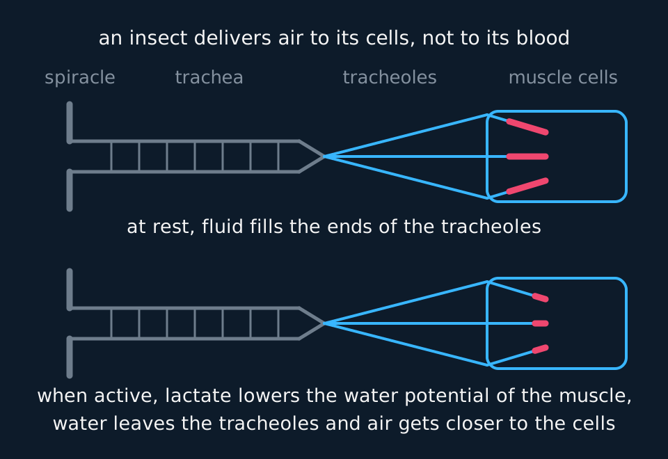
Describe the drawing
Two rows showing the same tracheal system. In each, a pore in the body wall leads into a wide ringed tube that branches into three fine tracheoles ending inside a block of muscle. In the upper row a thick coloured length of fluid fills most of each tracheole tip; in the lower row that fluid has shrunk to a short stub near the very end.
The fish gill, and recording both
- Use a fish head from a fishmonger.
- Lift the operculum with forceps and cut it away at its base to expose the four gill arches.
- Cut one arch free at both ends and put it in a dish of water.
- Under a hand lens or stereo microscope the filaments spread out in two rows, with the lamellae as fine plates across each filament.
- Lift the arch out of the water for a moment and watch the filaments, then return it.
- Make a biological drawing: a sharp pencil, single unbroken lines, no shading or colour, and labels on ruled lines that do not cross and carry no arrowheads.
- Keep the parts in proportion.
- Draw the branches you found, not the textbook pattern.
Sample data
These measurements are idealised. They were chosen to show how a drawing's magnification is worked out, not measured on a specimen, and a real locust or gill will differ. Nothing here is a record of a dissection.
| Structure | How it was measured | Real size | Drawn size | Magnification of the drawing |
|---|---|---|---|---|
| Locust, head to tip of abdomen | Ruler | 50 mm | 150 mm | ×3.0 |
| One gill arch | Ruler | 38 mm | 114 mm | ×3.0 |
| One gill filament | Ruler, under the hand lens | 6.0 mm | 90 mm | ×15 |
| Width of a trachea | Eyepiece graticule, ×10 objective, 1 division = 10 µm, 16 divisions | 160 µm | 80 mm | ×500 |
Magnification is the drawn length divided by the real length, in the same units. For the gill filament that is 90 mm ÷ 6.0 mm = 15, so the drawing is at ×15.
The trachea needs a change of unit first. 16 divisions × 10 µm = 160 µm, which is 0.16 mm, and 80 mm ÷ 0.16 mm = 500. Divide 80 by 160 without converting and the answer, 0.5, is a thousand times too small.
A scale bar does the same job and survives the drawing being photocopied larger or smaller. On the trachea drawing at ×500, a bar standing for 100 µm is 100 µm × 500 = 50 000 µm long, which is 50 mm.
Reading the drawing
Measure a real structure, such as one filament, and add a scale bar or the magnification, which is drawn length divided by real length.
What the drawing should show. On the locust: the main tracheae running along each side, the branches from them into the tissues, and the spiracles where they open. On the gill: the arch, the two rows of filaments, and, on a drawing of one filament at higher magnification, the lamellae across it. Each label names a structure; an annotation says what it does.
The trachea on the slide. The spiral thickening is the feature to draw and label. It is what keeps the tube open when the air pressure inside falls, which a tube of soft tissue alone could not do.
The filaments in air. Lifted out of water, the filaments cling together into a mass. That is the observation that explains why a fish out of water dies of lack of oxygen even though air holds more oxygen than water does.
Lifted into air, the filaments stick together: water no longer holds them apart, and the film of water between them draws them together. This is the loss of surface area that suffocates a stranded fish.
What it cannot show. Neither specimen is alive, so nothing about ventilation, the flow of water or the movement of fluid in the tracheoles can be seen. A drawing of the dead tissue records structure only.
Evaluation
Each of these is a real source of error in this investigation, and the middle column is the part an answer earns marks for: what it does to the result, and in which direction.
| Source of error | What it does to the result | What to do about it |
|---|---|---|
| Tracheoles are under a micrometre across and end among the muscle fibres | Only the larger tracheae can be traced | Say on the drawing that the finest branches were not seen, rather than drawing them from memory, and view a prepared slide of flight muscle for the tracheoles. |
| The specimen is dead | No ventilation or tracheal fluid can be observed | Watch a living locust's abdomen pump before the dissection, or use video of ventilation, and record that as a separate observation. |
| Tracheae tear, and gills are damaged in gutting | The drawing undercounts branches and lamellae | Keep the cut shallow and the point of the scissors up, keep the specimen under water, and choose an undamaged arch. |
| A drawing made from the textbook diagram | It shows the expected branching pattern, not this specimen's, so it records nothing that was found. | Draw only what is in front of you, and label any structure that could not be found as not seen. |
| Magnification worked out in mixed units | Dividing millimetres by micrometres gives an answer wrong by a factor of a thousand, in either direction. | Convert both lengths to one unit before dividing, and check the answer is about the size expected. |
Questions
Question 1 (2 marks)
Explain why the opened locust is covered with water.
Check your answer
the water supports the tracheae so they float apart and can be traced;
and stops them drying out and collapsing.
Question 2 (2 marks)
The tracheae look silvery in the dissection. Explain why.
Check your answer
they are full of air;
and the air in the tubes reflects light.
Question 3 (3 marks)
A trachea measured 16 eyepiece divisions wide, where one division is 10 µm. It was drawn 80 mm wide. Calculate the magnification of the drawing.
Check your answer
real width = 16 × 10 = 160 µm;
= 0.16 mm (or 80 mm = 80 000 µm);
magnification = 80 ÷ 0.16 = ×500.
Question 4 (3 marks)
Explain why gill filaments stick together when a gill is lifted out of water, and why this matters to a fish.
Check your answer
out of water there is nothing to hold the filaments apart, and the film of water between them draws them together;
so the surface area of the lamellae exposed for gas exchange is greatly reduced;
too little oxygen diffuses into the blood and the fish suffocates.
Question 5 (2 marks)
State two features of a good biological drawing of the gill filaments.
Check your answer
single, unbroken pencil lines with no shading;
labels on ruled lines that do not cross (accept: a scale bar or magnification; parts drawn in proportion).
Coverage maps are available for AQA 7401/7402, OCR Biology A H020/H420, Cambridge International 9700, Pearson Edexcel Biology A (Salters-Nuffield) 8BN0/9BN0 and Pearson Edexcel Biology B 8BI0/9BI0. Use the relevant route map to check what InkBiology covers, and your awarding organisation’s current specification as the final authority on examinable content.