Biology › Hormonal communication, plant responses and homeostasis › Regulation of blood glucose and diabetes mellitus
Regulation of blood glucose and diabetes mellitus
Blood glucose is held near 5 mmol dm⁻³ through meals, fasting and exercise. Insulin from beta cells and glucagon from alpha cells act antagonistically on the liver to maintain it, and diabetes mellitus is a persistent failure of that control, with distinct causes in its two common forms.
Before this Negative feedback and antagonistic effectors · Adrenaline, cyclic AMP and the second messenger model · Carbohydrates and glycogen
COMMON MISCONCEPTION
Insulin converts glucose into glycogen.
Insulin binds to receptors and changes enzyme activity. In muscle and adipose tissue it increases insertion of GLUT4 transporters; in liver and muscle it promotes glycogen synthesis, and it also increases glucose use. Insulin is the signal rather than the enzyme that converts glucose.
What you should be able to do
- State the normal blood glucose concentration and name the three places glucose in the blood comes from.
- Describe the effects of insulin and of glucagon, naming the cells that secrete each.
- Use glycogenesis, glycogenolysis and gluconeogenesis correctly.
- Describe how a beta cell secretes insulin, from glucose entering to vesicles fusing.
- Distinguish type 1 from type 2 diabetes by cause, and say how each is managed.
- Explain how a glucose test strip and a biosensor work, naming glucose oxidase and peroxidase.
- Read a glucose tolerance test curve and justify a diagnosis from it.
The variable and the limits it is held within
Blood glucose sits at about 90 mg per 100 cm³, which is roughly 5 mmol dm⁻³, and a healthy person stays between about 4 and 6 between meals. Glucose reaches the blood by three routes: absorption from the gut after a meal, glycogenolysis in the liver, and gluconeogenesis, the making of glucose from non-carbohydrate sources such as amino acids, lactate and glycerol.
Both directions of error are dangerous, and for different reasons. Too low, and the brain is in trouble within minutes: nervous tissue respires glucose almost exclusively and stores none, so confusion, unconsciousness and death follow quickly. Too high, and the blood's water potential falls, water leaves cells by osmosis, and glucose appears in the urine because the kidney's carrier proteins are saturated. The glucose drags water out with it, causing the thirst and constant urination that give diabetes its name.
Three terms are used throughout this topic and differ by only a few letters.
- Glycogenesis
- The synthesis of glycogen from glucose, in liver and muscle cells. Promoted by insulin.
- Glycogenolysis
- The breakdown of glycogen to glucose. Promoted by glucagon and by adrenaline.
- Gluconeogenesis
- The production of glucose from non-carbohydrate sources such as amino acids and glycerol, in the liver. Promoted by glucagon.
Two cell types in the same tissue
The islets of Langerhans are patches of endocrine tissue scattered through the pancreas, and each contains two kinds of cell that matter here. Beta cells, the larger group and mostly central in the islet, detect a rise in blood glucose and secrete insulin. Alpha cells, fewer and towards the edge, detect a fall and secrete glucagon. Both hormones go straight into the blood, and both find their main target in the liver.
In a stained section of the pancreas the islets are easy to pick out. Each is a rounded, paler-staining cluster of small cells with capillaries running between them, surrounded by the darker-staining acini, the groups of exocrine cells arranged round tiny ducts that make up most of the tissue.
Describe the drawing
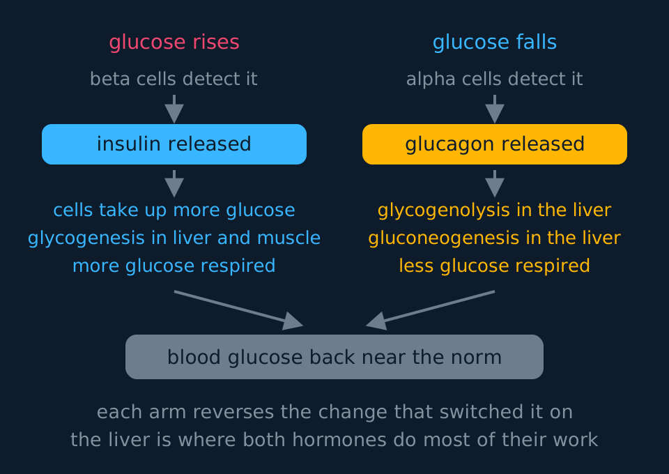
A control loop with two symmetrical arms. On the left a rise in blood glucose is detected by beta cells, insulin is released, cells take up more glucose, glycogenesis stores it and more is respired. On the right a fall is detected by alpha cells, glucagon is released, glycogenolysis and gluconeogenesis in the liver add glucose to the blood and less is respired. Both arms converge on a bar showing the concentration returned to its norm.
Insulin lowers blood glucose in several ways at once. It binds to receptors on liver, muscle and adipose cells. In muscle and adipose cells, vesicles carrying glucose transporter proteins fuse with the cell surface membrane, so more glucose can enter by facilitated diffusion; liver cells keep their transporters in place and respond by trapping and using the glucose that enters. It activates the enzymes of glycogenesis, so glucose entering the liver and muscle is stored as glycogen. It raises the rate of respiration of glucose, and it promotes the conversion of glucose into fat. At the same time it inhibits the alpha cells, so the opposing signal is switched off.
In the A-level model of blood-glucose control, glucagon raises blood glucose mainly by acting on hepatocytes. Binding to receptors on hepatocytes triggers the cyclic AMP cascade, glycogen phosphorylase is activated and glycogenolysis releases glucose, and the enzymes of gluconeogenesis are activated as well. Adrenaline produces the same two effects through the same second messenger, in response to a different stimulus.
Skeletal muscle glycogen supplies the muscle itself. Muscle cells lack glucose-6-phosphatase and therefore do not release free glucose into the blood, so the organ that raises blood glucose is the liver.
How a beta cell counts glucose
A beta cell converts the glucose concentration outside it into a quantity of hormone released. Exam-board note: only OCR A examines this mechanism. The sequence accounts for insulin secretion being graded to the glucose concentration rather than switching on and off.
Describe the drawing
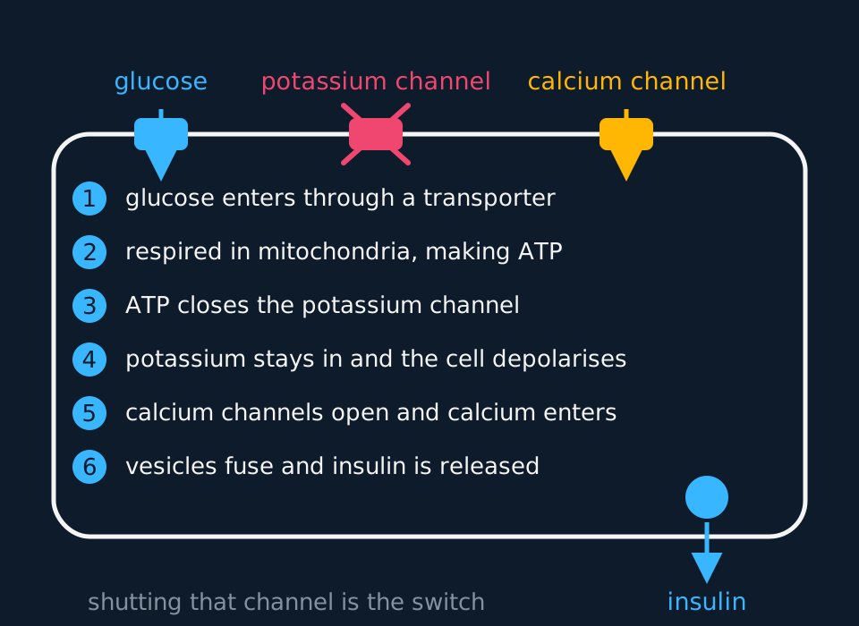
A beta cell drawn as a large outlined cell with three channels set into its upper membrane: a glucose transporter with glucose entering, a potassium channel crossed out to show it shut, and a calcium channel with calcium entering. Six numbered steps run down the inside of the cell from glucose entering to vesicles fusing, and a vesicle at the lower right releases insulin through the membrane into the blood.
At rest, potassium channels in the beta cell membrane are open and potassium ions diffuse out, holding the inside at about −70 mV. When blood glucose rises, more glucose enters the cell through its transporter, is phosphorylated and respired, and the concentration of ATP in the cytoplasm rises.
ATP binds to the potassium channel and closes it. Potassium can no longer leave, so positive charge builds up inside and the membrane depolarises. The depolarisation opens voltage-gated calcium channels, calcium ions diffuse in, and, exactly as at a synapse, the calcium causes vesicles of insulin to fuse with the cell surface membrane and release their contents by exocytosis.
Because ATP is produced in proportion to the glucose the cell is respiring, the quantity of insulin secreted is graded to the concentration outside. The cell does not detect glucose with a surface receptor; it respires the glucose that enters, and the resulting ATP concentration controls the potassium channel.
Type 1 and type 2 diabetes
Diabetes mellitus is a persistently raised blood glucose concentration, and its two common forms share that feature but differ in cause. Distinguish them by cause rather than by age at onset: age is a tendency and not a definition, since type 2 is diagnosed in teenagers and type 1 can begin in middle age.
| Type 1 | Type 2 | |
|---|---|---|
| Cause | Beta cells destroyed by the body's own immune system | Cells respond poorly to insulin; secretion may also fall |
| Insulin present? | Little or none produced | Produced, sometimes in excess, but the receptors respond weakly |
| Onset | Usually rapid, often in childhood or adolescence | Usually gradual, more often later in life |
| Risk factors | Genetic susceptibility; some viral triggers proposed | Obesity, especially around the abdomen; inactivity; diet; age; family history |
| Management | Insulin injections matched to meals and exercise | Diet, weight loss and exercise first; drugs; sometimes insulin |
| Diet alone? | Never sufficient | Often sufficient at first |
Two points about treatment follow from the biology. Insulin must be injected because it is a protein: swallowed, it would be hydrolysed by proteases in the stomach and small intestine to amino acids. And an injected dose is fixed in advance, whereas a beta cell adjusts its secretion continuously, which is why doses must be matched to meals and exercise.
Most insulin used now is human insulin made by genetically modified bacteria carrying the human gene. It has the same amino acid sequence as the body's own, so it causes fewer immune reactions than the pig or cattle insulin used before, and it can be made in large amounts at a steady quality. A possible future treatment for type 1 is to grow beta cells from stem cells and transplant them. The obstacles are rejection of the new cells, the same autoimmune attack destroying them again, the risk that undifferentiated cells form tumours and, for embryonic stem cells, ethical objections.
Type 2 shows why insulin must be described as a signal rather than as an enzyme. If insulin converted glucose into glycogen directly, insulin resistance could not occur. Because insulin is a signal, a cell can have plenty of the hormone around it and still fail to respond: the receptors and the signalling chain downstream of them respond weakly, so the transporters are not brought to the membrane, and over the years the beta cells themselves begin to fail. Glucose therefore stays in the blood while the pancreas secretes increasing amounts of insulin.
Exam-board note: AQA asks you to evaluate the positions of health advisers and the food industry on the rising number of people with type 2 diabetes. Health advisers link the rise to obesity, to diets high in sugar and refined starch, and to inactivity, and argue for less sugar in processed food, smaller portions, clearer labels and taxes on sugary drinks. Food companies have argued that what people eat is their own choice, that inactivity matters as much as diet, and that changing recipes and labels is enough. To evaluate either side, ask who paid for the evidence, whether it shows a correlation or a cause, and whether that side gains from its conclusion.
Measuring the glucose: strips and biosensors
Managing diabetes depends on measuring blood glucose, and a person with type 1 diabetes may need to do so several times a day. The measurement must be quick, must work on a drop of blood from a fingertip, and must respond to glucose specifically. Benedict's test meets none of those requirements: it takes minutes, requires heating, is at best semi-quantitative, and responds to any reducing sugar, so fructose or lactose would also give a positive result.
Specificity is obtained from an enzyme, whose active site is complementary to glucose alone. A test strip carries two immobilised enzymes and a colourless dye. Glucose oxidase catalyses the oxidation of glucose to gluconic acid, and that reaction also produces hydrogen peroxide. Peroxidase, the second enzyme, then catalyses a reaction between the hydrogen peroxide and the colourless dye, called a chromogen, which turns it into a coloured product. The more glucose there was, the more hydrogen peroxide is made and the deeper the colour, so the strip is compared against a printed chart and read as a range. The same chemistry on a different strip tests urine for glucose, which is how a clinic screens quickly for diabetes.
Two properties of that chain are worth stating in an answer. It is specific, because only glucose fits the active site of glucose oxidase, so no other sugar in the sample produces any colour at all. And it is only semi-quantitative, because a colour matched by eye against a chart gives a band rather than a number, and two people can read the same strip differently.
A biosensor removes the eye from the process. The same glucose oxidase is immobilised on an electrode inside a small meter; the oxidation of glucose releases electrons, the electrode collects them, and the current that flows is proportional to the concentration of glucose in the sample. A processor converts the current into a number on a display, in millimoles per cubic decimetre, within a few seconds and from a drop of blood a millimetre across. A numerical reading, available as often as required, is what allows an insulin dose to be matched to a meal.
- Biosensor
- A device that uses a biological molecule, usually an immobilised enzyme, to detect a specific substance and convert its concentration into an electrical signal.
- Chromogen
- A colourless substance that is converted into a coloured product, so that a reaction can be followed by eye.
Reading a glucose tolerance test
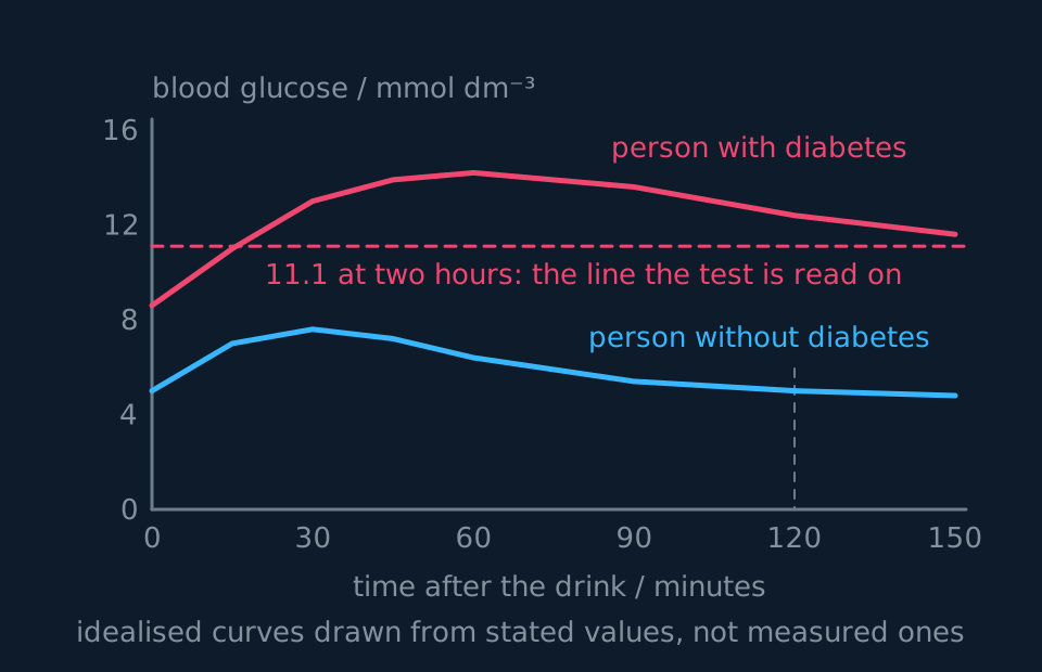
The test is standardised so that curves can be compared. The person fasts overnight, a fasting blood glucose is measured, they drink a solution containing 75 g of glucose, and blood glucose is measured at intervals for two hours. Diagnosis rests on two numbers: a fasting concentration of 7.0 mmol dm⁻³ or more, or a two-hour concentration of 11.1 mmol dm⁻³ or more. These are the World Health Organization diagnostic thresholds. They apply to venous plasma measured in a laboratory, and in a person without symptoms diagnosis requires a repeat test on another day rather than a single reading.
Describe the drawing
Two blood glucose curves plotted against time after a glucose drink. The lower cyan curve starts near five, peaks under eight at about half an hour and is back to its starting value by two hours. The upper coral curve starts near nine, climbs past fourteen and is still above twelve at two hours, well above the dashed diagnostic line drawn at eleven point one.
Rate of fall against the two-hour value
Between 60 and 120 minutes the person without diabetes falls from 6.4 to 5.0 mmol dm⁻³, and the person with diabetes falls from 14.2 to 12.4. A student concludes that the diabetic pancreas is clearing glucose faster. Calculate both rates, and say what is wrong with the conclusion.
Show the working
For the person without diabetes: a fall of 6.4 − 5.0 = 1.4 mmol dm⁻³ in 60 minutes, so the rate is 1.4 ÷ 60 = 0.023 mmol dm⁻³ min⁻¹.
For the person with diabetes: 14.2 − 12.4 = 1.8 mmol dm⁻³ in 60 minutes, so the rate is 1.8 ÷ 60 = 0.030 mmol dm⁻³ min⁻¹. The arithmetic supports the student: that is the faster fall.
The conclusion is still wrong, because rate of fall is not what the test measures. Some of the loss is glucose leaving in the urine rather than being taken into cells, and a concentration that starts at 14.2 has further to fall. What matters is where the curve ends up: at 120 minutes the diabetic value is 12.4, above the 11.1 threshold, while the other has returned to its fasting value.
A tolerance test measures whether the concentration is brought back under control within two hours, so quote the two-hour value and compare it with the threshold.
TRY IT: Explaining a symptom from first principles
An untreated person with type 1 diabetes produces large volumes of urine containing glucose, feels constantly thirsty and loses weight despite eating normally. Explain all three observations.
Check your answer
Glucose in the urine comes from saturation of the reabsorption mechanism. Glucose is filtered at the glomerulus and normally reabsorbed completely at the proximal convoluted tubule by carrier proteins. When the filtrate contains far more glucose than usual, every carrier is occupied, and the glucose that cannot be reabsorbed passes on into the urine.
The large volume follows from that glucose. Glucose left in the tubule lowers the water potential of the filtrate, so less water is reabsorbed by osmosis along the tubule and collecting duct, and a greater volume of urine is produced. Call it copious rather than dilute: the extra water is being held in the tubule by dissolved glucose, so the urine is glucose-rich and its overall concentration can still be high.
The thirst is the consequence of losing that water. Blood water potential falls, osmoreceptors in the hypothalamus detect it, and thirst is triggered, while ADH release also rises, which is why the urine is not as dilute as the volume alone would suggest.
The weight loss comes from the cells being unable to use the glucose. With little or no insulin, glucose transporters are not brought into the membranes of muscle and fat cells, so glucose cannot be taken up and respired. The body respires fat and protein instead, so mass is lost even though glucose is abundant in the blood.
In the exam
- Name the cells rather than the pancreas alone. Beta cells secrete insulin, alpha cells secrete glucagon, and both are in the islets of Langerhans.
- Insulin does not convert glucose to glycogen. It binds to receptors and activates the enzymes that do, and in muscle and adipose cells it increases the number of glucose transporters in the membrane.
- Spell the three 'glyco' terms carefully and use each in the correct direction: glycogenesis and glycogenolysis are opposite processes.
- Glucagon raises blood glucose by acting on hepatocytes. Muscle does not release glucose into the blood, so it cannot raise the blood glucose concentration.
- Separate the two diabetes types by cause, not by age: destroyed beta cells against a poor response to insulin.
- In a tolerance test question, quote figures from the graph with their units, and give the two-hour value against the 11.1 mmol dm⁻³ threshold rather than describing the shape of the line.
- For a test strip, name both enzymes and the order in which they act: glucose oxidase produces hydrogen peroxide, then peroxidase uses it to colour the chromogen. State that the specificity comes from the active site of glucose oxidase.
Check yourself
A person has a fasting blood glucose concentration of 8.2 mmol dm⁻³ and a two-hour value of 13.5 in a glucose tolerance test. Their blood contains a higher concentration of insulin than a person without diabetes. Which type of diabetes do they have? Justify your answer from both pieces of evidence, and explain why their pancreas is secreting more insulin than normal.
Answer
Both test values are diagnostic. A fasting concentration of 8.2 is above the 7.0 threshold, and a two-hour value of 13.5 is above 11.1, so the person has diabetes.
The high insulin concentration identifies it as type 2. In type 1 the beta cells have been destroyed by the immune system, so there would be little or no insulin in the blood. Insulin is present here in more than the usual amount, so the beta cells are working.
The cause is therefore a poor response to insulin rather than a shortage of it. The target cells have fewer receptors, or receptors that respond weakly, so fewer glucose transporters are moved into their membranes and less glucose is taken up and stored, and the concentration in the blood stays high.
That persistently high concentration is what drives the extra secretion. Beta cells respond to the glucose around them: more glucose entering means more ATP, the potassium channels stay shut, the cell stays depolarised and more insulin is secreted. The negative feedback loop is intact at the pancreas and broken at the target cell, so the correction is not made.
Questions
Question 15 marks
Describe how a rise in blood glucose concentration causes a beta cell in an islet of Langerhans to secrete insulin.
Mark scheme
- B1 more glucose enters the beta cell through its transporter protein and is phosphorylated and respired
- B1 the concentration of ATP in the cytoplasm therefore rises, in proportion to how much glucose the cell is respiring
- B1 ATP binds to the potassium channels in the cell surface membrane and closes them, so potassium ions can no longer diffuse out
- B1 positive charge builds up inside and the membrane depolarises, which opens voltage-gated calcium channels and calcium ions diffuse in
- A1 the calcium causes vesicles of insulin to fuse with the cell surface membrane and release their contents by exocytosis
Question 25 marks
A person has a single fasting blood glucose reading of 7.3 mmol dm⁻³, just above the 7.0 mmol dm⁻³ diagnostic threshold, and no symptoms. Their doctor immediately diagnoses type 2 diabetes on this result alone. Evaluate this diagnosis.
Mark scheme
- B1 in support: 7.3 mmol dm⁻³ fasting is above the World Health Organization threshold of 7.0 mmol dm⁻³, the internationally used cut-off for a raised fasting glucose
- B1 in support: blood glucose is normally held within a narrow range by insulin and glucagon acting antagonistically, so a reading persistently above that range does suggest the control mechanism has failed
- B1 against: in a person without symptoms, diagnosis should rest on a repeat test on a different day rather than a single reading, because glucose concentration varies with recent food, illness, stress and timing
- B1 against: a reading only just above the threshold, with no symptoms such as thirst or weight loss reported, gives less certainty than a reading far above it or one accompanied by symptoms
- B1 judgement: the diagnosis is premature; the result is concerning enough to warrant repeating the test on another day before diabetes is confirmed, rather than diagnosing from one borderline reading alone
Question 34 marks
Explain how insulin lowers the concentration of glucose in the blood, making clear why it is wrong to say that insulin converts glucose into glycogen.
Mark scheme
- B1 insulin binds to receptors on liver, muscle and adipose cells; it is a signal and never touches a glucose molecule, and the conversions are done by enzymes inside those cells
- B1 vesicles carrying glucose transporter proteins fuse with the cell surface membrane, so more glucose enters by facilitated diffusion
- B1 insulin activates the enzymes of glycogenesis, so glucose entering liver and muscle cells is stored as glycogen, and it also raises the rate of respiration of glucose and promotes its conversion into fat
- A1 it inhibits the alpha cells at the same time, so the opposing glucagon signal is switched off and less glucose is released from the liver
Question 44 marks
Compare type 1 diabetes with type 2 diabetes, referring to the cause of each, the insulin present in the blood, how each is usually managed and why age at onset does not separate them.
Mark scheme
- B1 in type 1 the beta cells have been destroyed by the person's own immune system, whereas in type 2 the target cells respond poorly to insulin and secretion may fall later
- B1 in type 1 little or no insulin is produced, whereas in type 2 insulin is produced, sometimes in excess, but the receptors respond weakly to it
- B1 type 1 must be managed with insulin injections matched to meals and exercise, whereas type 2 is usually managed first by diet, weight loss and exercise, with drugs and sometimes insulin later
- B1 the two are separated by cause and not by age: type 2 is regularly diagnosed in teenagers and type 1 can begin at fifty
Question 54 marks
In a glucose tolerance test a person's blood glucose concentration is 5.6 mmol dm⁻³ when fasting, rises to 9.8 mmol dm⁻³ at 30 minutes, and has returned to 5.6 mmol dm⁻³ at 120 minutes. Calculate the percentage increase in concentration between fasting and 30 minutes, and calculate the mean rate of fall between 30 and 120 minutes.
Mark scheme
- M1 the increase is 9.8 − 5.6 = 4.2 mmol dm⁻³
- M1 percentage increase is the increase divided by the starting value and multiplied by 100, so 4.2 ÷ 5.6 × 100
- A1 75 per cent
- A1 the fall of 4.2 mmol dm⁻³ takes 90 minutes, so the mean rate is 4.2 ÷ 90 = 0.047 mmol dm⁻³ min⁻¹
Question 64 marks
Insulin has to be injected rather than swallowed. Suggest why, and suggest why a person with type 2 diabetes can often control their blood glucose by diet and exercise alone while a person with type 1 cannot.
Mark scheme
- B1 insulin is a protein, and a protein that is swallowed is hydrolysed by proteases in the stomach and small intestine
- B1 it would be broken down into amino acids, which are absorbed but have none of the effects of the hormone, so it must enter the blood without passing through the gut
- B1 in type 2 diabetes the beta cells still secrete insulin, so reducing the glucose load of meals and losing weight can improve how well the target cells respond and let the person's own insulin cope
- A1 in type 1 the beta cells have been destroyed, so there is no insulin to improve the response to, and the hormone itself has to be replaced
Question 74 marks
Explain how a test strip carrying immobilised glucose oxidase and peroxidase measures the concentration of glucose in a drop of blood, and explain why the strip does not respond to other sugars.
Mark scheme
- B1 glucose oxidase catalyses the oxidation of glucose to gluconic acid, and hydrogen peroxide is also produced
- B1 peroxidase then catalyses a reaction between that hydrogen peroxide and a colourless chromogen, turning it into a coloured product
- B1 the more glucose there was, the more hydrogen peroxide is made and the deeper the colour, which is compared with a printed chart, so the reading is a range rather than an exact value
- A1 only glucose is complementary to the active site of glucose oxidase, so no other sugar in the sample is oxidised and none produces any colour
Question 84 marks
Compare the two responses in the table, quoting figures, and explain what person B's results suggest about their control of blood glucose.
| Time after the glucose drink / min | Person A: blood glucose / mmol dm⁻³ | Person B: blood glucose / mmol dm⁻³ |
|---|---|---|
| 0 | 4.8 | 8.2 |
| 30 | 7.6 | 13.4 |
| 60 | 6.4 | 14.6 |
| 90 | 5.4 | 13.8 |
| 120 | 4.9 | 12.6 |
| 150 | 4.8 | 11.4 |
Mark scheme
- B1 person B starts at 8.2 mmol dm⁻³ against 4.8 for person A, so B's fasting concentration is already well above the normal range
- B1 B peaks higher and later, at 14.6 mmol dm⁻³ at 60 min against 7.6 at 30 min for A
- B1 A is back to its starting concentration by 150 min, whereas B is still at 11.4 mmol dm⁻³ and has fallen by only 3.2 from its peak
- A1 B's glucose is removed from the blood slowly, so B either secretes too little insulin or B's cells respond poorly to it; glucose values alone cannot tell type 1 from type 2
Question 94 marks
Explain why a blood glucose concentration far below normal is dangerous within minutes, and explain why a blood glucose concentration far above normal is dangerous only over a longer period.
Mark scheme
- B1 nervous tissue respires almost exclusively on glucose and stores none of its own, so a severe fall quickly starves neurones of respiratory substrate
- B1 that danger develops within minutes because the brain has no store of its own to fall back on while blood glucose is being corrected
- B1 a very high concentration lowers the water potential of the blood, so water moves out of cells by osmosis, and once the kidney's reabsorption carriers are saturated glucose appears in the urine
- A1 that glucose in the urine drags water out with it by osmosis, so a persistently high concentration causes dehydration, thirst and copious urination over hours to days rather than minutes
Question 104 marks
Describe how glucagon raises the concentration of glucose in the blood, and describe why a rise in muscle glycogen breakdown does not have the same effect.
Mark scheme
- B1 glucagon binds to receptors on hepatocytes and, through the same cyclic AMP cascade as adrenaline, activates glycogen phosphorylase, so glycogen is broken down to glucose
- B1 glucagon also activates the enzymes of gluconeogenesis in the liver, so glucose is made from non-carbohydrate sources as well as released from stores
- B1 muscle cells also break down glycogen when activated by adrenaline, but they lack the enzyme glucose-6-phosphatase
- A1 without that enzyme, muscle cells cannot convert the phosphorylated glucose produced back into free glucose able to leave the cell, so muscle glycogen supplies the muscle itself rather than raising blood glucose
Question 114 marks
A person has fasted for three days. Suggest what has happened to their liver glycogen stores by this point, and suggest why their blood glucose concentration can still be close to normal.
Mark scheme
- B1 liver glycogen stores are largely depleted well before three days, since they are exhausted within about a day of fasting beginning
- B1 with little glycogen left, glycogenolysis can no longer supply much glucose to the blood
- B1 gluconeogenesis in the liver becomes the main source instead, converting amino acids from protein breakdown and glycerol from fat breakdown into glucose
- A1 glucagon secretion rises as blood glucose starts to fall, and it promotes both glycogenolysis while it lasts and gluconeogenesis, which is what keeps the concentration close to normal despite no food being eaten
Question 123 marks
Name the cells that secrete insulin and the cells that secrete glucagon, and name the patches of tissue in the pancreas that contain both kinds of cell.
Mark scheme
- B1 beta cells secrete insulin
- B1 alpha cells secrete glucagon
- B1 both are found in the islets of Langerhans, the endocrine tissue scattered through the pancreas
Question 133 marks
A biosensor for blood glucose gives a numerical reading rather than a colour to compare against a chart. Explain how the biosensor converts the concentration of glucose in a blood sample into that number.
Mark scheme
- B1 glucose oxidase immobilised on the electrode catalyses the oxidation of glucose, and this reaction releases electrons
- B1 the electrode collects those electrons, so an electric current flows that is proportional to the concentration of glucose in the sample
- A1 a processor built into the meter converts that current into a number displayed in millimoles per cubic decimetre
Question 143 marks
Complete each statement below with the correct term from glycogenesis, glycogenolysis and gluconeogenesis: (a) the synthesis of glycogen from glucose is called ___; (b) the breakdown of glycogen to glucose is called ___; (c) the production of glucose from amino acids or glycerol is called ___.
Mark scheme
- B1 (a) glycogenesis, the synthesis of glycogen from glucose
- B1 (b) glycogenolysis, the breakdown of glycogen to glucose
- B1 (c) gluconeogenesis, the production of glucose from non-carbohydrate sources
Question 152 marks
State the normal blood glucose concentration in mmol dm⁻³, and state one source, other than a meal, from which glucose can enter the blood.
Mark scheme
- B1 about 4 to 6 mmol dm⁻³, commonly quoted as roughly 5 mmol dm⁻³
- B1 glycogenolysis in the liver, or gluconeogenesis from non-carbohydrate sources such as amino acids or glycerol
Worth remembering
- About 5 mmol dm⁻³, held there by insulin from beta cells and glucagon from alpha cells.
- Insulin is a signal, not an enzyme: receptors, transporters, and the enzymes of glycogenesis.
- Glucagon and adrenaline act on the liver through cyclic AMP; the liver is the organ that puts glucose back.
- In a beta cell: glucose in, ATP up, potassium channel shut, depolarised, calcium in, vesicles fuse.
- Type 1 is caused by destruction of the beta cells; type 2 by a poor response of target cells to insulin. Distinguish them by cause rather than by age.
- A test strip is glucose oxidase, then peroxidase, then a chromogen changing colour; a biosensor turns the same reaction into a current and reads a number off it.
CHECK YOUR PROGRESS
Rate how confident you are with each objective for this lesson. Ratings are saved in this browser, on this device, unless you sign in.
- State the normal blood glucose concentration and name the three places glucose in the blood comes from.
- Describe the effects of insulin and of glucagon, naming the cells that secrete each.
- Use glycogenesis, glycogenolysis and gluconeogenesis correctly.
- Describe how a beta cell secretes insulin, from glucose entering to vesicles fusing.
- Distinguish type 1 from type 2 diabetes by cause, and say how each is managed.
- Explain how a glucose test strip and a biosensor work, naming glucose oxidase and peroxidase.
- Read a glucose tolerance test curve and justify a diagnosis from it.
Open the full revision checklist to see every objective in the curriculum in one place.
Practise this lesson
WORKBOOK
The written questions from this topic, on paper with room to work, and a separate book of mark schemes. The question that is set on a diagram stays on this page, where the diagram can be drawn. Free to use; please do not redistribute or sell.