Biology › Hormonal communication, plant responses and homeostasis › Osmoregulation and kidney function
Osmoregulation and kidney function
Each nephron ultrafilters blood plasma at the renal capsule, selectively reabsorbs useful substances along the tubule and varies water reabsorption in the collecting duct. Nephron structure, the medullary water-potential gradient, the action of ADH, kidney failure and dialysis are set out here.
Before this Diffusion and osmosis: water potential and the movement of water down a gradient · Active transport: co-transport, carrier proteins and the sodium-potassium pump
COMMON MISCONCEPTION
The loop of Henle concentrates the urine by reabsorbing water from it.
Water does leave the descending limb, but the ascending limb pumps out sodium and chloride ions and is impermeable to water, so the fluid leaving the loop is more dilute than plasma. The loop builds the medullary gradient; the urine is concentrated in the collecting duct when ADH makes it permeable to water.
What you should be able to do
- Label a kidney in section, from the fibrous capsule to the renal pelvis.
- Label a nephron and name the parts in the order the filtrate meets them.
- Explain ultrafiltration in terms of hydrostatic pressure and the three layers of the barrier.
- Describe selective reabsorption at the proximal convoluted tubule, including co-transport.
- Explain how the loop of Henle builds a concentration gradient in the medulla.
- Describe the control of water potential by osmoreceptors, ADH and aquaporins.
- State what a dialysis fluid must contain, and justify each component.
A million filters in each kidney
Blood arrives at each kidney down a renal artery, and about a fifth of everything your heart pumps goes through the pair of them. Inside, the work is done by roughly a million nephrons, each a tube about 3 cm long with its own blood supply.
The gross structure comes first, since a kidney in section is labelled from these regions. Each one is wrapped in a tough fibrous capsule that holds its shape and protects it. Inside, the outer band is the cortex, which looks granular because it is packed with renal capsules and convoluted tubules. Beneath it the medulla looks striped, because what runs through it are loops of Henle, collecting ducts and blood vessels lying parallel to one another, and it is gathered into cone-shaped renal pyramids whose tips point inwards. Those tips drain into the renal pelvis, the funnel-shaped space at the centre of the organ, and the pelvis narrows into the ureter that carries urine to the bladder. Artery, vein and ureter all meet the kidney at the same notch on its inner curve.
The overall design accounts for the structure of the organ. A nephron does not select what to excrete: it forces a large volume of plasma through a barrier that retains blood cells and plasma proteins, then selectively reabsorbs useful substances as the filtrate passes along the tubule. About 180 dm³ of filtrate is produced a day and 1.5 dm³ leaves as urine, so more than 99 per cent is reabsorbed. A consequence is that a substance the body has no reabsorption mechanism for, such as a drug or a novel metabolite, is filtered out and not reclaimed.
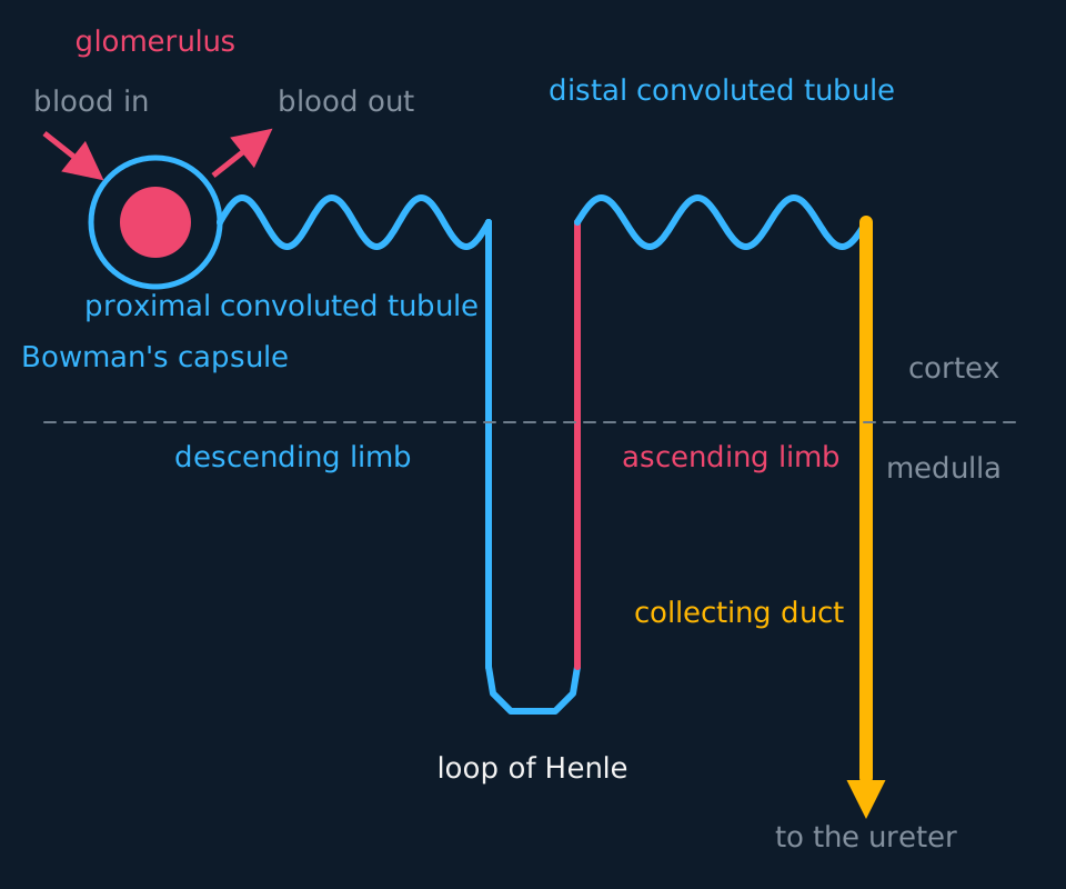
Describe the drawing
A single nephron. At the top left a glomerulus sits inside a Bowman's capsule, fed by one arteriole and drained by another. The filtrate passes into a coiled proximal convoluted tubule, down a straight descending limb into the medulla, round a hairpin, back up the ascending limb into the cortex, through a coiled distal convoluted tubule, and into a thick collecting duct running down through the medulla to the ureter. A dashed line separates cortex from medulla.
- Nephron
- The functional unit of the kidney: a renal capsule and its tubule, running from the cortex into the medulla and back.
- Ultrafiltration
- Filtration under pressure through a partially permeable barrier, separating small molecules from large ones.
- Selective reabsorption
- The return of useful substances from the filtrate to the blood, by transport proteins in the tubule wall.
- Osmoregulation
- The control of the water potential of the blood and body fluids.
- Fibrous capsule
- The tough outer layer enclosing a kidney, which protects it and holds its shape.
- Renal pelvis
- The funnel-shaped space at the centre of a kidney, into which the collecting ducts drain and which narrows into the ureter.
Exam-board note. OCR A treats this material as excretion, so the liver and the formation of urea are examined with it. AQA treats it as control of the water potential of the blood and ends at ADH. Cambridge International 9700 requires more detail of the cells themselves: the microvilli, the mitochondria and the basement membrane.
Ultrafiltration: pressure, and a sieve made of protein
The glomerulus is a knot of capillaries inside the cup of the renal capsule, also called Bowman's capsule. Blood reaches it along an afferent arteriole and leaves along an efferent arteriole that is narrower. Because the outflow vessel is narrower than the inflow vessel, the hydrostatic pressure inside the glomerular capillaries is far higher than in an ordinary capillary bed, and fluid is forced out through the capillary wall.
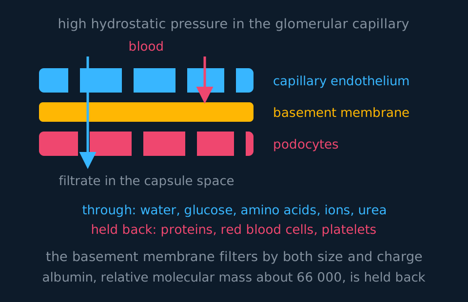
Describe the drawing
The filtration barrier drawn as three stacked bands with blood above and capsule space below. The top band is the capillary endothelium, broken by wide pores; the middle band is the basement membrane, drawn unbroken; the bottom band is the layer of podocytes, broken by narrow slits. One arrow passes right through all three, while a second stops dead at the basement membrane. Lists beneath name what crosses and what does not.
Three layers lie between the blood and the capsule space. The capillary endothelium is fenestrated, with pores between and through its cells that are far too wide to sort molecules. The podocytes lining the capsule have foot processes with filtration slits between them, each slit bridged by a thin protein sheet called the slit diaphragm. Between the two lies the basement membrane, a mesh of collagen and glycoproteins, and at A-level this is the layer named as the filter. In full, all three layers contribute, and the selectivity is graded by size and charge rather than a single sharp cut-off: albumin, at about 66 000 relative molecular mass, is almost entirely held back, which means plasma proteins stay in the blood while water, glucose, amino acids, mineral ions and urea pass through.
Not all of that pressure ends up as filtration. Two things oppose it: the hydrostatic pressure of the fluid already in the capsule, and, more importantly, the water potential of the plasma, made more negative by the proteins left behind. Filtration continues only while the outward pressure exceeds their sum, which is why it stops before the blood leaves the glomerulus.
Working out how much is taken back
The glomerular filtration rate of a healthy adult is about 125 cm³ min⁻¹. If 1.5 dm³ of urine is produced in a day, what percentage of the filtrate is reabsorbed?
Show the working
First get both quantities into the same units and the same time. 125 cm³ min⁻¹ × 60 × 24 = 180 000 cm³ per day, which is 180 dm³.
Reabsorbed volume is what was filtered minus what left: 180 − 1.5 = 178.5 dm³.
As a percentage, 178.5 ÷ 180 × 100 = 99.2 per cent.
Small changes in that percentage have large effects on urine volume: reabsorbing 99.0 per cent instead of 99.2 per cent would raise the urine volume from 1.5 to 1.8 dm³ a day.
Selective reabsorption, and the cells built for it
The proximal convoluted tubule is where the bulk of the reclamation happens. All of the glucose and all of the amino acids are reabsorbed here, along with most of the water and sodium; published figures for the proportion of water range from about two thirds to 85 per cent.
Glucose is taken back by co-transport. Prerequisite: co-transport of glucose in the ileum. Sodium ions are actively pumped out of the tubule cell at the far side, into the blood, which lowers the sodium concentration inside the cell. Sodium in the filtrate then diffuses into the cell down that gradient through a co-transporter protein, and glucose is dragged in with it, against its own concentration gradient. Glucose then leaves the cell into the blood by facilitated diffusion. The ATP is spent on the sodium pump, not on the movement of glucose.
The cells lining this part of the tubule are adapted in three ways. Their surface facing the filtrate carries microvilli, which give a large surface area for carrier and channel proteins. Their membranes are packed with those transport proteins. And their cytoplasm is full of mitochondria, supplying the ATP the sodium pumps consume. In an electron micrograph of a tubule cell, link each of those three features to its function.
Water follows by osmosis wherever the tubule wall is permeable to it. As solutes are reabsorbed, the water potential of the filtrate rises above that of the surrounding tissue and blood, and water moves out. Water is not actively transported at any point in the nephron.
The loop of Henle: building a gradient for the collecting duct
The loop dips from the cortex into the medulla and comes back, and the two limbs have different properties. The descending limb is permeable to water; the ascending limb is not, and its thick upper section actively transports sodium and chloride ions out into the surrounding tissue.
Those two facts, plus the fact that fluid keeps flowing, are enough to build a gradient far steeper than the pump could manage on its own. The pump can only ever create a difference of about 200 arbitrary units between the ascending limb and the tissue around it, which is the single effect. Because the two limbs run alongside each other with fluid flowing in opposite directions, that small difference is applied again and again at successive levels, and the effect is multiplied down the loop.

Describe the drawing
The loop of Henle building its gradient in five steps, with concentrations in arbitrary units shown in the descending limb, the ascending limb and the surrounding medulla. First every column reads 300 and there is no gradient. Then the ascending limb pumps sodium and chloride out, so the tissue reads 400 while the ascending limb reads 200: a difference of 200, the same at every level. Water then leaves the descending limb by osmosis until it too reads 400. Flow carries that fluid deeper and the pump repeats, so the difference multiplies down the loop until the tip reads 1200 and the top still reads 300. Finally the collecting duct is drawn running back down through the gradient, with water leaving it along its whole length.
The sequence in words. Ions are pumped out of the ascending limb, so the tissue becomes concentrated and the ascending limb dilute. Water then leaves the descending limb by osmosis, since it is permeable and the tissue outside it is now more concentrated, and the fluid in the descending limb becomes as concentrated as the tissue. That concentrated fluid flows round the hairpin into the ascending limb, where the pump acts on it again, starting this time from a higher concentration. Repeat, and the tip of the medulla reaches about four times the concentration at the top.
A straight blood vessel running through the medulla would remove the gradient. The vasa recta are hairpin vessels running alongside the loop, so blood descending takes up solutes and blood ascending returns them, and the gradient is maintained.
The gradient is used by the collecting duct. The collecting duct carries filtrate from the distal tubule back down through the whole depth of the medulla. At every millimetre of that descent, the tissue outside is more concentrated than the fluid inside, so water leaves by osmosis wherever the duct wall is permeable to water. The loop establishes the gradient; water leaves the collecting duct down it.
One prediction follows from that. Longer loops build a steeper gradient, so more water leaves the collecting ducts and the urine is more concentrated. Desert mammals such as the kangaroo rat have exceptionally long loops and thick medullas; a beaver has short ones. Apply the same reasoning to an unfamiliar species given a measurement of medulla thickness.
ADH: changing what the duct will let through
The volume of water leaving the collecting duct is regulated by a negative feedback loop acting on the permeability of the duct wall.
Osmoreceptors in the hypothalamus monitor the water potential of the blood. After sweating, a salt intake or a low water intake, the water potential of the blood falls, water leaves the osmoreceptor cells by osmosis and they shrink, and they send impulses along neurones to the posterior pituitary. The pituitary releases antidiuretic hormone into the blood.
ADH binds to receptors on the cells of the collecting duct wall, and on those of the last part of the distal convoluted tubule, and acts through cyclic AMP, exactly as adrenaline does at the liver. The cascade causes vesicles carrying aquaporins, which are water channel proteins, to fuse with the membrane facing the filtrate. More aquaporins means the wall is more permeable to water, so more water leaves down the gradient the loop has built, and a small volume of concentrated urine is produced. The reabsorbed water raises the water potential of the blood, the osmoreceptors are no longer stimulated, and ADH secretion falls.
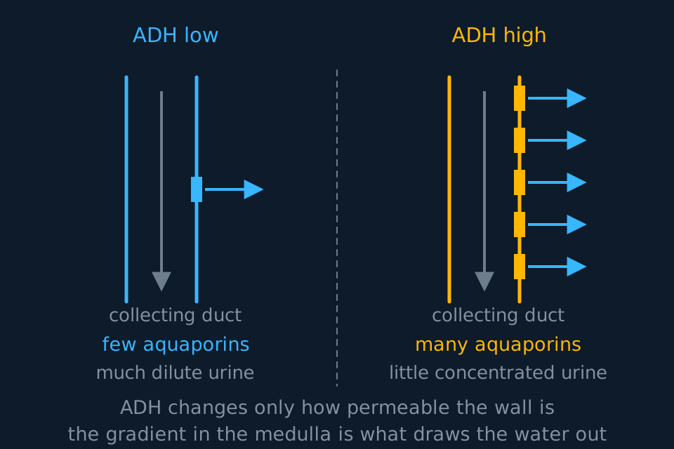
Describe the drawing
Two drawings of the same collecting duct side by side. On the left, with little antidiuretic hormone, a single aquaporin sits in the wall and one arrow of water leaves, so a large volume of dilute urine passes on. On the right, with a great deal of the hormone, five aquaporins sit in the same length of wall and five arrows of water leave, so a small volume of concentrated urine passes on.
After a large intake of water the sequence reverses: the water potential of the blood rises, less ADH is released, aquaporins are withdrawn into vesicles, the duct becomes less permeable and a large volume of dilute urine is produced. Alcohol inhibits ADH release, so it increases urine volume and can cause dehydration despite the volume of liquid consumed.
When the kidney fails, and what dialysis has to replace
Diabetes and high blood pressure cause most kidney failure, with infections, autoimmune disease and inherited polycystic disease behind them, and what follows is the same whatever the cause: urea accumulates, ion concentrations drift, water is retained and blood pressure rises. Untreated it is fatal. Exam-board note: OCR A examines renal failure and dialysis; AQA's specification ends with ADH; Cambridge International 9700 examines urine analysis.
Kidney function is judged by the glomerular filtration rate (GFR), the volume of plasma filtered each minute, about 125 cm³ min⁻¹ in a healthy adult. It is estimated from the concentration of creatinine in the blood: creatinine is a waste product of muscle that is filtered and not reabsorbed, so as nephrons are lost the GFR falls and blood creatinine rises. A GFR below about 15 cm³ min⁻¹ is taken as kidney failure. By then the kidney cannot adjust how much sodium and potassium it excretes, and a high blood potassium disturbs the rhythm of the heart.
During haemodialysis, blood leaves and returns through vascular access, usually an arteriovenous fistula or graft, or a venous catheter. It passes along one side of a partially permeable membrane while dialysis fluid flows on the other side of that membrane, in the opposite direction. The composition of the fluid follows from a single principle: a substance diffuses across only where a concentration gradient exists, so the fluid establishes a gradient for substances to be removed and abolishes it for substances to be retained.
| Component of dialysis fluid | Concentration | Why |
|---|---|---|
| Urea | None | A steep gradient outwards, so urea diffuses from blood into the fluid |
| Glucose | Normal blood concentration | No gradient, so glucose is not lost from the blood |
| Mineral ions | Normal plasma concentration | Only an excess in the blood diffuses out; a normal level is unchanged |
| Water | Water potential close to the blood's | Excess water is drawn off by a pressure difference across the membrane (ultrafiltration), set for each patient |
| Proteins | None | They cannot cross the membrane in either direction anyway |
| Temperature and pH | Body temperature, blood pH | So that the blood returned is not damaged or chilled |
Two design features should be stated explicitly. The fluid is replaced continuously, so the gradient for urea is maintained. And blood and fluid flow in opposite directions, which maintains a difference in concentration along the whole length of the exchange surface. This is the same counter-current exchange as in a fish gill and in the vasa recta. The loop of Henle differs: it is a counter-current multiplier, because its ascending limb pumps ions actively.
Haemodialysis takes several hours, three times a week, and blood composition drifts between sessions. Peritoneal dialysis uses the patient's own peritoneum as the membrane and can be done at home. A transplant restores continuous function and a normal diet, at the cost of a shortage of donors and a lifetime of immunosuppressants, which leave the recipient open to infection.
Urine is also used in diagnosis, because substances in the plasma, or the products the liver turns them into, are filtered into it. A pregnancy test uses monoclonal antibodies to detect hCG, a hormone made by the placenta that passes into the urine, and many drug tests use test strips carrying monoclonal antibodies in the same way. Anabolic steroids, and any positive drug screen, are identified by gas chromatography and mass spectrometry, which separates the substances in the sample and identifies each by the masses of its fragments.
TRY IT: Reasoning about a change to the fluid
A dialysis machine is set up by mistake with dialysis fluid containing no glucose and no mineral ions. Predict the effect on the patient's blood, and explain each prediction.
Check your answer
Glucose would be lost from the blood. With none in the dialysis fluid there is a steep concentration gradient from blood to fluid across the partially permeable membrane, and glucose is small enough to cross, so it would diffuse out continuously and the patient's blood glucose concentration would fall.
Nervous tissue respires glucose and stores none, so the patient would become confused and could lose consciousness, and less glucose would be available for respiration everywhere else.
Mineral ions would be lost the same way. Sodium and potassium would diffuse down their gradients into the fluid, and a fall in blood potassium in particular disturbs the resting potential of excitable cells, so heart rhythm becomes unreliable.
The principle is that a dialysis fluid is not pure water. It matches normal blood in every substance that should be retained and differs from it only in those to be removed, which is why urea is the solute deliberately absent.
In the exam
- For ultrafiltration, state that the efferent arteriole is narrower and that this raises the hydrostatic pressure, and name the basement membrane as the filter rather than referring only to pores.
- State that water moves by osmosis down a water-potential gradient. Water is not actively transported anywhere in the kidney.
- For the proximal tubule, describe co-transport in the right order: sodium pumped out into the blood first, then sodium and glucose in together from the filtrate.
- The loop of Henle establishes the medullary gradient. Water is reabsorbed from the descending limb, and the regulated reabsorption that concentrates the urine occurs at the collecting duct.
- ADH changes the permeability of the collecting duct by inserting aquaporins. It does not pump water, and it does not act on the loop.
- In a dialysis answer, justify each component of the fluid by the gradient it creates or abolishes: give the concentration and what it achieves.
Check yourself
A person drinks two litres of water quickly. Describe and explain what happens to the volume and concentration of urine they produce over the following hour, naming the receptors, the hormone and the part of the nephron involved, and state what kind of feedback this is.
Answer
The water is absorbed from the gut into the blood, so the water potential of the blood rises, becoming less negative. Osmoreceptors in the hypothalamus detect the change: water enters them by osmosis and they swell, and they send fewer impulses to the posterior pituitary.
Less ADH is therefore released into the blood. With less ADH binding to receptors on the collecting duct cells, fewer aquaporins are held in the membrane facing the filtrate, because the vesicles carrying them are withdrawn, so the duct wall becomes much less permeable to water.
Less water can therefore leave the duct down the gradient the loop of Henle maintains in the medulla, and a large volume of dilute urine is produced.
This is negative feedback. The response, losing the excess water in the urine, lowers the blood's water potential back towards the norm, which removes the stimulus that caused the response. Notice that the medulla's gradient is unchanged throughout: what the hormone altered was the permeability of the duct, not the driving force acting on the water.
Questions
Question 15 marks
Describe how glucose in the filtrate is reabsorbed at the proximal convoluted tubule, and describe two features of the tubule cells that suit them to the job.
Mark scheme
- B1 sodium ions are actively pumped out of the tubule cell at the side facing the blood, which lowers the sodium concentration inside the cell
- B1 sodium ions in the filtrate then diffuse into the cell down that gradient through a co-transporter protein
- B1 glucose is carried in with the sodium, against its own concentration gradient, and then leaves the cell into the blood by facilitated diffusion
- B1 the surface facing the filtrate carries microvilli, giving a large surface area for the carrier and channel proteins the membranes are packed with
- A1 the cytoplasm contains many mitochondria, supplying the ATP that the sodium pumps consume
Question 25 marks
Explain how the loop of Henle produces a concentration gradient in the tissue of the medulla, and explain why it is wrong to say that the loop itself concentrates the urine.
Mark scheme
- B1 the thick section of the ascending limb actively transports sodium and chloride ions out into the surrounding tissue, and that limb is not permeable to water
- B1 this makes the tissue fluid of the medulla more concentrated than the ascending limb at every level, so the medulla has a lower water potential
- B1 the descending limb is permeable to water, so water leaves it by osmosis into the now more concentrated tissue and the fluid inside becomes more concentrated
- B1 because the two limbs run alongside each other with fluid flowing in opposite directions, that concentrated fluid is delivered to the pump again at a higher starting concentration, so the small difference is multiplied down the loop
- A1 the fluid leaving the ascending limb is more dilute than plasma; the urine is concentrated later, in the collecting duct, as water leaves it down the gradient the loop has built
Question 35 marks
A person has a glomerular filtration rate of 120 cm³ min⁻¹ and produces 1.8 dm³ of urine in a day. Calculate the volume of filtrate produced in a day, calculate the volume reabsorbed, and calculate the percentage of the filtrate that is reabsorbed.
Mark scheme
- M1 convert the glomerular filtration rate to a daily volume: 120 × 60 × 24 = 172 800 cm³ per day
- A1 172.8 dm³ of filtrate is produced per day
- M1 reabsorbed volume = filtered volume − urine volume = 172.8 − 1.8 = 171.0 dm³
- M1 percentage reabsorbed = reabsorbed volume ÷ filtered volume × 100 = 171.0 ÷ 172.8 × 100
- A1 99.0 per cent of the filtrate is reabsorbed
Question 44 marks
Explain how ultrafiltration produces a filtrate in the renal capsule that contains glucose and urea but no plasma protein.
Mark scheme
- B1 the efferent arteriole leaving the glomerulus is narrower than the afferent arteriole bringing blood in, so the blood is squeezed into a bottleneck
- B1 the hydrostatic pressure in the glomerular capillaries is therefore much higher than in an ordinary capillary bed, and fluid is forced out through the capillary wall
- B1 the pores of the fenestrated endothelium and the filtration slits between the podocytes are both far too wide to sort molecules, so the basement membrane between them does the sorting
- A1 water, glucose, amino acids, mineral ions and urea are small enough to pass through it, while plasma proteins are too large and stay in the blood
Question 54 marks
A person sweats heavily during a long run. Describe the sequence of events that raises the concentration of their urine, naming the receptors, the hormone and the part of the nephron involved.
Mark scheme
- B1 water is lost in sweat, so the water potential of the blood falls and becomes more negative
- B1 osmoreceptors in the hypothalamus lose water by osmosis and shrink, and send impulses along neurones to the posterior pituitary
- B1 the posterior pituitary releases more antidiuretic hormone into the blood, and it binds to receptors on the cells of the collecting duct wall
- A1 vesicles carrying aquaporins fuse with the membrane facing the filtrate, so the wall becomes more permeable and more water leaves by osmosis down the gradient in the medulla, giving a small volume of concentrated urine
Question 64 marks
Explain why a haemodialysis fluid contains no urea but contains glucose at the same concentration as normal blood plasma.
Mark scheme
- B1 a substance diffuses across the partially permeable membrane only where a concentration gradient exists between the blood and the fluid
- B1 urea is deliberately absent from the fluid so that a steep gradient exists from blood to fluid, and urea that has built up in the patient's blood diffuses out and is removed
- B1 glucose is included at the normal blood concentration so that no gradient exists for it
- A1 without a gradient, glucose already at the normal concentration does not diffuse out of the blood, so useful glucose is not lost during dialysis
Question 74 marks
Describe two common causes of kidney failure, and describe the resulting changes in blood urea concentration, ion concentration and blood pressure.
Mark scheme
- B1 diabetes and high blood pressure are the two most common causes of kidney failure
- B1 with fewer working nephrons, urea is filtered and excreted more slowly, so its concentration in the blood rises
- B1 ion concentrations in the blood drift away from normal, because the kidney's control of their reabsorption and excretion is lost
- A1 water that should have been excreted is retained instead, which raises blood volume and in turn raises blood pressure
Question 84 marks
Compare how the proximal convoluted tubule and the loop of Henle each affect the water content of the filtrate, making clear which one is responsible for the concentration gradient in the medulla.
Mark scheme
- B1 at the proximal convoluted tubule water leaves the filtrate by osmosis continuously, following the solutes reabsorbed there, so a large proportion of the filtered water is reclaimed at this point
- B1 the loop of Henle works differently: its descending limb does let water leave by osmosis, but only because the ascending limb has actively pumped ions into the surrounding tissue, not because the loop itself is reabsorbing water for its own sake
- B1 the ascending limb is impermeable to water altogether, so no water crosses there regardless of the ion pumping taking place
- A1 the loop's real function is to build the medullary concentration gradient that the collecting duct will later use, rather than to reabsorb a large fraction of the filtrate's water itself
Question 93 marks
A desert rodent produces urine four times as concentrated as human urine, and the medulla of its kidney is unusually thick. Suggest how the structure of its nephrons accounts for this.
Mark scheme
- B1 a thick medulla holds unusually long loops of Henle
- B1 sodium and chloride ions are pumped out of the ascending limb over a greater depth of tissue, so the single effect is multiplied over more levels and the gradient in the medulla is steeper
- A1 the collecting duct passes down through that steeper gradient, so water leaves it by osmosis all the way down and a smaller volume of more concentrated urine is produced
Question 103 marks
A kidney is cut in half lengthways. Name the tough outer layer enclosing the organ, name the region in which the renal capsules lie, and name the funnel-shaped space that the collecting ducts drain into.
Mark scheme
- B1 the fibrous capsule encloses the kidney
- B1 the renal capsules and convoluted tubules lie in the cortex, the outer region
- B1 the collecting ducts drain into the renal pelvis, which narrows into the ureter
Question 113 marks
Explain why blood and dialysis fluid are made to flow in opposite directions through a dialysis machine, rather than in the same direction.
Mark scheme
- B1 opposite, counter-current flow maintains a concentration difference between blood and fluid along the whole length of the exchange surface
- B1 if the two fluids flowed in the same direction, the difference would be largest at the start and would shrink to almost nothing by the far end, as the two fluids approached the same composition
- A1 with counter-current flow, fresh dialysis fluid is always meeting blood that has already lost some urea further back, so diffusion of urea out of the blood can continue effectively along the entire membrane
Question 123 marks
Drinking alcohol is followed by producing an unusually large volume of dilute urine, even though no extra water has been drunk. Suggest why, given that alcohol inhibits the release of ADH.
Mark scheme
- B1 with ADH release inhibited, less ADH reaches the collecting duct, so fewer aquaporins are inserted into the membrane facing the filtrate
- B1 the collecting duct wall is therefore less permeable to water than usual, so less water leaves it by osmosis down the gradient the loop of Henle has built
- A1 more water than usual stays in the filtrate and is passed as urine, so a large volume of dilute urine is produced despite no extra water having been taken in, which is why drinking alcohol can cause dehydration
Question 133 marks
Explain why the blood vessels running through the renal medulla, the vasa recta, are shaped as hairpin loops rather than running straight through like an ordinary capillary.
Mark scheme
- B1 a straight vessel would carry blood in one direction only, continuously washing solute out of the medulla and into the general circulation
- B1 that would remove the concentration gradient the loop of Henle has built, since diffusion between the vessel and the surrounding tissue would gradually even out the difference
- A1 because the vasa recta run as hairpins alongside the loop of Henle, blood descending into the medulla takes up solute and blood ascending back out returns most of it, so the gradient is maintained rather than washed away
Question 142 marks
State which of the three layers between the blood and the capsule space acts as the filter during ultrafiltration, and state one component of the blood that it holds back.
Mark scheme
- B1 the basement membrane, a mesh of collagen and glycoproteins, is the filter
- B1 it holds back plasma proteins, such as albumin at about 66 000 relative molecular mass
Question 152 marks
State what is meant by osmoregulation, and state what is meant by ultrafiltration.
Mark scheme
- B1 osmoregulation is the control of the water potential of the blood and body fluids
- B1 ultrafiltration is filtration under pressure through a partially permeable barrier, separating small molecules from large ones
Worth remembering
- Fibrous capsule outside, then cortex, then medulla, with the renal pelvis in the middle draining into the ureter.
- Small molecules are filtered under pressure, then useful substances are selectively reabsorbed.
- The basement membrane is the sieve; the efferent arteriole being narrower is why there is any pressure to filter with.
- The proximal tubule takes back all the glucose by co-transport, and the ATP is spent on the sodium pump.
- The loop establishes the medullary gradient; the regulated water reabsorption that concentrates urine happens at the collecting duct.
- ADH inserts aquaporins and changes permeability. Longer loops mean steeper gradients mean more concentrated urine.
CHECK YOUR PROGRESS
Rate how confident you are with each objective for this lesson. Ratings are saved in this browser, on this device, unless you sign in.
- Label a kidney in section, from the fibrous capsule to the renal pelvis.
- Label a nephron and name the parts in the order the filtrate meets them.
- Explain ultrafiltration in terms of hydrostatic pressure and the three layers of the barrier.
- Describe selective reabsorption at the proximal convoluted tubule, including co-transport.
- Explain how the loop of Henle builds a concentration gradient in the medulla.
- Describe the control of water potential by osmoreceptors, ADH and aquaporins.
- State what a dialysis fluid must contain, and justify each component.
Open the full revision checklist to see every objective in the curriculum in one place.
Practise this lesson
WORKBOOK
The questions from this topic, on paper with room to work, and a separate book of mark schemes. Free to use; please do not redistribute or sell.