Biology › Practicals › Required practical 6: aseptic technique and antibiotic discs
Required practical 6: aseptic technique and antibiotic discs
Spread bacteria evenly over agar, lay a paper disc carrying an antibiotic on top, and the antibiotic diffuses out into the agar. Where it is concentrated enough to stop growth, the lawn stays clear. The size of that clear zone compares antibiotics, or plant extracts, against one bacterium, and aseptic technique is what keeps the plate a test of that bacterium and nothing else.
The capability this develops
This guide is one investigation under capability P09: Aseptic technique and the effect of antimicrobials, which sets out the techniques the investigation belongs to, the calculations it carries and the errors that cost marks in all of them. The notes behind the biology are in Evolution and speciation.
Where this sits on the specifications
This library maps a qualification to its own specification one numbered heading at a time, against a document whose sha256 is recorded, and publishes the result as a route map. These are the headings whose practical work this investigation serves, on the five qualifications mapped so far. Each links to the route map the row belongs to.
| Qualification | Heading |
|---|---|
| AQA 7401/7402 | 3.4.4 Genetic diversity and adaptation |
| OCR H020/H420 | 6.2.1 Cloning and biotechnology |
| Cambridge International 9700 | 10.2 Antibiotics |
| Pearson Edexcel Biology A (Salters-Nuffield) 8BN0/9BN0 | 4.14 Conditions required for bacterial growth |
| Pearson Edexcel Biology A (Salters-Nuffield) 8BN0/9BN0 | 6.14 Bacteriostatic and bactericidal antibiotics |
| Pearson Edexcel Biology B 8BI0/9BI0 | 6.3 i Action of antibiotics |
Some boards number their practical activities, and those lists were read here from the same specifications as the headings above. This investigation is numbered on them as:
- AQA 7401/7402 required practical 6
- OCR Biology A H020/H420 PAG 7
- Pearson Edexcel Biology A (Salters-Nuffield) 8BN0/9BN0 core practical 9
- Pearson Edexcel Biology A (Salters-Nuffield) 8BN0/9BN0 core practical 15
The other qualifications above are matched by specification heading only, because none of them numbers an activity that is this investigation. Use your awarding organisation’s current specification and its practical handbook as the authority on which activities your course requires and how they are assessed.
The principle
Paper discs, each carrying one antibiotic, are laid on a lawn of bacteria spread over agar. Each antibiotic diffuses out of its disc, and where it is concentrated enough to stop growth a clear zone of inhibition forms. The sizes of the zones compare how well the antibiotics stop that bacterium growing under the conditions of the plate.
Aseptic technique is the set of habits that keeps unwanted microorganisms out of the culture and keeps the culture off you: a disinfected bench, sterile instruments, a lid lifted only as far as the work needs, and a plate that is never opened once it has grown. A contaminant that grows on the plate is a second organism being tested without anyone knowing.
The comparison is made by area. Area goes as the square of the radius, so a zone twice as wide has four times the area, and a ranking by area spreads the same discs further apart than a ranking by diameter. The order is the same either way, but the ratio between two antibiotics is not.
- Zone of inhibition
- The clear area round a disc on a lawn of bacteria, where a substance diffusing from the disc has stopped the bacteria growing.
- Antibiotic disc
- A small disc of absorbent paper carrying a fixed amount of one antibiotic, laid on a lawn of bacteria to test whether that antibiotic stops them growing.
- Bacterial lawn
- An even layer of bacteria grown over the whole surface of an agar plate from a spread liquid culture.
Apparatus
- A broth culture of a bacterium your school's risk assessment approves, from a recognised supplier
- Sterile nutrient agar plates, a sterile pipette and a sterile spreader
- Antibiotic discs, each printed with its code, and plain sterile paper discs
- Sterile forceps, a Bunsen burner and a heat-proof mat
- Tape, a marker pen and a ruler
- An incubator and disinfectant
Safety
Risk assessment. Treat every culture as a possible pathogen, use only the culture your school's risk assessment approves, and never culture organisms from yourself or your surroundings. Anyone allergic to penicillin or another antibiotic says so before the practical. Handle discs only with forceps, and wash your hands afterwards. The plate selects for resistant bacteria. That is one more reason it stays taped once incubated and is examined through the lid, never opened unless the risk assessment allows it. Report a spill at once and leave it to be dealt with by the school's procedure. Tie long hair back near the Bunsen burner. Every plate and culture is killed by autoclaving or pressure-cooking before disposal. Check the CLEAPSS guidance and your school's risk assessment before starting; where they differ from this page, follow them.
Variables
Independent variable
The antibiotic, one per disc, with a plain paper disc beside them. In the plant version it is the plant the extract was made from.
Dependent variable
The area of the zone of inhibition, from the mean of two diameters measured at right angles across the disc, halved for the radius r, and πr².
Control variables
- The bacterium and the lawn. One culture, of one age, and the same volume spread over every plate.
- The agar. One batch, poured to the same depth, because the antibiotic spreads further through thinner agar.
- The timing. Every disc placed within a few minutes of spreading, so no bacteria get a head start.
- The amount on each disc. Fixed by the manufacturer for antibiotic discs; for extracts, the same volume pipetted onto every disc.
- The spacing. Discs well apart, so zones do not merge.
- Incubation. The same temperature and the same time for every plate.
Method
Before the plate is opened
- Disinfect the bench, wash your hands, and label the base of each plate round its edge. Work beside a lit Bunsen burner, or as your school's practice directs. The flame is usually said to keep airborne microorganisms away from an open plate; lids can be swapped, bases cannot.
Spreading the lawn and placing the discs
- Flame the neck of the culture bottle, take the volume the method gives with a sterile pipette, and flame the neck again before replacing the cap. Lift the lid only far enough to add the culture, spread it over the whole surface, and replace the lid. An even lawn gives every disc the same density of bacteria.
- With sterile forceps, place the antibiotic discs and one plain disc well apart, press each gently into full contact with the agar, and do not move a disc once it is down. Mark each position on the base. The plain disc is the negative control; a disc that is moved leaves antibiotic in two places.
Incubating and measuring
- Tape the lid with two or four short strips, never all the way round. Invert the plate and incubate it for two to three days at no higher than the temperature the school's risk assessment sets, well below body temperature. Sealing all round can make the plate anaerobic and favour anaerobic bacteria; a lower temperature avoids favouring organisms that grow well in people.
- Without opening the plate, measure the diameter of each clear zone across its disc, twice at right angles. Zones are rarely perfect circles.
- Take the mean of the two diameters for each zone and then across the plates, halve it for the radius r, and calculate the area of the zone as πr². Where bacteria grow up to the edge of a disc, record no zone.
- Set up at least three plates, each with every disc on it.
Describe the drawing
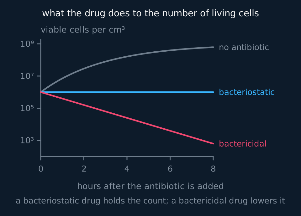
A graph of the number of living bacteria per cubic centimetre, on a logarithmic scale from ten cubed to ten to the ninth, against hours after an antibiotic is added. Three cultures start at ten to the sixth. The grey culture with no antibiotic rises towards ten to the ninth. The cyan culture given a bacteriostatic antibiotic stays level at ten to the sixth. The coral culture given a bactericidal antibiotic falls steadily to below ten cubed after eight hours.
The plant-extract version
- Crush the same mass of each plant with the same volume of ethanol for the same time, and filter off the liquid. Only the plant differs between extracts.
- Soak discs in each extract, and some in ethanol alone. Dry them well away from any flame until the ethanol has evaporated. Ethanol is flammable and kills bacteria itself.
- Then spread, place, incubate and measure exactly as above. The ethanol-only disc is the negative control, and an antibiotic disc on the same plate is the positive control, which shows that a zone could form on that lawn.
Sample data
These results are idealised. They are the four antibiotics the lesson works, P, Q, R and S, with the plate-by-plate diameters made up to give its means, and real zones scatter more. Nothing here is a record of an experiment.
| Disc | Zone diameter on plates 1, 2 and 3 / mm | Mean diameter / mm | Radius / mm | Area / mm² |
|---|---|---|---|---|
| P | 21, 23, 22 | 22.0 | 11.0 | 380 |
| Q | 13, 15, 14 | 14.0 | 7.0 | 154 |
| R | no zone on any plate | no zone | 0 | |
| S | 17, 19, 18 | 18.0 | 9.0 | 254 |
| Plain paper | no zone on any plate | no zone | 0 |
Each diameter on a plate is already the mean of two measured at right angles. For P the mean across the plates is (21 + 23 + 22) ÷ 3 = 22.0 mm, so the radius is 11.0 mm and the area is π × 11.0² = 380 mm².
Ranked by zone area: P, S, Q, then R.
P's zone has 380 ÷ 154 = 2.5 times the area of Q's, though its diameter is only 22 ÷ 14 = 1.6 times as large. State whether a comparison uses diameters or areas.
The plain disc had bacteria growing up to its edge on every plate, so the paper itself does not stop growth and the zones round P, Q and S came from the antibiotics.
Reading the graph
The antibiotics are categories, so the chart is a bar chart: one bar per disc, the mean area of its zone up the vertical axis, and a bar at the top of each showing the range across the three plates. R and the plain disc are plotted as zero, not left off.
The order. P's zone is largest, then S, then Q. R gave no zone at all, so at the amount on its disc it did not inhibit this bacterium, which is resistant to it under these conditions.
The ranking holds for these discs on this agar, and it does not decide which antibiotic a doctor should prescribe. Different discs carry different amounts of drug, and antibiotics diffuse through agar at different rates. So a large zone does not by itself show a more effective antibiotic.
A clear zone also cannot show whether the bacteria were killed or only stopped from dividing.
Colonies inside a zone. A few colonies growing inside a clear zone are either resistant cells the antibiotic has selected or contaminants. Either way they are a reason to keep the plate shut, and they are recorded, not ignored.
The plant extracts. The same reading applies, with one more caution. A crude extract has an unknown concentration, so a large zone round garlic shows that something in the extract diffused out and stopped growth, not that garlic's compound is the most potent.
Evaluation
Each of these is a real source of error in this investigation, and the middle column is the part an answer earns marks for: what it does to the result, and in which direction.
| Source of error | What it does to the result | What to do about it |
|---|---|---|
| Discs placed at different times after spreading | Bacteria have a head start round later discs, so their zones are smaller | Place every disc within a few minutes and treat every plate alike |
| A disc not in full contact with the agar | Little antibiotic diffuses out, giving a small or lopsided zone | Press each disc down gently with sterile forceps |
| Discs out of date or stored warm | Some antibiotic has broken down, so zones are too small | Use discs within date, stored as the supplier directs |
| Arms of a multi-antibiotic disc close together | Zones merge and cannot be measured | Use single discs spaced well apart |
| Contamination from the air or hands | Colonies that are not the culture | Open the lid as little as possible beside the flame; incubate an uninoculated plate alongside, which should grow nothing |
| Zone edges judged by eye | The edge of a zone fades rather than stops, so two people measure different diameters. Squaring the radius doubles the percentage error: ±1 mm on a 14 mm zone is ±7% on the diameter and about ±14% on the area. | Measure against a dark background with a ruler held under the base, one person measuring every plate. |
Questions
Question 1 (2 marks)
Explain why the lid is taped with short strips rather than sealed all the way round.
Check your answer
sealing all round would stop oxygen reaching the plate, making conditions anaerobic;
which would favour the growth of anaerobic bacteria, which include many pathogens.
Question 2 (2 marks)
The zone round disc S had a mean diameter of 18.0 mm. Calculate its area.
Check your answer
radius = 18.0 ÷ 2 = 9.0 mm;
area = π × 9.0² = 254 mm².
Question 3 (2 marks)
Explain the purpose of the plain paper disc.
Check your answer
it is a negative control;
it shows that the paper alone does not stop growth, so a zone round another disc was caused by its antibiotic.
Question 4 (2 marks)
Explain why the plates are incubated at a temperature well below 37 °C.
Check your answer
37 °C is human body temperature;
a lower temperature avoids favouring the growth of organisms that grow well in people, which may be pathogens.
Question 5 (3 marks)
A student concludes that P is the most effective antibiotic because it has the largest zone. Give three reasons why this conclusion is not justified.
Check your answer
the discs may carry different amounts of antibiotic;
antibiotics diffuse through agar at different rates;
the plate cannot show whether the bacteria were killed or only stopped from dividing (accept: only one bacterium was tested; conditions on agar differ from those in the body).
Coverage maps are available for AQA 7401/7402, OCR Biology A H020/H420, Cambridge International 9700, Pearson Edexcel Biology A (Salters-Nuffield) 8BN0/9BN0 and Pearson Edexcel Biology B 8BI0/9BI0. Use the relevant route map to check what InkBiology covers, and your awarding organisation’s current specification as the final authority on examinable content.