Biology › Practicals › Required practical 4: beetroot membranes and temperature
Required practical 4: beetroot membranes and temperature
The red pigment in beetroot sits inside the vacuole, behind two membranes. Hold equal discs in water at a series of temperatures, and the deeper the colour of the water, the more pigment got out and the more permeable the membranes had become. A colorimeter puts a number on that colour, and the curve it gives has two gradients for two separate reasons.
The capability this develops
This guide is one investigation under capability P06: Investigating membrane permeability, which sets out the techniques the investigation belongs to, the calculations it carries and the errors that cost marks in all of them. The notes behind the biology are in Membranes and transport across cells.
Where this sits on the specifications
This library maps a qualification to its own specification one numbered heading at a time, against a document whose sha256 is recorded, and publishes the result as a route map. These are the headings whose practical work this investigation serves, on four of the five qualifications mapped so far. Each links to the route map the row belongs to.
| Qualification | Heading |
|---|---|
| OCR H020/H420 | 2.1.5 Biological membranes |
| Cambridge International 9700 | 4.2 Movement into and out of cells |
| Pearson Edexcel Biology A (Salters-Nuffield) 8BN0/9BN0 | 2.2 Cell membranes and the fluid mosaic as a model |
| Pearson Edexcel Biology B 8BI0/9BI0 | 4.2 iv Cell transport mechanisms |
Some boards number their practical activities, and those lists were read here from the same specifications as the headings above. This investigation is numbered on them as:
- AQA 7401/7402 required practical 4
- OCR Biology A H020/H420 PAG 5
- Pearson Edexcel Biology A (Salters-Nuffield) 8BN0/9BN0 core practical 3
- Pearson Edexcel Biology B 8BI0/9BI0 core practical 5
The other qualifications above are matched by specification heading only, because none of them numbers an activity that is this investigation. Use your awarding organisation’s current specification and its practical handbook as the authority on which activities your course requires and how they are assessed.
The principle
The standard practical uses beetroot because its vacuoles contain betalain, a water-soluble red pigment that is straightforward to measure. The pigment is inside the vacuole, so it must cross two membranes to leave the cell: the tonoplast and the cell-surface membrane. Pigment in the surrounding water is therefore evidence that both membranes have become permeable to it.
A membrane is a phospholipid bilayer with proteins set into it. The bilayer keeps a large, charged molecule such as betalain in, and the proteins that span it are held in their shape by hydrogen and ionic bonds. Heat acts on both. Phospholipids with more kinetic energy move more, and the bilayer becomes more fluid and leakier. At high temperatures the proteins denature and leave gaps where they were.
The colorimeter shines green light through the water, because a red solution absorbs green. The more pigment in the water, the less light reaches the detector and the higher the absorbance. The instrument may also give percentage transmission, %T, which falls as absorbance rises: absorbance = log₁₀(100 ÷ %T).
- Tonoplast
- The membrane around the vacuole of a plant cell.
- Permeability
- How readily a substance can cross a membrane.
- Absorbance
- A colorimeter's measure of how much light a solution absorbs, with no unit; it rises as the solution becomes more concentrated.
Apparatus
- A fresh beetroot, a cork borer, a white tile, a scalpel and a ruler
- A beaker of distilled water for washing the discs, and a sieve or tea strainer
- Test tubes in a rack, and a graduated pipette or syringe for 5 cm³
- Water baths at 20, 30, 40, 50, 60 and 70 °C, or one bath reset between runs, and a thermometer
- Forceps for moving the discs
- A colorimeter with a green filter (about 530 nm) and cuvettes
- A stopwatch
- Eye protection
Safety
Work to your school’s risk assessment. Cut the beetroot on a tile with the blade moving away from you, and push the cork borer in with a twisting motion, never into beetroot held in the hand. Water baths at the top of the range scald, so lift tubes out with a holder and keep cables and plugs clear of the water. Beetroot juice stains skin and clothing. If ethanol is the variable instead of temperature, it is highly flammable, so there must be no naked flame in the room.
Variables
Independent variable
Temperature, set by the water bath and checked with a thermometer in the tube: 20, 30, 40, 50, 60 and 70 °C.
Dependent variable
The absorbance of the water the disc sat in, read through a green filter, as a measure of how much pigment leaked out.
Control variables
- The size of the discs. One cork borer and one thickness, so every disc has the same surface area and the same number of cut cells.
- The washing. Every disc rinsed the same way before it goes in, so the pigment from cut cells is gone from all of them.
- The volume of water. 5 cm³ of distilled water in every tube, since the same pigment in more water reads paler.
- The time in the bath. 15 minutes for every disc, timed from the moment it goes in.
- The beetroot. Every disc from the same root, because older tissue leaks more.
- The time before reading. The same short wait for every tube between taking the disc out and reading the water.
Method
Cutting and washing the discs
- Cut discs of equal size with a cork borer.
- Push the borer through the beetroot on the tile, slide out the cylinder, and cut it into discs 2 mm thick against a ruler. Cut eighteen, three for each temperature.
- Wash them thoroughly to remove pigment released by the cutting itself.
- Rinse them in a sieve under running water, then leave them in a beaker of distilled water, changing it until it stays clear.
Heating the discs
- Put 5 cm³ of distilled water in each tube and stand the tubes in the baths.
- Stand each tube of water in the bath for about five minutes before its disc goes in, so that the water starts at the bath temperature.
- Hold each one in its own tube containing a fixed volume of distilled water, in a water bath at a set temperature for a fixed time, such as 15 minutes.
- Start the discs at 1-minute intervals, so that each comes out on time.
Describe the drawing
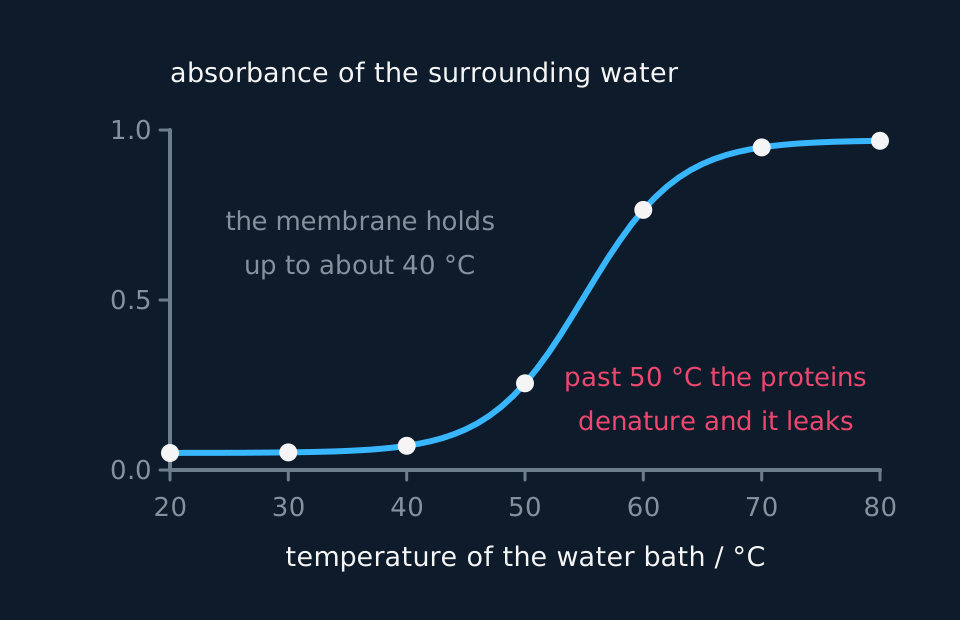
A line graph rising from left to right. Temperature from twenty to eighty degrees Celsius runs along the horizontal axis and absorbance from zero to one up the vertical axis. The curve stays almost flat and close to zero up to about forty degrees, turns sharply upward between fifty and sixty, then levels off near the top of the scale. Seven plotted points sit on the curve, one for each ten-degree step.
Reading the colour
- Remove the disc and measure the colour of the water it sat in with a colorimeter using a green filter of around 530 nm.
- Measure that water, not a fresh volume the disc is moved into afterwards.
- Zero the colorimeter on a cuvette of distilled water first. Shake each tube gently before pouring, so the pigment is evenly mixed, and read every tube after the same wait.
- Repeat with fresh discs to give three readings at each temperature, and take the mean.
Sample data
These readings are idealised. They were made up to show how a results table with repeats is processed, so the three readings at each temperature scatter by an invented amount, and real readings scatter more. Nothing here is a record of an experiment.
| Temperature / °C | Absorbance, disc 1 | Absorbance, disc 2 | Absorbance, disc 3 | Mean absorbance | Range |
|---|---|---|---|---|---|
| 20 | 0.07 | 0.09 | 0.08 | 0.08 | 0.02 |
| 30 | 0.10 | 0.12 | 0.11 | 0.11 | 0.02 |
| 40 | 0.14 | 0.17 | 0.14 | 0.15 | 0.03 |
| 50 | 0.35 | 0.41 | 0.38 | 0.38 | 0.06 |
| 60 | 0.88 | 0.97 | 0.91 | 0.92 | 0.09 |
| 70 | 1.15 | 1.21 | 1.18 | 1.18 | 0.06 |
Take the 60 °C row. The mean is (0.88 + 0.97 + 0.91) ÷ 3 = 2.76 ÷ 3 = 0.92, and the range is 0.97 − 0.88 = 0.09.
An absorbance of 0.92 is a percentage transmission of 100 ÷ 10^0.92 = 100 ÷ 8.3 = 12%. So at 60 °C only about an eighth of the green light got through the water.
The ranges grow as the absorbance grows, but none of the six overlaps its neighbour. The lowest reading at each temperature is above the highest at the temperature below, so every step in the table is a real rise and not scatter.
Reading the graph
Plot mean absorbance on the vertical axis against temperature on the horizontal axis, with a bar at each point showing the range, and draw a smooth curve through the means.
Higher absorbance means more pigment escaped, and so more permeable membranes.
The gentle rise, 20 °C to 40 °C. The absorbance goes from 0.08 to 0.15. The phospholipids are gaining kinetic energy and moving more, so the bilayer becomes slightly more fluid and slightly more pigment gets through, but the proteins still hold their shape.
The steep rise, 40 °C to 60 °C. The absorbance goes from 0.15 to 0.92, six times as much over the same 20 °C. Here the intrinsic proteins denature: hydrogen and ionic bonds break, they lose their tertiary structure and no longer fill the space they occupied in the membrane. Pigment leaves through the gaps. Name the two processes separately, because the graph has two gradients for two reasons.
The top of the curve. From 60 °C to 70 °C the rise slows, from 0.92 to 1.18. By then most of the membranes have lost their barrier and most of the pigment that can leave a disc in 15 minutes has left. The readings above about 1.0 are also the least reliable on most colorimeters, which is a second reason not to read too much into the last point.
Absorbance rises with concentration because ethanol is an organic solvent: it dissolves phospholipids out of the bilayer and disrupts the packing of those left behind.
That is the same curve with alcohol concentration on the horizontal axis and the bath at one temperature. The 0% tube is the control, and temperature becomes a control variable.
Evaluation
Each of these is a real source of error in this investigation, and the middle column is the part an answer earns marks for: what it does to the result, and in which direction.
| Source of error | What it does to the result | What to do about it |
|---|---|---|
| Discs not washed, or washed unevenly | Pigment from cells cut open by the borer is read as if the temperature had released it. It adds most, as a share, at the low temperatures, where the real leakage is small, and so flattens the start of the curve. | Wash every disc the same way until the water stays clear. |
| Discs of different thickness | A thicker disc has more cells, and a disc cut at an angle has more cut surface, so it leaks more at any temperature. The readings scatter, and the ranges widen. | Cut against a ruler, or use a slicing guide, so every disc is 2 mm thick. |
| Water not at the bath temperature when the disc goes in | The disc spends the first minutes cooler than the bath, so it leaks less than the bath temperature would give. The error is largest at the hottest baths, which pulls the top of the curve down. | Stand each tube in its bath for about five minutes first, and check it with a thermometer. |
| Times in the bath that differ | Leakage goes on for as long as the disc is in the water, so a disc left a minute longer reads higher for a reason that is not temperature. | Start the discs at timed intervals and take each out at exactly 15 minutes. |
| Absorbances above about 1.0 | Little light reaches the detector, so a small error in the reading is a large error in the concentration. The 70 °C point is the least trustworthy in the table. | Dilute the water from the hottest tubes by a known factor, for example 1 cm³ with 1 cm³ of distilled water, and multiply the reading back up. |
| Discs from different beetroots | Older tissue leaks more, so a set of discs from two roots measures two sets of membranes. | Cut every disc from one beetroot, and compare results only within one session. |
Questions
Question 1 (2 marks)
Explain why the beetroot discs are washed before they are put into the water baths.
Check your answer
cutting breaks open cells at the surface of each disc, releasing pigment;
washing removes it, so the pigment measured is only what leaked out because of the temperature.
Question 2 (2 marks)
Explain why pigment in the water shows that two membranes have become more permeable.
Check your answer
the pigment is in the vacuole, so it must cross the tonoplast;
and then the cell-surface membrane to reach the water.
Question 3 (4 marks)
Use the table to describe and explain the effect of temperature on the absorbance between 20 °C and 60 °C.
Check your answer
absorbance rises slowly from 0.08 at 20 °C to 0.15 at 40 °C, then steeply to 0.92 at 60 °C;
at the lower temperatures the phospholipids gain kinetic energy and move more, so the bilayer becomes more fluid and a little more pigment leaks;
above about 40 °C the membrane proteins denature: hydrogen and ionic bonds break and the tertiary structure is lost;
leaving gaps in the membranes, so much more pigment leaves and the absorbance climbs steeply.
Question 4 (2 marks)
Explain why a green filter is used in the colorimeter.
Check your answer
the pigment is red, so it absorbs green light (green is its complementary colour);
so the absorbance changes most with the amount of pigment, making the reading most sensitive.
Question 5 (2 marks)
The mean absorbance at 60 °C was 0.92. Calculate the percentage transmission, using absorbance = log₁₀(100 ÷ %T).
Check your answer
100 ÷ %T = 10^0.92 = 8.3;
%T = 100 ÷ 8.3 = 12%.
Coverage maps are available for AQA 7401/7402, OCR Biology A H020/H420, Cambridge International 9700, Pearson Edexcel Biology A (Salters-Nuffield) 8BN0/9BN0 and Pearson Edexcel Biology B 8BI0/9BI0. Use the relevant route map to check what InkBiology covers, and your awarding organisation’s current specification as the final authority on examinable content.