Biology › Practicals › Required practical 2: root tip squash and mitotic index
Required practical 2: root tip squash and mitotic index
The tip of a growing root is a meristem, where cells are still dividing. Soften it in warm acid, stain its DNA and squash it to a single layer of cells, and every cell on the slide is fixed at the stage it had reached. Counting them gives the mitotic index, and the share of cells in each stage gives how long that stage lasts.
The capability this develops
This guide is one investigation under capability P02: Preparing biological material: squashes, sections and mounts, which sets out the techniques the investigation belongs to, the calculations it carries and the errors that cost marks in all of them. The notes behind the biology are in Cell cycles, reproduction and development.
Where this sits on the specifications
This library maps a qualification to its own specification one numbered heading at a time, against a document whose sha256 is recorded, and publishes the result as a route map. These are the headings whose practical work this investigation serves, on four of the five qualifications mapped so far. Each links to the route map the row belongs to.
| Qualification | Heading |
|---|---|
| OCR H020/H420 | 2.1.6 Cell division, cell diversity and cellular organisation |
| Cambridge International 9700 | 5.2 Chromosome behaviour in mitosis |
| Pearson Edexcel Biology A (Salters-Nuffield) 8BN0/9BN0 | 3.10 Mitosis and the cell cycle |
| Pearson Edexcel Biology B 8BI0/9BI0 | 2.3 ii Eukaryotic cell cycle and division |
Some boards number their practical activities, and those lists were read here from the same specifications as the headings above. This investigation is numbered on them as:
- AQA 7401/7402 required practical 2
- OCR Biology A H020/H420 PAG 1
- Pearson Edexcel Biology A (Salters-Nuffield) 8BN0/9BN0 core practical 5
- Pearson Edexcel Biology B 8BI0/9BI0 core practical 3
The other qualifications above are matched by specification heading only, because none of them numbers an activity that is this investigation. Use your awarding organisation’s current specification and its practical handbook as the authority on which activities your course requires and how they are assessed.
The principle
The mitotic index is the calculation this topic requires, and it is obtained from a root-tip squash. The tip of a root is a meristem: a few millimetres in which every cell is still dividing. Tissue cut more than 5 mm back contains cells that have left the cycle and begun elongating.
Three treatments turn a root tip into something that can be counted. Warm hydrochloric acid breaks down the middle lamella, the layer of pectin that holds neighbouring plant cells together, so the cells can be pushed apart, and it kills the cells, so each stays at the stage it had reached. A stain that binds DNA makes the chromosomes dark. Pressing the coverslip straight down spreads the cells into one layer, where each one can be seen and scored.
The slide is a snapshot of cells dividing out of step with each other. So the proportion of cells caught in a stage is the proportion of the cycle that stage takes up. Multiply that proportion by the length of the whole cycle and you have the length of the stage. The cycle length itself cannot be read from a slide and has to be given.
- Mitotic index
- The number of cells in mitosis divided by the total number of cells counted, given as a proportion or a percentage.
- Meristem
- A region of a plant, such as a root tip, where the cells are still dividing by mitosis.
- Middle lamella
- The layer between the walls of neighbouring plant cells that holds them together.
Apparatus
- Garlic cloves or onions with roots grown over water to 1 to 2 cm
- Hydrochloric acid, 1 mol dm⁻³, in a tube in a water bath at about 60 °C
- A stain that binds DNA: toluidine blue or acetic orcein
- A scalpel and a white tile, fine forceps and a mounted needle
- Microscope slides, coverslips, filter paper and a watch glass of cold water
- A light microscope with ×10 and ×40 objectives
- A tally counter or a prepared tally sheet
- Eye protection
Safety
Risk assessment. Hydrochloric acid can harm the eyes and skin, and its classification depends on its concentration. Stains mark skin and clothing, and acetic orcein is made up in ethanoic acid. Check the CLEAPSS Hazcard and your school's risk assessment for the acid and the stain, and where they differ from this page, follow them. Wear eye protection, warm the acid in a water bath, never over a flame, and put broken glass in the broken-glass container.
Variables
Independent variable
None in the basic practical, which measures one quantity, the mitotic index, rather than testing the effect of one thing on another. Extended into an investigation, the independent variable is whatever is changed between root tips: the distance from the tip at which the tissue is taken, the temperature the roots grew at, or a substance in the water they grew in.
Dependent variable
The mitotic index, the number of cells in prophase, metaphase, anaphase or telophase divided by the total number of cells counted. A count of the cells in each stage gives the length of each stage as well.
Control variables
- Which tissue is counted. The terminal 5 mm of the root only, because further back the cells have stopped dividing.
- The acid treatment. The same concentration, temperature and time for every tip, since more softening spreads the cells further and changes what can be scored.
- Which cells are counted. Fields reached by a fixed movement of the stage, not chosen for the dividing cells in them, and every cell in each field scored.
- The number counted. At least 200 cells, decided before counting starts.
- What counts as dividing. One rule, agreed before counting, for a cell in very early prophase.
Method
Preparing the squash
- Grow garlic or onion roots over water until they are 1 to 2 cm long.
- Cut off the terminal 5 mm or so of a root on a tile. This is the meristem; further back, cells have stopped dividing.
- Put the tip in warm hydrochloric acid, typically 1 mol dm⁻³ at about 60 °C in a water bath, for about 5 minutes.
- The acid breaks down the middle lamella so the cells separate. The hot acid also kills the cells, so each stays at the stage it had reached.
- Rinse the tip in cold water and blot it. Leftover acid would go on softening the tissue and interfere with the stain.
Staining and squashing
- On a slide, add a drop of stain and break the tip up with a mounted needle; leave it for the time the method gives. The stain binds DNA, so condensed chromosomes show darkly.
- Lower a coverslip, fold filter paper over it and press straight down with a thumb, without twisting.
- Pressing straight down spreads the cells into one layer. Sideways movement rolls cells together and smears the chromosomes.
- Find the meristem at low power, then count at ×400. Stages can be told apart only at high power.
Describe the drawing
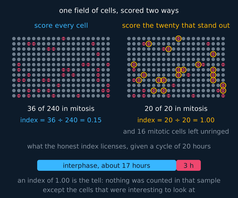
The same field of two hundred and forty cells drawn twice, with thirty-six of them shown in a visibly mitotic state. On the left every cell in the field is counted, giving thirty-six in mitosis out of two hundred and forty and an index of nought point one five. On the right the twenty most conspicuous cells are ringed, and because a cell with condensed chromosomes is what stands out down a microscope, all twenty of them are in mitosis and the index from that sample is one point nought nought, with sixteen mitotic cells left unringed and two hundred and twenty cells never counted at all. Beneath, a bar for a twenty-hour cycle is divided so that mitosis takes the honest index of it, which is about three hours, because the fraction of a population caught in a stage is the fraction of the cycle that stage occupies.
Counting
- Counting rules. Reach each field by moving the stage a fixed distance, not by looking for dividing cells. Decide the number of cells in advance, at least 200 over several fields. Count a cell crossing the top or left edge and ignore one crossing the bottom or right, so none is counted twice. Score each cell as interphase or one of the four stages, and decide beforehand whether very early prophase counts as dividing.
- Record the counts field by field in a table, total each column, and calculate the index from the totals.
Sample data
These counts are idealised. They were made up so that the fields add to the totals the lesson works, 200 cells with 40 of them in mitosis, and a real slide would not divide so evenly between fields. Nothing here is a record of an experiment.
| Field | Interphase | Prophase | Metaphase | Anaphase | Telophase | Total cells | Mitotic index |
|---|---|---|---|---|---|---|---|
| 1 | 30 | 4 | 2 | 1 | 1 | 38 | 0.21 |
| 2 | 35 | 5 | 2 | 1 | 1 | 44 | 0.20 |
| 3 | 33 | 5 | 1 | 2 | 0 | 41 | 0.20 |
| 4 | 29 | 4 | 1 | 1 | 1 | 36 | 0.19 |
| 5 | 33 | 4 | 2 | 1 | 1 | 41 | 0.20 |
| All five | 160 | 22 | 8 | 6 | 4 | 200 | 0.20 |
Cells in mitosis: 22 + 8 + 6 + 4 = 40. Mitotic index = 40 ÷ 200 = 0.20.
The index is calculated from the totals, not by averaging the five field indices, because the fields hold different numbers of cells. Here the two give the same answer; with fields of very different sizes they would not.
The cell cycle in these roots takes 24 hours. Metaphase holds 8 ÷ 200 = 0.04 of the cells, so it takes 0.04 of the cycle: 0.04 × 24 h = 0.96 h, about 58 minutes. The same arithmetic gives prophase 22 ÷ 200 × 24 h = 2.64 h, anaphase 0.72 h, about 43 minutes, telophase 0.48 h, about 29 minutes, and interphase 19.2 h.
The five field indices run from 0.19 to 0.21. That spread is the variation between fields, and it is why one field is not a count: a field placed on a patch of dividing cells could have given 0.30.
Reading the graph
The stages are categories, not points on a scale, so the chart is a bar chart: the five stages along the horizontal axis, in the order they happen, and the time in each stage, in hours, up the vertical axis. The bars do not touch.
Interphase dominates. Its bar is 19.2 h of a 24 h cycle, 80% of it. Even a tissue dividing as fast as it can spends most of its time not dividing, so an index of 0.20 means an active meristem, not a slow one.
Prophase is the longest stage of mitosis at 2.64 h, and telophase the shortest at 0.48 h. That is the reading the snapshot licenses. The stage with the most cells in it is the stage that lasts longest, not the stage that matters most.
What the counts cannot show. How fast the tissue divides. Two tissues with the same index divide at different rates if their cycles are different lengths, so a comparison of indices between tissues is a comparison of proportions only.
The small stages are the uncertain ones. Four cells in telophase is a count where one cell more or less changes the answer by a quarter. A longer count narrows that far more for the short stages than for interphase.
Evaluation
Each of these is a real source of error in this investigation, and the middle column is the part an answer earns marks for: what it does to the result, and in which direction.
| Source of error | What it does to the result | What to do about it |
|---|---|---|
| Tissue from behind the meristem | Cells that have stopped dividing are counted, so the index comes out too low. | Cut only the terminal few millimetres |
| Squash too light, or too heavy | Overlapping cells hide stages, and burst cells scatter their chromosomes, so some cells cannot be scored and the ones that can are not a fair sample of the field. | Press once, firmly and vertically |
| Stages judged differently | Late prophase is easily scored as metaphase, and early prophase as interphase, so counts differ between students in both directions. | Agree the criteria; have a second person recount the same fields |
| Fields chosen for their dividing cells | A field picked because it looks interesting holds more cells in mitosis than the tissue does, so the index comes out too high. | Move the stage a fixed distance between fields, and count every cell in each. |
| Too few cells counted | The short stages hold a handful of cells each, so the index and every stage length carry a large uncertainty. | Count at least 200 cells, and more when the stage lengths are wanted, and state the number counted beside the index. |
Questions
Question 1 (2 marks)
Explain why the root tip is put in warm hydrochloric acid before it is stained.
Check your answer
the acid breaks down the middle lamella, so the cells separate and can be squashed into a single layer;
it kills the cells, so each stops at the stage of mitosis it had reached.
Question 2 (2 marks)
Of 200 cells counted, 160 were in interphase. Calculate the mitotic index.
Check your answer
cells in mitosis = 200 − 160 = 40;
mitotic index = 40 ÷ 200 = 0.20 (or 20%).
Question 3 (3 marks)
The cell cycle lasts 24 hours and 8 of the 200 cells were in metaphase. Calculate the length of metaphase in minutes, and state the assumption the calculation makes.
Check your answer
8 ÷ 200 = 0.04 of the cycle;
0.04 × 24 × 60 = 57.6, about 58 minutes;
the cells divide out of step, so the proportion in a stage equals the proportion of the cycle that stage takes.
Question 4 (2 marks)
Explain why the coverslip is pressed straight down rather than pushed sideways.
Check your answer
pressing straight down spreads the cells into one layer;
sideways movement rolls cells over each other and smears the chromosomes, so stages cannot be identified.
Question 5 (3 marks)
A student chooses fields that contain many dividing cells. Explain the effect on the mitotic index, and how the fields should be chosen instead.
Check your answer
the fields are not representative of the meristem;
so the index is too high;
move the stage by a fixed distance between fields (or use random coordinates) and count every cell in each field.
Coverage maps are available for AQA 7401/7402, OCR Biology A H020/H420, Cambridge International 9700, Pearson Edexcel Biology A (Salters-Nuffield) 8BN0/9BN0 and Pearson Edexcel Biology B 8BI0/9BI0. Use the relevant route map to check what InkBiology covers, and your awarding organisation’s current specification as the final authority on examinable content.