Biology › Cell cycles, reproduction and development › Homeobox genes, Hox genes and apoptosis
Homeobox genes, Hox genes and apoptosis
A homeobox is a conserved DNA sequence of 180 base pairs that codes for a 60-amino-acid homeodomain. Homeodomain proteins act as transcription factors during development. Hox genes help specify regions along the anterior-posterior axis, while apoptosis removes selected cells during development and tissue maintenance.
Before this The cell cycle and mitosis · Control of gene expression · Gametes and fertilisation
COMMON MISCONCEPTION
Cell death is damage. A cell dies when something has gone wrong with it, so an organism that is developing normally is not losing cells.
Apoptosis is controlled cell death used in development and tissue maintenance. During digit development, mitosis forms the tissue and apoptosis removes cells between the developing digits. Membrane-bound fragments are then removed by phagocytes.
What you should be able to do
- State what the homeobox is, how long it is, and what the homeodomain it codes for does.
- Explain why a homeobox gene can change a whole body region although it codes for only one protein.
- Describe the colinear arrangement of a Hox cluster, and say what colinearity means.
- Describe the stages of apoptosis in order, and give the signals that control it.
- Explain how mitosis and apoptosis together produce a structure, using the separation of the digits.
- Explain what follows when the balance between mitosis and apoptosis is lost, in either direction.
The homeobox and the homeodomain
Development depends on controlled gene expression as well as cell division. Homeobox genes code for transcription factors that regulate groups of other genes, allowing a single regulatory gene to influence the identity and development of a body region.
The homeobox is a sequence of 180 base pairs. It lies within a gene rather than constituting the whole of one: the genes that carry it are thousands of bases long. Three bases code for one amino acid, so 180 bases code for 60, and those 60 amino acids in the finished protein are the homeodomain.
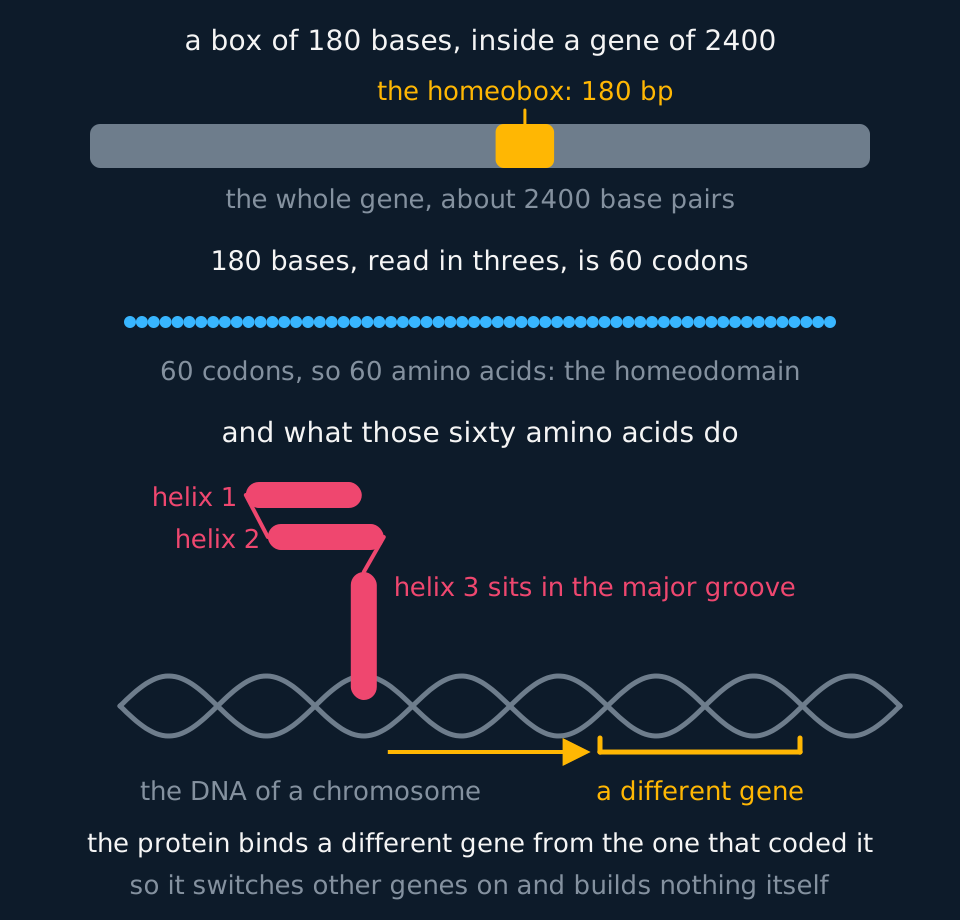
The homeodomain folds into a compact shape, three short helices with a turn between two of them, and one of those helices sits into the major groove of a DNA double helix. A protein that binds to DNA at a particular sequence and alters whether the gene there is transcribed is a transcription factor, and that is what a homeobox gene codes for. A homeobox gene therefore contributes no structural component itself; it regulates the transcription of other genes, whose products build the structure.
The sequence is also extraordinarily conserved. The homeobox of a mouse gene and the homeobox of the fruit fly gene that answers to it differ by only a handful of bases, across animals whose ancestors parted company well over five hundred million years ago, and the mouse version can be put into a fly embryo and do the fly gene's job. Conservation on that scale is evidence of two things at once: that the animals share a common ancestor which already had these genes, and that almost any change to them is fatal, so almost none of the changes that arose was ever passed on.
- Homeobox
- A sequence of 180 base pairs found within many genes that control development, coding for the part of the protein that binds to DNA.
- Homeodomain
- The 60 amino acid section of a protein coded for by a homeobox, which folds into a shape that binds to a specific DNA sequence.
- Transcription factor
- A protein that binds to DNA near a gene and controls whether that gene is transcribed into mRNA.
- Conserved sequence
- A sequence of bases that has changed very little between species over long periods, because nearly every mutation in it is harmful and is not passed on.
Hox genes and colinearity
Homeobox genes form a large family, and Hox genes are the group that helps specify what each region along the anterior-posterior axis of an animal becomes. They are not scattered through the genome; they lie together in a cluster, and the order within that cluster is examinable.
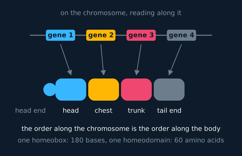
This match between the two orders is called colinearity. The gene at one end of the cluster is expressed in and specifies the most anterior region, the next gene along specifies the next region back, and the gene at the far end specifies the tail end. Nothing about a gene's chemistry requires it to sit where it does, so the arrangement is a piece of history: the cluster arose by one ancestral gene being duplicated along a chromosome, and each copy took on the region next along.
The fruit fly has one such cluster, in two pieces. A mouse and a human have four clusters, on four different chromosomes, holding thirty-nine Hox genes between them, which is the same cluster duplicated whole twice over in an early vertebrate. Each of the four keeps its internal order, so colinearity survived the duplications.
A Hox gene acts as a transcription factor within its region. Its protein binds to the control sequences of many other genes and increases their transcription, and the products of those genes determine that the region develops as a thorax rather than a head: the cells there divide on a particular schedule, differentiate into particular types and form particular structures. A mutation in one such gene can therefore have a large effect, because it alters the expression of every gene downstream of it.
The clearest evidence comes from mutations that give a region the identity of another region. A fly carrying a particular mutation in the gene called Antennapedia grows a pair of legs on its head where its antennae should be. The legs are complete and normally formed, so the genes that build a leg are intact; they were expressed in the wrong place. A mutation with that effect is a homeotic mutation, and homeotic is the word the homeobox is named after.
| Word | What it names | The mistake it prevents |
|---|---|---|
| Homeobox | A 180 base pair sequence inside a gene | Calling the whole gene the homeobox |
| Homeodomain | The 60 amino acid part of the protein it codes for | Confusing the piece of DNA with the piece of protein |
| Homeobox gene | Any gene containing a homeobox | Assuming every one of them is a Hox gene |
| Hox gene | A homeobox gene of the clustered family that specifies regions along the head to tail axis | Thinking they are the only genes with a homeobox |
| Homeotic mutation | A mutation that makes one region develop with the identity of another | Describing it as a structure being deleted |
Reading a colinear map
A short cluster of four Hox genes is arranged along a chromosome in the order W, X, Y, Z. Gene W is expressed in the most anterior region. An embryo is found in which gene Y is switched on throughout the region normally specified by gene X, as well as in its own. Predict what this embryo will look like, and explain what colinearity implies about where gene Z acts.
Show the working
The region normally specified by X will develop as though it were the region specified by Y, because the identity of a region is set by the Hox protein present in it and gene Y's protein is now there. The embryo has two copies of one region and lacks the region X would have specified. Nothing is missing from the genome and no structure is deleted; one region has developed with the identity of another.
Colinearity says gene Z acts furthest back, at the tail end, because it lies last in the cluster and the two orders match along their whole length.
Do not describe the embryo as having lost gene X. The gene is present and intact; the change is in which region expresses which Hox protein, which is what a homeotic mutation does.
Apoptosis
Apoptosis is programmed cell death. Intracellular enzymes dismantle the cell in a controlled sequence while the cell-surface membrane remains intact. The cell fragments into membrane-bound apoptotic bodies, which are removed by phagocytosis without releasing their contents into the surrounding tissue.
Apoptosis occurs on a large scale in healthy tissue. Estimates for an adult human are of the order of ten thousand million cells a day, with mitosis replacing them.
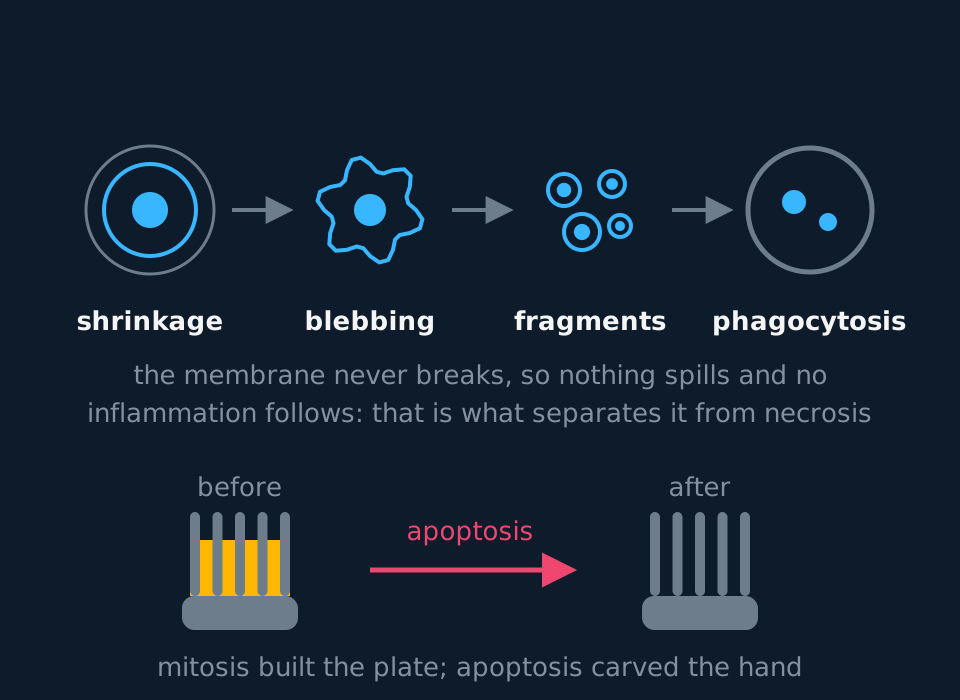
The order of the steps is examinable. Enzymes inside the cell break down its cytoskeleton, so the cell loses its shape and shrinks, and the cytoplasm grows dense with organelles packed close together. The chromatin condenses, the nuclear envelope breaks down, and the DNA is cut into fragments. The cell surface membrane changes and begins to bleb, bulging outwards without tearing. The cell then breaks up into apoptotic bodies, each one a piece of the cell still wrapped in membrane. Finally those bodies are recognised and taken in by phagocytosis, and their contents are digested and the materials reused.
Everything in that sequence turns on the membrane staying whole. A cell killed by injury, by poison or by a shortage of oxygen swells instead, bursts, and spills hydrolytic enzymes and other contents into the tissue around it, which damages neighbouring cells and provokes inflammation. That is necrosis. State the contrast in those terms: apoptosis is controlled and does not release cell contents, whereas necrosis is uncontrolled and causes inflammation.
| Apoptosis | Necrosis | |
|---|---|---|
| What starts it | A signal the cell responds to | Injury, toxin, or lack of oxygen or nutrients |
| The cell's volume | Shrinks | Swells |
| The membrane | Stays intact, blebs, then wraps each fragment | Breaks, and the contents escape |
| Effect on neighbours | None; the fragments are engulfed | Damage to nearby cells and inflammation |
| Is it controlled? | Yes, by signals and by the cell's own enzymes | No |
Apoptosis is switched on and off by cell signalling, from outside the cell and from inside it. Signals from outside include cytokines released by cells of the immune system, hormones, growth factors and nitric oxide, and the same signal can point either way depending on the cell that receives it: withdrawal of a growth factor initiates apoptosis as effectively as delivery of a cytokine does. Internal signals arise from the cell's own condition, and DNA damage that the repair systems cannot correct is the example to learn. Both routes converge on activation of the enzymes that carry out the sequence, so initiation is regulated while the sequence itself is stereotyped.
- Apoptosis
- Programmed cell death, in which a cell is broken down in an ordered sequence by its own enzymes and the fragments are removed by phagocytosis, without the release of the cell's contents.
- Necrosis
- Death of a cell caused by injury or by conditions it cannot survive, in which the cell swells, the membrane breaks and the contents escape and cause inflammation.
- Apoptotic body
- A membrane-bound fragment of a cell undergoing apoptosis, engulfed whole by a phagocyte.
- Blebbing
- The bulging of the cell surface membrane during apoptosis, which changes the shape of the cell without the membrane breaking.
Mitosis and apoptosis in digit development
Mitosis adds cells and apoptosis removes them, and a structure develops where the two are regulated relative to each other in space. Mitosis alone would produce a mass of tissue; apoptosis alone would remove it. The developing hand is the standard example of the two acting together.
In a human embryo the hand begins as a paddle. Cell division across the whole plate builds a flat, rounded lump of tissue with five thickenings inside it, and at that stage there are no separate fingers because the tissue between them is still there. Apoptosis then removes that tissue, cell by cell, along four strips running out from the wrist. No tissue is cut or reabsorbed by a neighbouring structure; the cells of the interdigital tissue receive a signal that initiates apoptosis, and phagocytes remove the fragments. What remains is five separate digits.
Mitosis forms the digit tissue; apoptosis removes cells between the developing digits. The digit count is the same before and after, because the digits were already specified by the regulatory genes described above; apoptosis removed the tissue connecting them.
The same pairing does the same job elsewhere. A tadpole's tail is removed by apoptosis during metamorphosis rather than being shed or digested from outside. A developing nervous system produces far more neurones than it keeps, and those that fail to make a working connection with a target cell no longer receive the growth factor that connection supplies, and die. In the immune system, lymphocytes that would react against the body's own tissue are removed by apoptosis before release. Next topic for that last example: the specific immune response.
Explaining a webbed hand
In a small number of babies two or more fingers are joined by a web of skin at birth, a condition called syndactyly. Explain how this arises, and state what has not gone wrong.
Show the working
The tissue between the developing digits is normally removed by apoptosis, in strips, during the weeks in which the hand plate is taking shape. If the cells of that tissue do not receive the signal to undergo apoptosis, or cannot respond to it, they are not removed. They stay where they are, and the digits stay joined by them.
What has not gone wrong is the number of digits or the building of them. Mitosis produced the whole plate as it should have done, and the five digits are specified and present. The fault is in the removal step alone.
State it as 'apoptosis of the tissue between the digits did not take place'. Describing the fingers as having failed to grow apart, or as having fused together later, reverses the mechanism: the tissue between them was present from the start and was not removed.
Consequences of an imbalance between the two
A tissue maintains its size because the rate of mitosis and the rate of apoptosis are matched. A sustained difference between the two rates changes the tissue, and the two directions lead to different kinds of disease.
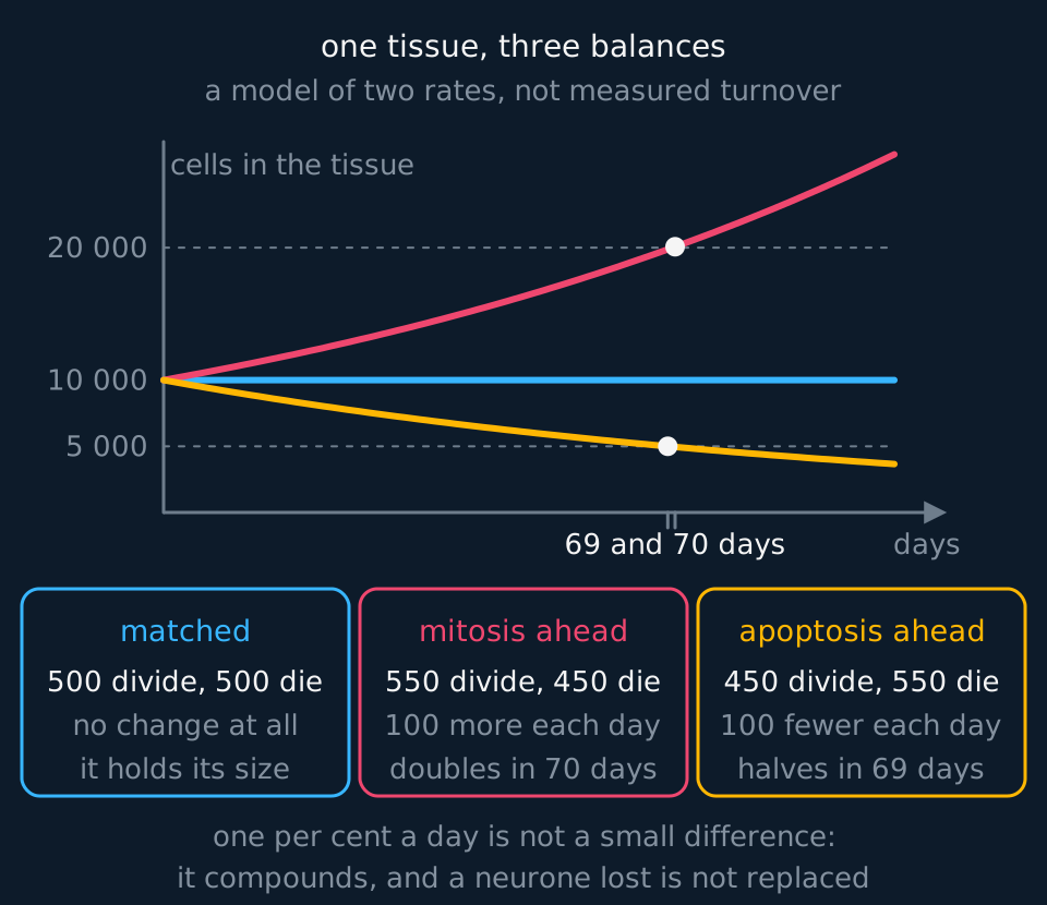
Consider too little apoptosis first. Prerequisite: the cell-cycle checkpoints. A cell held at a checkpoint with damage it cannot repair does not remain there indefinitely. It is normally removed by apoptosis, and the protein most often responsible is the product of a tumour suppressor gene called p53, which halts the cycle while repair is attempted and initiates apoptosis when repair fails. A cell therefore has two safeguards against passing on damaged DNA: it can stop dividing, and it can be removed. A mutation in the gene providing both removes both. The damaged cell divides, its daughters inherit the damage and the same defect, and further mutations accumulate in that lineage, which is one route to a tumour. Next topic: mutation, gene expression and cancer.
In the other direction, excessive apoptosis destroys tissue that is healthy and cannot be replaced. Neurones are the clearest case, because most of them have left the cell cycle for good, so a neurone lost is a neurone gone; excessive apoptosis of neurones is part of the damage in neurodegenerative disease and in the tissue around a stroke. The same logic explains why a drug that switched apoptosis off everywhere would be a poor idea however useful it sounded for protecting neurones, and why one that switched it on everywhere would be worse.
Two general points follow. Development is controlled by genes that regulate other genes, which is why relatively few are required and why one mutation can have such a large effect. And a body plan is produced by the removal of cells as well as by their proliferation, with the removal regulated as closely as the growth.
TRY IT: Linking apoptosis to a tumour
Cells taken from a tumour are found to carry a mutation in the p53 gene. Explain how this mutation contributes to the growth of the tumour, referring to both the cell cycle and apoptosis. (4 marks)
Check your answer
The protein coded for by p53 normally halts the cell cycle at a checkpoint when DNA damage is detected, so that the damage can be repaired before the DNA is replicated and passed on.
It also triggers apoptosis in a cell whose damage cannot be repaired, so that the cell is broken down and removed rather than dividing.
A mutation in the gene means the protein is not produced, or is produced in a form that does not work, so neither the halt nor the removal happens.
Cells with damaged DNA therefore continue through the cycle and divide. Mutations accumulate in their descendants, the rate of division exceeds the rate of apoptosis, and a mass of cells builds up.
Include the apoptosis step as well as the checkpoint: the question names both.
In the exam
- Quote both numbers together: 180 base pairs of homeobox, 60 amino acids of homeodomain. State which is DNA and which is protein.
- State that a homeobox gene codes for a transcription factor, then explain what a transcription factor does to the genes it regulates.
- Colinearity is a match between two orders: the order of the genes along the chromosome and the order of the regions along the body. Name both orders.
- Describe apoptosis in sequence and state that the cell-surface membrane remains intact throughout. In a comparison with necrosis, the release of cell contents is the distinguishing feature.
- For the digits, state what each process contributed: mitosis formed the hand plate, apoptosis removed the tissue between the digits. The number of digits does not change.
- For cancer, give both parts: increased division and reduced apoptosis, naming the checkpoint and the removal step.
Check yourself
A homeobox gene in a mouse and the equivalent gene in a fruit fly have homeobox sequences that differ at only a few bases. Explain what a homeobox codes for, why a mutation in one can alter a whole region of an animal, and what the similarity between the two species suggests.
Answer
A homeobox is 180 base pairs long and codes for a homeodomain of 60 amino acids. The homeodomain is the part of the protein that binds to DNA, so the protein is a transcription factor.
A mutation in it can alter a whole region because the protein does not build any structure itself. It binds near a long list of other genes and controls whether they are transcribed, so changing it changes the whole list, and every cell of that region develops according to the wrong list. A region can end up with the identity of a different region, which is a homeotic mutation.
The similarity suggests that the two species inherited the sequence from a common ancestor, and that the sequence has been conserved because almost any change to it is harmful and so was not passed on. Genes this important to development have very little room to vary.
The similarity is in the regulatory sequence, not in the body plan. A fly and a mouse develop very different body plans from homeodomain proteins that bind DNA in the same way.
Questions
Question 14 marks
Describe the changes that take place in a cell during apoptosis, in the order in which they occur.
Mark scheme
- B1 enzymes break down the cytoskeleton, so the cell shrinks and the cytoplasm becomes dense with closely packed organelles
- B1 the chromatin condenses, the nuclear envelope breaks down and the DNA is cut into fragments
- B1 the cell surface membrane blebs, bulging outwards without breaking, and the cell separates into apoptotic bodies each wrapped in membrane
- B1 the apoptotic bodies are engulfed by phagocytosis and their contents are digested and reused
Question 24 marks
A homeobox of 180 base pairs lies within a gene 1200 base pairs long. Calculate the number of amino acids in the homeodomain, and the percentage of the gene that the homeobox occupies.
Mark scheme
- M1 three bases code for one amino acid, so divide 180 by 3
- A1 60 amino acids
- M1 180 divided by 1200, multiplied by 100
- A1 15 per cent
Question 34 marks
In a fly carrying a mutation in the gene Antennapedia, a pair of legs grows on the head where the antennae should be, and the legs are complete and normally formed. Explain what this shows about how homeotic mutations work, and explain why this is called a homeotic mutation rather than a loss of a structure.
Mark scheme
- B1 the legs are complete and normally formed, so the genes that build a leg are intact and functioning correctly
- B1 what has changed is where those leg-building genes are expressed: they are switched on in the head region instead of the region that normally specifies legs
- B1 this shows that a Hox gene's role is to specify the identity of a region, activating the developmental programme appropriate to it, rather than to build any structure directly
- A1 it is called a homeotic mutation because one region develops with the identity of another region, rather than because any structure has been lost or deleted
Question 43 marks
Explain why a mutation in a single homeobox gene can alter the development of a whole region of an animal.
Mark scheme
- B1 the homeodomain binds to DNA, so the protein coded for by a homeobox gene is a transcription factor
- B1 it controls the transcription of many other genes, and those genes are the ones that build the structures of the region
- A1 a change to the transcription factor therefore changes which set of genes is switched on in every cell of that region, so the region can develop with the identity of a different region
Question 53 marks
Compare the events of apoptosis with the events of necrosis in a tissue.
Mark scheme
- B1 a cell undergoing apoptosis shrinks, whereas a cell undergoing necrosis swells
- B1 in apoptosis the cell surface membrane stays intact and wraps every fragment, whereas in necrosis the membrane breaks and the cell contents escape
- B1 apoptosis is controlled by signals and by the cell's own enzymes and causes no inflammation, whereas necrosis follows injury or a shortage of oxygen or nutrients and damages neighbouring cells, causing inflammation
Question 63 marks
The hand of a human embryo is at first a flat plate of tissue with no separate fingers. Explain how mitosis and apoptosis together produce five separate digits.
Mark scheme
- B1 mitosis produces the cells of the whole hand plate, including the tissue lying between the developing digits
- B1 the cells of the tissue between the digits receive a signal and undergo apoptosis, and the apoptotic bodies are removed by phagocytosis
- A1 the five digits are left free of one another; the number of digits is unchanged, because it is the webbing between them that is removed
Question 73 marks
Cells in some tumours fail to undergo apoptosis even though their DNA is damaged. Suggest how this failure contributes to the growth of the tumour.
Mark scheme
- B1 a cell with DNA damage that cannot be repaired is normally removed by apoptosis, so that the damage is not passed on
- B1 if apoptosis does not happen the damaged cell survives and continues through the cell cycle, and its daughter cells inherit the damage
- B1 further mutations accumulate in those descendants, the rate of division exceeds the rate of cell death, and a mass of cells builds up
Question 83 marks
A homeobox sequence from a mouse gene can be inserted into a fruit fly embryo and successfully carry out the fly gene's own job, even though the two species' ancestors diverged over five hundred million years ago. Suggest what this shows about the two species, and suggest why almost no changes to this sequence have been passed on over that time.
Mark scheme
- B1 it shows that the two species inherited the homeobox sequence from a shared common ancestor that already possessed it
- B1 it shows the sequence and its function have been conserved across an enormous span of evolutionary time, since a fly protein and its distant mammalian relative can still substitute for one another
- A1 almost any mutation to a sequence this central to development is likely to be harmful, so individuals carrying it are strongly selected against and the mutation is rarely passed on
Question 93 marks
Withdrawal of a growth factor from a cell can trigger apoptosis just as effectively as delivery of a cytokine can. Explain how a signal from outside a cell and a signal from inside a cell can both lead to apoptosis, giving one example of each.
Mark scheme
- B1 external signals include cytokines released by cells of the immune system, hormones, growth factors and nitric oxide, and either the delivery of one of these or, for a growth factor, its withdrawal can initiate apoptosis
- B1 internal signals arise from the cell's own condition, and the example to give is DNA damage that the cell's repair systems cannot correct
- A1 both kinds of signal converge on activating the same enzymes, which then carry out the same ordered sequence of apoptosis regardless of which signal started it
Question 103 marks
Outline how a failure of apoptosis between the developing digits can result in a baby being born with fingers joined by a web of skin, a condition called syndactyly.
Mark scheme
- B1 the tissue between the developing digits is normally removed by apoptosis, in strips, as the hand plate takes shape
- B1 if the cells of that tissue do not receive, or cannot respond to, the signal to undergo apoptosis, they are not removed and remain in place
- A1 the digits themselves have formed normally and are present in the correct number; only the removal of the tissue joining them has failed
Question 113 marks
In a healthy adult, apoptosis removes cells at a rate of the order of ten thousand million cells a day, and mitosis replaces them at a matching rate. Calculate the approximate number of cells replaced in one year, and give your answer in standard form to two significant figures.
Mark scheme
- M1 number per year = number per day × 365
- M1 1 × 1010 × 365
- A1 3.7 × 10¹² cells replaced in a year (accept 3.65 × 10¹²)
Question 123 marks
Compare the consequence of too little apoptosis in a cell with damaged DNA with the consequence of too much apoptosis in neurones, referring to a named condition each can lead to.
Mark scheme
- B1 too little apoptosis lets a cell with DNA damage it cannot repair go on dividing, whereas too much apoptosis destroys cells, such as neurones, that are healthy and often cannot be replaced
- B1 too little apoptosis, combined with unchecked division, is one route to a tumour building up from the damaged cell's descendants, whereas too much apoptosis of neurones contributes to neurodegenerative disease and to the tissue damage seen around a stroke
- A1 both show that apoptosis must be matched to mitosis: neither too little nor too much keeps a tissue at a stable, healthy size
Question 132 marks
State the length of a homeobox in base pairs, and the length in amino acids of the homeodomain it codes for.
Mark scheme
- B1 the homeobox is 180 base pairs long
- B1 the homeodomain is 60 amino acids long, because three bases code for one amino acid
Question 142 marks
State what colinearity means in a Hox gene cluster, and state how many Hox gene clusters a human has.
Mark scheme
- B1 colinearity is the match between the order of the Hox genes along the chromosome and the order of the body regions they specify, from head to tail
- B1 a human has four Hox gene clusters, on four different chromosomes
Question 152 marks
State the name of the gene whose protein product halts the cell cycle at a checkpoint and triggers apoptosis when DNA damage cannot be repaired, and state what type of gene this is.
Mark scheme
- B1 the gene is p53
- B1 it is a tumour suppressor gene
Worth remembering
- The homeobox is 180 base pairs of DNA; the homeodomain is the 60 amino acids it codes for.
- Homeobox genes code for transcription factors, so they regulate other genes rather than contributing a structural product.
- Hox genes lie in a cluster whose order along the chromosome is the order of the regions they specify from head to tail, which is colinearity.
- Apoptosis runs in order: the cell shrinks and the chromatin condenses, the membrane blebs, the cell breaks into membrane-wrapped apoptotic bodies, and a phagocyte engulfs them.
- The membrane stays intact in apoptosis and breaks in necrosis, which is why one causes inflammation and the other does not.
- Mitosis and apoptosis act together in development: mitosis forms the digit tissue, apoptosis removes cells between the developing digits, and the digit count does not change.
- Too little apoptosis alongside unchecked division gives a tumour; too much destroys healthy tissue that may not be replaceable.
CHECK YOUR PROGRESS
Rate how confident you are with each objective for this lesson. Ratings are saved in this browser, on this device, unless you sign in.
- State what the homeobox is, how long it is, and what the homeodomain it codes for does.
- Explain why a homeobox gene can change a whole body region although it codes for only one protein.
- Describe the colinear arrangement of a Hox cluster, and say what colinearity means.
- Describe the stages of apoptosis in order, and give the signals that control it.
- Explain how mitosis and apoptosis together produce a structure, using the separation of the digits.
- Explain what follows when the balance between mitosis and apoptosis is lost, in either direction.
Open the full revision checklist to see every objective in the curriculum in one place.
Practise this lesson
WORKBOOK
The same questions as the player, on paper with room to work, and a separate book of mark schemes. Free to use; please do not redistribute or sell.