Biology › Cell cycles, reproduction and development › The cell cycle and mitosis
The cell cycle and mitosis
The cell cycle takes about a day in a rapidly dividing cell, and mitosis occupies roughly a tenth of that time. The whole cycle is covered here: the interphase stages in which the DNA is replicated and checked, the four stages of mitosis, and cytokinesis.
Before this DNA replication · Eukaryotic cell ultrastructure
COMMON MISCONCEPTION
Cells spend most of their lives dividing, and interphase is the resting phase between divisions.
Interphase is the busiest part of the cycle, not a rest. The cell grows, replicates its whole genome and checks the copy, and this takes most of the cycle. Mitosis is the short part at the end.
What you should be able to do
- Describe what happens in each phase of the cell cycle, and say which phase takes longest.
- State what each of the three checkpoints checks, and what becomes of a cell that fails one.
- Explain why a chromosome has two chromatids before mitosis and one after it.
- Explain why replication leaves the end of the lagging strand uncopied, and say what a telomere therefore protects and what telomerase does about it.
- Put the four stages of mitosis in order and say what defines each one, including what the spindle does.
- Calculate a mitotic index from a count of cells and interpret what it means.
- Describe binary fission in a prokaryote and set it against mitosis in a eukaryote.
- Explain how uncontrolled cell division leads to a tumour, using the cycle's checkpoints.
- Prepare a stained root tip squash, count its cells by fixed rules, and work out how long each stage of mitosis lasts from the counts.
Three phases before anything divides
A cell that is going to divide works through the same sequence every time. In G1 it grows and makes more of the organelles it will have to share out. In S phase it replicates its DNA, semi-conservatively, so that every chromosome ends up as two identical sister chromatids held together at a centromere. In G2 it keeps growing and checks the copy for errors. Only then does mitosis begin.
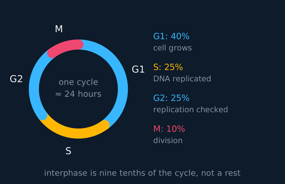
That ring is drawn for a cell cycling in about 24 hours, and the proportions are a typical case rather than a constant of nature. G1 stretches and shrinks: a root-tip meristem cell runs the whole cycle in less than a day, and mitosis then takes a larger share of a smaller total. Keep the ordering, which never changes, and treat the exact tenth as a figure attached to a 24-hour cycle.
Some cells leave the cycle. A neurone or a mature muscle fibre stops at the end of G1 and enters G0, which is less a phase than the exit from one. Neither of those cells will replicate its DNA again, but other cells in G0, such as liver cells and resting lymphocytes, can be signalled back into the cycle. Most of your cells are in G0 now.
The chromosome count does not change during S phase, which trips people up. A human cell in G1 has 46 chromosomes; after S phase it still has 46 chromosomes, but each one is now two chromatids, so there is twice as much DNA. Count centromeres, not strands, and the arithmetic behaves.
- Chromatid
- One of the two identical copies of a chromosome, joined to its sister at the centromere after DNA replication.
- Homologous pair
- Two chromosomes of the same size carrying the same genes at the same loci, one inherited from each parent. Not the same thing as a pair of chromatids.
- Spindle
- The framework of protein microtubules built across the cell before the chromosomes are moved, running from pole to pole and attaching to every centromere; shortening the fibres is what separates the chromatids.
- Mitotic index
- The proportion of cells in a sample that are visibly in mitosis.
Telomeres and the end-replication problem
S phase copies almost the whole chromosome. The exception is at the two ends, and it follows from the mechanism of replication rather than from a fault in any one enzyme.
Two constraints on DNA polymerase apply. It builds a new strand only in the 5 prime to 3 prime direction, and it can only add nucleotides to the 3 prime end of something that is already there, which is why a short RNA primer has to be laid down first. On the leading strand that is no trouble: one primer at the start, then the polymerase runs continuously to the far end of the template. The lagging strand is built backwards in fragments, each with its own primer, and the primers are afterwards removed and the gaps filled by extending the fragment next door. The last primer at the very end of the chromosome has no fragment beyond it. When it is removed there is no 3 prime end for the polymerase to work from, so that gap stays open and a short stretch of the end is never copied.
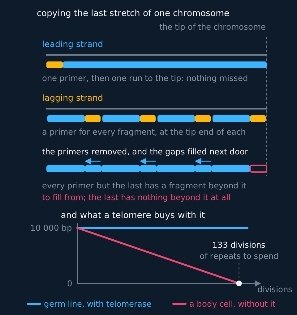
Describe the drawing
Two panels. In the upper one the leading strand at one end of a chromosome is drawn as a single primer followed by an unbroken run of new DNA reaching the tip, so every base of the template is copied. The lagging strand below it is drawn as four Okazaki fragments, each with its own primer at the end nearer the tip. When the primers are taken out, three of the gaps are filled by the fragment beyond them extending into the space, and the fourth, at the very tip, has no fragment beyond it to be extended from and stays open, so the tip goes uncopied and no better enzyme could reach it. The lower panel plots telomere length against division number from ten thousand base pairs of non-coding repeat: a body cell loses seventy-five base pairs a division and so has a hundred and thirty-three divisions of repeats to spend before the erosion reaches a gene, while a germ line cell, in which telomerase adds the repeats back, holds a flat line and can divide without limit, which is what most cancers switch back on.
A chromosome therefore becomes slightly shorter at every division. In a human cell the loss is of the order of fifty to a hundred base pairs per division, and it is cumulative: it continues at every division and is inherited by every descendant cell.
This is what the ends of chromosomes are for. Each one is capped by a telomere: a long run of a short sequence repeated over and over, TTAGGG in humans and something similar in most other eukaryote, thousands of copies of it, and coding for nothing. Because the repeats are non-coding, the shortening erodes repeats rather than genes, and a cell can divide many dozens of times before the erosion reaches a transcribed sequence.
The telomere has a second function. An uncapped double-stranded end resembles a chromosome broken in half. Repair enzymes join a free end to any other free end available, and the damage checkpoints halt the cycle until they have. Two chromosomes fused end to end are then torn apart at the next anaphase and the damage compounds. A telomere, with the proteins bound along it, is folded back and capped so that the repair enzymes do not act on it as though it were a break. A telomere therefore protects the coding sequence from erosion and protects the end itself from inappropriate repair.
Cells that divide indefinitely require their telomeres to be maintained. Telomerase is an enzyme that carries a short RNA molecule of its own and uses it as a template to add telomere repeats back onto the end, restoring what replication takes. It is active in the cells of the germ line, which make gametes and must hand a full-length chromosome to the next generation, and in stem cells and some cells of the immune system. In most ordinary body cells it is switched off, telomeres shorten with every division, and a cell whose telomeres have grown too short stops dividing altogether or dies. Most cancer cells express telomerase again, which is one factor allowing a tumour to continue dividing beyond the usual replicative limit.
- Telomere
- A cap of short non-coding sequences repeated many times at each end of a chromosome, which is eroded instead of the genes when replication fails to copy the end, and which stops the end being treated as a break in the DNA.
- Telomerase
- An enzyme carrying its own RNA template, which adds telomere repeats back onto the end of a chromosome; it is active in germ line cells, stem cells and most cancer cells.
The three checkpoints
Between the phases lie checkpoints. Learn each by what it checks as well as by name. The cycle does not proceed past a checkpoint until the relevant condition is met, and the checking is carried out by the cell's own regulatory proteins, which is why a mutation in one of those proteins has such large consequences.
| Checkpoint | Where it sits | The question it asks |
|---|---|---|
| G1 checkpoint | End of G1, before S phase | Is the cell big enough, are nutrients there, is the DNA undamaged? |
| G2 checkpoint | End of G2, before mitosis | Did replication finish, and finish without errors left in it? |
| Spindle checkpoint | Metaphase, before anaphase | Is every centromere attached to fibres from both poles? |
The G1 checkpoint is the point of no return. A cell that passes it is committed to copying its entire genome whether conditions stay favourable or not, so cell size, nutrients, signals from neighbours and DNA damage all get their say here. Failing it has more than one outcome: most cells pause, repair the fault and try again, some withdraw into G0, and a cell that cannot be mended may stop dividing permanently or be removed by apoptosis.
The G2 checkpoint is a proofreading stop. S phase copies about six thousand million base pairs in a human cell and does not do it flawlessly, so this checkpoint halts a cell with unrepaired breaks. Damage that gets past is handed to both daughters.
The spindle checkpoint sits inside mitosis rather than between two phases, and it is why anaphase is so abrupt: one chromosome not yet attached to both poles holds the whole cell at metaphase, and when the last attachment is made every centromere divides at once.
Distinguish two outcomes. A checkpoint delays the cycle, and most cells held at one repair the fault and proceed. Apoptosis ends the cell and occurs when repair fails.
The four stages of mitosis
Mitosis is conventionally divided into four stages. The division is a convention, since the process is continuous, but each stage is defined by features visible under a microscope, which is why a stage can be identified from a photograph.

Describe the drawing
Mitosis animated through its four stages in order. Prophase: the chromosomes condense inside the nucleus, each already two chromatids joined at a centromere, and the nuclear envelope begins to break down. Metaphase: the chromosomes line up along the equator with spindle fibres running to them from poles on each side. Anaphase: the centromeres divide and the chromatids are pulled to opposite poles. Telophase: a nuclear envelope re-forms around each group and the cytoplasm divides, giving two genetically identical daughter cells.
| Stage | What defines it | What to look for |
|---|---|---|
| Prophase | Chromosomes condense; the spindle forms; nuclear envelope breaks down | Visible chromosomes, no clear nuclear edge |
| Metaphase | Chromosomes line up on the equator, each centromere attached to spindle fibres from both poles | A single line of chromosomes across the middle |
| Anaphase | Centromeres divide; chromatids pulled to poles | Two groups moving apart, chromatids V-shaped as they are dragged |
| Telophase | Nuclear envelopes re-form; chromosomes decondense | Two clusters, each acquiring an envelope |
Two of the four stages are defined by the behaviour of the spindle. It is a framework of protein microtubules, assembled during prophase and running from one pole of the cell to the other. In an animal cell the poles are organised by a pair of centrioles; a plant cell assembles the same spindle without them. Fibres attach to every centromere from both sides, and anaphase consists of those fibres shortening.
Cytokinesis follows, and it is not part of mitosis proper: mitosis divides the nucleus, cytokinesis divides the cytoplasm. In animal cells a ring of protein filaments under the membrane contracts and pinches it inwards. In plant cells it cannot, because of the cell wall, so vesicles line up along the middle, fuse into a cell plate, and a new wall is built outwards from it. Organelles are distributed approximately rather than in equal numbers.
The mitotic index
The mitotic index is the calculation this topic requires, and it is obtained from a root-tip squash. The tip of a root is a meristem: a few millimetres in which every cell is still dividing. Tissue cut more than 5 mm back contains cells that have left the cycle and begun elongating.
The preparation determines whether the count is reliable. The tip is warmed in dilute hydrochloric acid, which breaks down the middle lamella holding the cells together, then stained with toluidine blue or acetic orcein, both of which bind to DNA, and squashed under a coverslip to a single layer. Overlapping layers cannot be counted reliably, because a chromosome in one plane of focus is not readily distinguished from one in another.
Then count. Score every cell in the field rather than a selection, because a sample chosen by eye is biased towards cells in mitosis. Count several fields and add the totals, and state the total number of cells counted alongside the index.
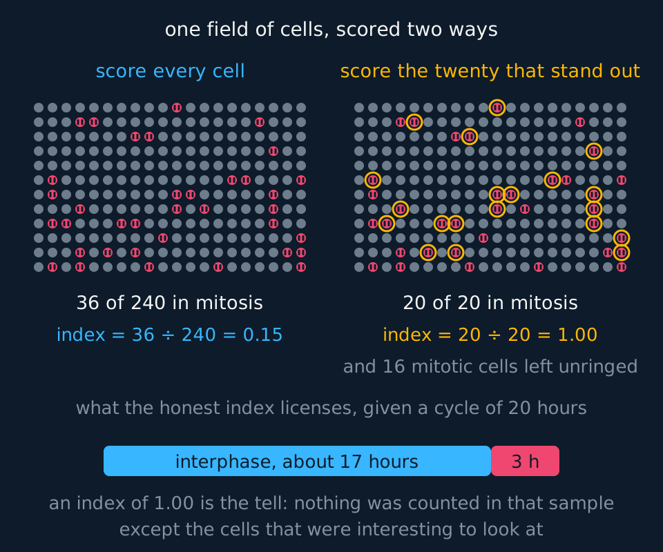
Describe the drawing
The same field of two hundred and forty cells drawn twice, with thirty-six of them shown in a visibly mitotic state. On the left every cell in the field is counted, giving thirty-six in mitosis out of two hundred and forty and an index of nought point one five. On the right the twenty most conspicuous cells are ringed, and because a cell with condensed chromosomes is what stands out down a microscope, all twenty of them are in mitosis and the index from that sample is one point nought nought, with sixteen mitotic cells left unringed and two hundred and twenty cells never counted at all. Beneath, a bar for a twenty-hour cycle is divided so that mitosis takes the honest index of it, which is about three hours, because the fraction of a population caught in a stage is the fraction of the cycle that stage occupies.
Reading a root tip
A student examines a stained root tip squash and counts 240 cells, of which 36 are visibly in mitosis. Calculate the mitotic index, and suggest what a much lower value in a second sample would indicate.
Show the working
The mitotic index is the proportion in mitosis: 36 ÷ 240 = 0.15, or 15%. Give it as a proportion or a percentage, but say which.
A much lower value means fewer cells are dividing at the moment of fixing. In a root tip that would suggest the sample came from further back from the tip, away from the meristem where division happens.
One caution about what the number means. Mitosis is a small fraction of any cycle, so even tissue dividing as fast as it can shows most of its cells in interphase, and 0.15 is a dividing tissue rather than a sluggish one. Published values for onion root tips vary with the temperature, the time of day the roots were fixed and how much of the tip went into the squash. Tissue from behind the meristem, where cells have stopped dividing, pulls the index down.
One inference the index licenses, and one it does not. A fixed slide is a snapshot of a population, so the fraction caught in a stage is the fraction of the cycle that stage occupies: cycle 20 hours, index 0.15, mitosis about 3 hours. What it cannot show alone is how fast the tissue divides, since equal indices mean different rates when the cycles differ in length.
Root tip squash practical: preparing, staining and counting
optional
AQA sets this as required practical 2: Preparation of stained squashes of cells from plant root tips; set-up and use of an optical microscope to identify the stages of mitosis in these stained squashes and calculation of a mitotic index. Pearson Edexcel Biology A (9BN0) sets Core Practical 5: Prepare and stain a root tip squash to observe the stages of mitosis. Biology B (9BI0) sets Core Practical 3: Make a temporary squash preparation of a root tip to show stages of mitosis in the meristem under the light microscope. OCR A examines the same stages and counts, and its practical group for microscope work is PAG1. Cambridge International 9700 does not name the squash, but examines the stages on slides and photomicrographs.
Grow garlic or onion roots over water until they are 1 to 2 cm long. Toluidine blue, acetic orcein or Feulgen reagent will stain them; Feulgen has its own sequence, so follow the method supplied with it.
| Step | What to do | Why |
|---|---|---|
| 1 | Cut off the terminal 5 mm or so of a root on a tile | This is the meristem; further back, cells have stopped dividing |
| 2 | Put the tip in warm hydrochloric acid, typically 1 mol dm⁻³ at about 60 °C in a water bath, for about 5 minutes | Breaks down the middle lamella so the cells separate. The hot acid also kills the cells, so each stays at the stage it had reached |
| 3 | Rinse the tip in cold water and blot it | Leftover acid would go on softening the tissue and interfere with the stain |
| 4 | On a slide, add a drop of stain and break the tip up with a mounted needle; leave it for the time the method gives | The stain binds DNA, so condensed chromosomes show darkly |
| 5 | Lower a coverslip, fold filter paper over it and press straight down with a thumb, without twisting | Spreads the cells into one layer; sideways movement rolls cells together and smears the chromosomes |
| 6 | Find the meristem at low power, then count at ×400 | Stages can be told apart only at high power |
Counting rules. Reach each field by moving the stage a fixed distance, not by looking for dividing cells. Decide the number of cells in advance, at least 200 over several fields. Count a cell crossing the top or left edge and ignore one crossing the bottom or right, so none is counted twice. Score each cell as interphase or one of the four stages, and decide beforehand whether very early prophase counts as dividing.
Counts from one root tip
These counts are idealised. Of 200 cells, 160 were in interphase, 22 in prophase, 8 in metaphase, 6 in anaphase and 4 in telophase. The cell cycle takes 24 hours. Calculate the mitotic index and the length of metaphase.
Show the working
Cells in mitosis: 22 + 8 + 6 + 4 = 40. Mitotic index = 40 ÷ 200 = 0.20.
Metaphase holds 8 ÷ 200 = 0.04 of the cells, so it takes 0.04 of the cycle: 0.04 × 24 h = 0.96 h, about 58 minutes.
This assumes the cells divide out of step, so a slide catches each stage in proportion to its length. The cycle time cannot be read from the slide and has to be given.
| Source of error | Effect | Improvement |
|---|---|---|
| Tissue from behind the meristem | Non-dividing cells lower the index | Cut only the terminal few millimetres |
| Squash too light, or too heavy | Overlapping cells hide stages; burst cells scatter chromosomes | Press once, firmly and vertically |
| Stages judged differently | Counts differ between students | Agree the criteria; have a second person recount the same fields |
Risk assessment. Hydrochloric acid can harm the eyes and skin, and its classification depends on its concentration. Stains mark skin and clothing, and acetic orcein is made up in ethanoic acid. Check the CLEAPSS Hazcard and your school's risk assessment for the acid and the stain, and where they differ from this page, follow them. Wear eye protection, warm the acid in a water bath, never over a flame, and put broken glass in the broken-glass container.
Binary fission, and how meiosis differs
Mitosis addresses two features of eukaryotic cells: a nuclear envelope that must be disassembled and rebuilt, and several long linear chromosomes that must be separated without tangling. A prokaryote has neither, and divides by binary fission. The prokaryote's single circular chromosome is replicated, and dedicated proteins move the two copies into opposite halves of the growing cell. Older textbooks had the copies attached to the membrane and carried apart passively as the cell grew, but the segregation is now known to be an active process. Plasmids are replicated too, though not to a fixed number, and a new membrane and wall are laid down across the middle.
State three differences from mitosis: there is no spindle, there is no nuclear envelope to break down or re-form, and there is no alignment at an equator, so do not describe the chromosome as lining up. Binary fission is also rapid: Escherichia coli divides in twenty minutes in favourable conditions, against about a day for a human cell.
The other nuclear division on the specifications is meiosis, which halves the chromosome number and produces genetically different cells, and is taught in meiosis and the sources of genetic variation.
| Feature | Mitosis | Binary fission | Meiosis |
|---|---|---|---|
| Cells produced | Two | Two | Four |
| Chromosome number | Unchanged | Unchanged | Halved |
| Daughters identical? | Yes, barring rare copying errors | Yes, apart from plasmid number | No, every one different |
| Spindle used? | Yes | No | Yes, in both divisions |
| What it is for | Growth, repair, asexual reproduction | Reproduction of the whole organism | Making gametes |
Loss of checkpoint control and tumour formation
Every daughter cell from mitosis is genetically identical to the parent, barring the rare copying error or missorted chromosome that slips through the checkpoints, which is what makes mitosis the right tool for growth, for repair, and for asexual reproduction: a yeast cell buds off a daughter cell, a Hydra grows a bud that breaks away, and a strawberry plant sends out runners, all by mitosis. The same property makes a fault in the process serious: an error is copied faithfully into everything descended from that cell.
Mutations in the genes controlling the checkpoints can leave a cell dividing when it should have stopped. Two categories matter. Proto-oncogenes normally stimulate division, and a mutation can leave one permanently switched on. Tumour suppressor genes normally halt the cycle at a checkpoint, and a mutation can prevent one being expressed. In either case the cycle is no longer restrained, division continues unchecked, and a mass of cells accumulates as a tumour.
That also explains why many chemotherapy drugs work the way they do. Some prevent DNA replication in S phase, some disrupt spindle formation so metaphase cannot complete. Neither targets cancer cells specifically. Both target dividing cells, which is why hair follicles and the gut lining, both dividing rapidly, suffer the familiar side effects.
TRY IT: Explaining a drug's side effects
A drug used in chemotherapy binds to tubulin and prevents spindle fibres forming. Explain how this stops a tumour growing, and why the patient loses their hair.
Check your answer
Without spindle fibres, chromosomes cannot be attached and lined up on the equator, so the cell cannot pass from metaphase into anaphase. Division stops, and the tumour stops growing.
The drug cannot distinguish a tumour cell from any other dividing cell. Cells in hair follicles divide rapidly, so they are blocked as well and hair is not replaced as it is shed.
Name the stage: 'chromosomes cannot align at metaphase, so anaphase never begins', rather than 'it stops mitosis'.
In the exam
- Name the phase, not just 'interphase'. G1, S and G2 are distinguished in questions.
- After replication a cell has the same number of chromosomes and twice the DNA. Apply that to graphs of DNA mass against time.
- Anaphase begins when the centromeres divide. Use that observable event to identify a stage from a photograph.
- State that cytokinesis is not a stage of mitosis, and describe it separately for plant and animal cells.
- The mitotic index is a proportion of the cells counted, so state the total counted alongside it.
- Name the spindle: 'the fibres cannot attach to the centromeres, so metaphase never completes', rather than 'the chromosomes cannot separate'.
- Give the reason for telomere shortening, not only the fact. The last primer on the lagging strand leaves a gap with no 3 prime end to extend from, so the very end is not copied and a short length is lost at each division.
- Give both functions of a telomere where the marks allow: the repeats are non-coding, so genes are not eroded, and the capped end is not treated as a broken chromosome.
Check yourself
A cell from a mouse has 40 chromosomes. State the number of chromosomes and the number of chromatids present in that cell at the end of G2, and in each daughter cell immediately after mitosis. Explain your answers.
Answer
At the end of G2 the cell has 40 chromosomes and 80 chromatids. S phase has replicated every chromosome, so each one is now two sister chromatids joined at a centromere, but the centromere count, and so the chromosome count, has not changed.
Immediately after mitosis each daughter cell has 40 chromosomes and 40 chromatids, or equivalently 40 single-chromatid chromosomes. Anaphase separated the sisters, and once separated each is a chromosome in its own right.
The difficulty is terminological rather than biological. A chromatid becomes a chromosome at the moment its centromere divides, so the same structure has two names depending on the stage.
Questions
Question 14 marks
Describe what happens to the chromosomes during each of the four stages of mitosis, in the order the stages occur.
Mark scheme
- B1 prophase: chromosomes condense and become visible, each already two sister chromatids
- B1 metaphase: chromosomes line up along the equator, attached to spindle fibres at their centromeres
- B1 anaphase: centromeres divide and the sister chromatids are pulled to opposite poles
- B1 telophase: chromosomes reach the poles, decondense, and a nuclear envelope re-forms around each group
Question 24 marks
Explain how a mutation in a tumour suppressor gene can lead to the formation of a tumour.
Mark scheme
- B1 tumour suppressor genes normally halt the cell cycle at a checkpoint
- B1 a mutation can switch the gene off, so the protein it codes for is not produced or does not function
- B1 the checkpoint no longer stops cells with damaged DNA or incomplete replication from dividing
- A1 division continues unchecked and a mass of cells accumulates, which is a tumour
Question 34 marks
Each chromosome carries a telomere at each of its ends. Explain why a short length of DNA is lost from the end of a chromosome every time a cell divides, and explain the two things a telomere protects.
Mark scheme
- B1 DNA polymerase builds a new strand only in the 5 prime to 3 prime direction and can only add nucleotides to an existing 3 prime end, so each fragment of the lagging strand has to be started with an RNA primer
- B1 when the last primer at the very end of the lagging strand is removed there is no fragment beyond it to extend from, so that gap cannot be filled and the end is left uncopied
- B1 a telomere is a short sequence repeated many times and codes for nothing, so the shortening removes repeats rather than genes and the cell can divide many times before a transcribed sequence is reached
- A1 the capped telomere also stops the end being treated as a broken chromosome, which would otherwise be joined to another end by repair enzymes or would halt the cycle at a damage checkpoint
Question 44 marks
Compare cytokinesis in an animal cell with cytokinesis in a plant cell, referring to what forms in each case.
Mark scheme
- B1 in an animal cell a ring of protein filaments contracts beneath the cell surface membrane and pinches the membrane inwards, whereas in a plant cell the rigid cell wall prevents this pinching
- B1 in the plant cell, instead, vesicles line up along the middle of the cell and fuse to form a cell plate, which does not happen in an animal cell
- B1 the cell plate is where a new cell wall is built outwards from, separating the two daughter cells, whereas the animal cell simply pinches apart with no wall formed between them
- A1 in both cases cytokinesis divides the cytoplasm and follows the division of the nucleus in mitosis, rather than being part of mitosis itself
Question 54 marks
Compare binary fission in a prokaryote with mitosis in a eukaryote, referring to the spindle, the nuclear envelope and the alignment of the chromosomes.
Mark scheme
- B1 mitosis uses a spindle of protein microtubules to move the chromosomes, whereas binary fission uses no spindle at all
- B1 mitosis breaks down and re-forms a nuclear envelope around each set of chromosomes, whereas a prokaryote has no nuclear envelope to break down or re-form
- B1 in mitosis the chromosomes line up at an equator before separating, whereas in binary fission the circular chromosome is not aligned at an equator, and dedicated proteins move the two copies apart
- A1 both processes produce two daughter cells, each carrying a complete copy of the parental DNA, but binary fission is far faster, dividing in about twenty minutes rather than about a day
Question 64 marks
Outline how a root tip is prepared and stained so that its mitotic index can be counted reliably.
Mark scheme
- B1 a root tip within about 5 mm of the tip is used, because that is the meristem where cells are still dividing
- B1 the tip is warmed in dilute hydrochloric acid, which breaks down the middle lamella holding the cells together
- B1 it is stained with a dye such as toluidine blue or acetic orcein, which binds to DNA and makes the chromosomes visible
- B1 it is then squashed under a coverslip to a single layer of cells, because overlapping layers cannot be counted reliably
Question 74 marks
A class prepared root tip squashes from one batch of garlic roots. Every student but one obtained a mitotic index between 0.15 and 0.20; that student obtained 0.05, having cut 15 mm from the end of each root, pressed the coverslip down lightly, and scored cells in early prophase as interphase. Explain how each of these three choices would lower the mitotic index, and suggest how the student could show that the low value was not caused by the way the cells were scored.
Mark scheme
- B1 a 15 mm piece includes tissue behind the meristem, where cells have stopped dividing and are elongating, so non-dividing cells are added to the total
- B1 a light squash leaves cells in overlapping layers, so chromosomes cannot be distinguished and dividing cells are missed or scored as interphase
- B1 cells in early prophase are in mitosis, so scoring them as interphase removes them from the number in mitosis; prophase is the longest stage, so many cells are affected
- B1 agree the criteria for each stage with the class and have a second person recount the same fields; accept scoring against reference photographs of each stage
Question 83 marks
A cell from a mouse has 40 chromosomes. Explain why the cell still has 40 chromosomes at the end of G2, even though the quantity of DNA in it has doubled.
Mark scheme
- B1 DNA is replicated during S phase, so each chromosome is copied
- B1 the two copies stay joined at a single centromere as sister chromatids
- A1 chromosome number is counted by centromeres, so it is unchanged at 40 while the DNA quantity is doubled
Question 93 marks
A student counts 240 cells in a stained root tip squash and finds 36 of them are visibly in mitosis. Calculate the mitotic index, and give your answer as a proportion.
Mark scheme
- M1 mitotic index = number of cells in mitosis divided by the total number of cells counted
- M1 36 divided by 240
- A1 0.15, or equivalently 15 per cent
Question 103 marks
A drug being tested as a cancer treatment binds to tubulin and prevents spindle fibres from forming. Suggest why this stops a tumour growing, and why the patient loses their hair.
Mark scheme
- B1 without spindle fibres the chromosomes cannot be attached and aligned at the equator, so the cell cannot pass from metaphase into anaphase
- B1 division stops, so the tumour cannot grow by producing more cells
- B1 the drug cannot distinguish tumour cells from any other dividing cells, and hair follicle cells divide rapidly, so hair is not replaced as it is shed
Question 113 marks
Telomerase is active in cells of the germ line and in stem cells, but is switched off in most other body cells. Most cancer cells switch it back on. Suggest why telomerase activity is needed in the germ line, and suggest how switching it back on helps a tumour to grow.
Mark scheme
- B1 telomerase adds telomere repeats back onto the end of a chromosome, using an RNA template it carries, so it restores what replication fails to copy
- B1 germ line cells give rise to gametes and so to the next generation, which must receive full-length chromosomes; without telomerase the telomeres would be shorter in every generation
- A1 a body cell whose telomeres become too short stops dividing or dies, so a tumour cell with telomerase restored can go on dividing past the point where its divisions would otherwise have run out
Question 123 marks
A mature muscle fibre stops dividing and enters G0. Explain what happens to a cell that enters G0, and explain why the fibre does not replicate its DNA again.
Mark scheme
- B1 a cell in G0 has left the cell cycle at the end of G1 and is not preparing to divide
- B1 it does not pass through S phase while it remains in G0, so its DNA is not replicated
- A1 many cells, such as neurones and mature muscle fibres, remain in G0 for the rest of their lives
Question 133 marks
Explain how a mutation in a proto-oncogene can contribute to the formation of a tumour.
Mark scheme
- B1 a proto-oncogene normally codes for a protein that stimulates the cell cycle to proceed
- B1 a mutation can leave the gene, or the protein it codes for, permanently switched on rather than active only when division is appropriate
- A1 the cell cycle is no longer restrained by the normal signals, so the cell divides repeatedly and a mass of cells accumulates as a tumour
Question 143 marks
A tissue has a cell cycle lasting 20 hours and a mitotic index of 0.15. Calculate the length of time, in hours, that a cell in this tissue spends in mitosis.
Mark scheme
- M1 the mitotic index gives the fraction of the cycle spent in mitosis, since it is the fraction of cells caught in that stage in a snapshot
- M1 multiply the cycle length by the index: 20 × 0.15
- A1 3.0 hours (accept 3 hours) spent in mitosis
Question 153 marks
Suggest why counting every cell in the microscope field, rather than a sample of cells chosen by eye, gives a more reliable mitotic index.
Mark scheme
- B1 a sample chosen by eye is likely to be biased towards the cells that are easiest to notice, which are the ones visibly in mitosis with condensed chromosomes
- B1 counting every cell in the field, dividing and non-dividing alike, avoids that bias and gives a count representative of the whole tissue
- B1 several fields should be counted and the totals added, because a single field may not be a representative sample of the whole root tip
Question 163 marks
A student scored 250 cells in a stained root tip squash: 205 were in interphase, 20 in prophase, 8 in metaphase, 10 in anaphase and 7 in telophase. The counts are idealised for this question. The cell cycle in this root tip lasts 18 hours. Calculate how long anaphase lasts, in minutes.
Mark scheme
- M1 proportion of cells in anaphase = 10 ÷ 250 = 0.04, which is the fraction of the cycle spent in anaphase
- M1 0.04 × 18 hours = 0.72 hours
- A1 0.72 × 60 = 43 minutes; accept 43.2
Question 172 marks
State what the G1 checkpoint checks and what the G2 checkpoint checks, before each allows the cell cycle to proceed.
Mark scheme
- B1 the G1 checkpoint checks that the cell is large enough, that nutrients are available and that the DNA is undamaged, before allowing entry to S phase
- B1 the G2 checkpoint checks that DNA replication is complete and that no errors or damage remain, before allowing entry to mitosis
Worth remembering
- Interphase occupies about nine tenths of a 24-hour cycle, and the cell is metabolically active throughout it.
- S phase doubles the DNA and leaves the chromosome number alone.
- Prophase, metaphase, anaphase, telophase: the spindle forms in the first, and anaphase starts when the centromeres divide.
- Cytokinesis divides the cytoplasm and is separate from mitosis.
- A telomere is repeated non-coding sequence capping each chromosome end; replication cannot copy the very end of the lagging strand, so the repeats are lost instead of genes.
- Telomerase rebuilds the repeats and is active in germ line cells, stem cells and most cancer cells, but not in most body cells.
- Loss of checkpoint control allows unrestrained division, which is how a tumour forms.
CHECK YOUR PROGRESS
Rate how confident you are with each objective for this lesson. Ratings are saved in this browser, on this device, unless you sign in.
- Describe what happens in each phase of the cell cycle, and say which phase takes longest.
- State what each of the three checkpoints checks, and what becomes of a cell that fails one.
- Explain why a chromosome has two chromatids before mitosis and one after it.
- Explain why replication leaves the end of the lagging strand uncopied, and say what a telomere therefore protects and what telomerase does about it.
- Put the four stages of mitosis in order and say what defines each one, including what the spindle does.
- Calculate a mitotic index from a count of cells and interpret what it means.
- Describe binary fission in a prokaryote and set it against mitosis in a eukaryote.
- Explain how uncontrolled cell division leads to a tumour, using the cycle's checkpoints.
- Prepare a stained root tip squash, count its cells by fixed rules, and work out how long each stage of mitosis lasts from the counts.
Open the full revision checklist to see every objective in the curriculum in one place.
Practise this lesson
Practise the cell cycle and mitosis with 17 original questions and point-by-point mark schemes
WORKBOOK
The questions from this topic, on paper with room to work, and a separate book of mark schemes. Free to use; please do not redistribute or sell.
Used later in Gametes, fertilisation and early development · Homeobox genes, Hox genes and apoptosis · The specific immune response · Meiosis and the sources of genetic variation