Biology › Cells, microscopy and biological organisation › Microscopy, magnification and resolution
Microscopy, magnification and resolution
Magnification and resolution are separate quantities: magnification is the ratio of image size to actual size, while resolution is the smallest separation at which two points remain distinguishable and is limited by the wavelength used.
Before this Cell ultrastructure and the organelles · Quantitative data: standard form and SI prefixes
COMMON MISCONCEPTION
A microscope that magnifies more shows you more.
Magnification is how much bigger; resolution is how much detail there is to enlarge. A light microscope cannot separate two points closer than about 0.2 µm, so enlarging past that gives a bigger blur and nothing new.
What you should be able to do
- Define magnification and resolution and explain why they are not the same thing.
- Explain why an electron microscope resolves finer detail than a light microscope.
- Compare a light microscope, a TEM and an SEM by beam, resolution, specimen and the kind of image each produces.
- Rearrange and use magnification = image size ÷ actual size, converting units correctly.
- Describe how an eyepiece graticule is calibrated with a stage micrometer, and explain why artefacts appear in electron micrographs.
- Describe cell fractionation and ultracentrifugation, and justify the cold, isotonic, buffered solution and the order the organelles pellet in.
Magnification and resolution
Magnification is the number of times larger the image is than the object. It is a ratio, so it has no units. An image can be enlarged without limit, whether or not it contains any further detail.
Resolution is the shortest distance between two points that can still be seen as two points rather than one. It is a length, so it does have units, and the smaller the number the better the instrument. A resolution of 0.1 nm is therefore better than a resolution of 200 nm, because 0.1 is the smaller number.
Magnification beyond what the resolution supports is called empty magnification: the image is larger but contains no additional detail. Resolution sets the limit on the detail available; magnification only sets the size at which it is displayed.
Resolution is limited by wavelength. Two points closer together than roughly half a wavelength of the illuminating radiation cannot be distinguished, because the waves spread and overlap. The half is more precisely a half divided by the lens's numerical aperture, a measure of how wide a cone of light it gathers, which for the best oil-immersion objectives is a little over one.
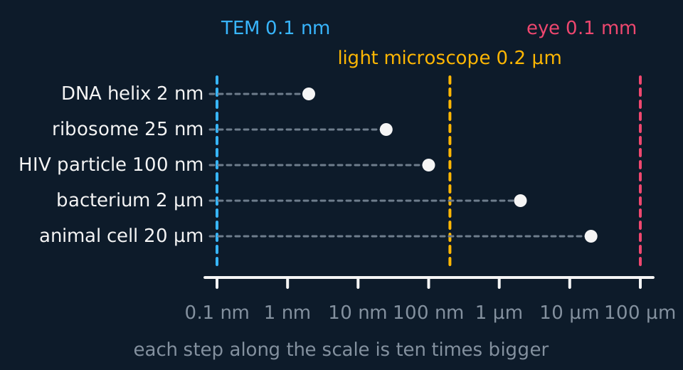
Visible light runs from about 400 to 700 nm, which places the best light microscope at around 0.2 µm, or 200 nm. A beam of electrons behaves as a wave too, and at the accelerating voltages a transmission electron microscope uses, that wavelength is about 0.004 nm: roughly a hundred thousand times shorter than visible light. A shorter wavelength gives a finer resolution, which is why a TEM resolves about 0.1 nm where light stops at 200 nm.
The half-wavelength rule alone would predict a far finer limit than 0.1 nm. A wave 0.004 nm long would separate points a few thousandths of a nanometre apart, and no transmission electron microscope of this kind achieves that. The limit is set by the lenses rather than by the wavelength. Electrons are focused by magnetic fields, which are cruder than a shaped piece of glass, and the aberrations they introduce blur the image long before the wavelength would.
Describe the drawing
A logarithmic size scale running from 0.1 nanometres to 100 micrometres, one decade per step. Five structures are plotted against it by dashed horizontal lines: a DNA helix at 2 nanometres, a ribosome at 25 nanometres, an HIV particle at 100 nanometres, a bacterium at 2 micrometres and an animal cell at 20 micrometres. Three dashed vertical lines mark the resolving limits of the transmission electron microscope at 0.1 nanometres, the light microscope at 0.2 micrometres and the unaided eye at 0.1 millimetres.
- Magnification
- How many times larger the image is than the actual object. A ratio, with no units.
- Resolution
- The minimum distance between two points at which they can still be distinguished as separate. A length, and smaller is better.
- Empty magnification
- Enlarging an image beyond the detail its resolution contains, so nothing further becomes visible.
Light microscopes, TEM and SEM
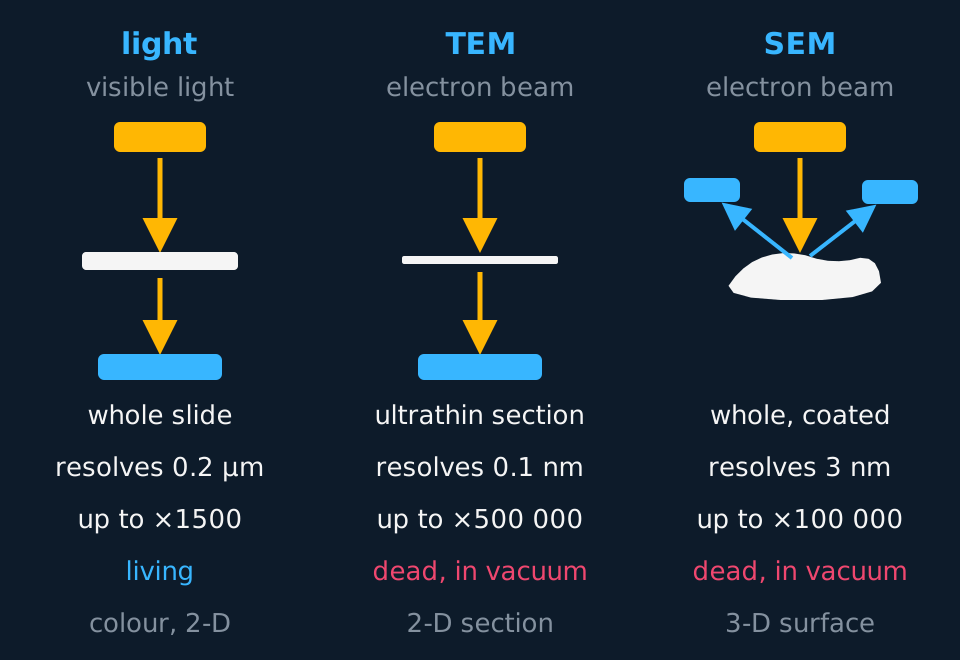
The light microscope uses visible light and glass lenses, magnifies usefully to about ×1500 and resolves to about 0.2 µm. It is the only one of the three that can image living material, so a live protoctist can be observed swimming, in colour, in a drop of pond water.
The transmission electron microscope fires a beam of electrons through a very thin section of specimen. Electrons that pass straight through land on a screen or detector below; parts of the specimen stained with heavy metals scatter electrons and come out dark. The result is a flat, two-dimensional section at resolutions down to about 0.1 nm on the best instruments under ideal conditions, and magnifications of half a million times. Most published images of cell ultrastructure are TEM images.
The scanning electron microscope sweeps a beam across the surface of a whole specimen coated in a thin film of metal, and collects the electrons knocked off that surface with detectors placed above it. Because the signal comes from the surface and depends on its shape, the image looks three-dimensional. The cost is resolution: an SEM typically manages a few nanometres, worse than a TEM, though still far beyond anything light can do.
Describe the drawing
Three microscopes drawn side by side as columns. In the light microscope column a lamp sends light down through a whole specimen on a slide to an image below. In the transmission electron microscope column an electron gun sends electrons down through an ultrathin section to an image below. In the scanning electron microscope column the beam strikes the surface of a whole coated specimen and electrons bounce back up to detectors placed either side above it. Under each column are its resolution, its greatest useful magnification, whether the specimen can be alive, and whether the image is two or three dimensional.
| Light | TEM | SEM | |
|---|---|---|---|
| Radiation | Visible light | Beam of electrons | Beam of electrons |
| Focused by | Glass lenses | Electromagnets | Electromagnets |
| Resolution | ≈ 0.2 µm | ≈ 0.1 nm | ≈ 3–10 nm |
| Useful magnification | ×1500 | ×500 000 | ×100 000 |
| Specimen | Whole or sectioned, may be alive | Ultrathin section, dead | Whole, coated in metal, dead |
| Image | Colour, two-dimensional | Two-dimensional section | Three-dimensional surface |
Both electron microscopes have to work in a vacuum, because air molecules deflect electrons and the beam would scatter before it reached anything. A vacuum removes the water from a living cell instantly, so in the standard TEM and SEM preparations this course describes the specimen must be dead, fixed and dehydrated before it goes in. Electron micrographs are therefore images of dead material, and any colour on one was added after the image was recorded.
The magnification equation
One relationship links the three quantities, and the units must be converted before the division is carried out.
magnification = image size ÷ actual sizerearranged: actual size = image size ÷ magnification
The image size is measured with a ruler, so it is in millimetres. The actual size of a cell or an organelle is stated in micrometres or nanometres. Convert one to match the other before dividing, so that both lengths are in the same unit. Magnification itself has no units.
1 mm = 1000 µm 1 µm = 1000 nm 1 mm = 1 000 000 nmthe three conversions this topic requires
Many micrographs carry a scale bar instead of a magnification. Measure the bar with a ruler and divide by the length it represents to get the magnification, or compare an object with the bar directly: an organelle three bar-lengths long beside a 1 µm bar is 3 µm long.
From a micrograph to a real length
A mitochondrion on an electron micrograph measures 42 mm from end to end. The micrograph is labelled ×15 000. Calculate the actual length of the mitochondrion in micrometres.
Show the working
Convert first. 42 mm × 1000 = 42 000 µm, so the image is 42 000 µm long in the units the answer wants.
Now divide: actual = image ÷ magnification = 42 000 ÷ 15 000 = 2.8 µm.
Check the result against a known range. Mitochondria are about 1 to 10 µm long, so 2.8 µm is a plausible value. A result of 2800 µm is nearly three millimetres, which would be visible without a microscope and indicates a conversion error.
TRY IT: The same equation the other way up
A chloroplast is 5 µm long. On a printed micrograph it measures 60 mm. Calculate the magnification of the micrograph.
Check your answer
×12 000.
Convert to a common unit first: the image is 60 mm, and 60 × 1000 = 60 000 µm.
Then magnification = image ÷ actual = 60 000 ÷ 5 = 12 000.
Write it as ×12 000 with no unit after it. A ratio of two lengths cannot carry a length as its unit.
Calibrating an eyepiece graticule
A micrograph carries its magnification printed on it. A microscope on the bench does not, so the value of one eyepiece division must be established before any measurement is taken.
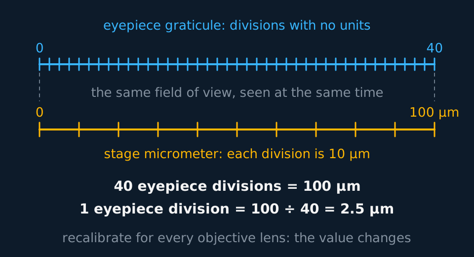
There are two scales involved. The eyepiece graticule is a transparent disc with a ruled scale, sitting in the eyepiece. Its divisions are arbitrary, staying the same apparent size whichever objective lens you use, because they are not part of the image of the specimen. The stage micrometer is a slide with a scale of known length: usually 1 mm ruled into 100 parts, so each division is 10 µm. It goes on the stage, where the objective magnifies it along with everything else.
Describe the drawing
Two scales drawn one above the other over the same width. The upper scale is an eyepiece graticule ruled into forty equal divisions numbered zero to forty with no units. The lower scale is a stage micrometer ruled into ten divisions of ten micrometres each, spanning one hundred micrometres in total. Dashed lines join the two ends to show that both scales cover the same field of view, and the arithmetic below divides one hundred micrometres by forty divisions to give 2.5 micrometres per eyepiece division.
The procedure is short. Put the stage micrometer on the stage, focus, and turn the eyepiece until the two scales lie parallel. Count how many eyepiece divisions cover a measured number of stage divisions. Divide the known length by the number of eyepiece divisions to get the value of one division. Then take the micrometer away, put your specimen on, and measure it in eyepiece divisions.
A calibration is valid for one objective lens only. The objective magnifies the stage micrometer but not the graticule, so changing from ×10 to ×40 makes each eyepiece division worth a quarter of its previous value. Calibrate separately for every lens to be used.
Reading a cell off the graticule
A student calibrates an eyepiece graticule against a stage micrometer and finds that 40 eyepiece divisions cover 10 stage divisions. The stage micrometer is 1 mm divided into 100 parts. An onion epidermis cell then measures 34 eyepiece divisions wide. Calculate its width in micrometres.
Show the working
Start with the stage micrometer, because it is the only thing here with real units. 1 mm ÷ 100 = 0.01 mm per division, which is 10 µm. Ten of those divisions is 100 µm.
That length is covered by 40 eyepiece divisions, so one eyepiece division = 100 ÷ 40 = 2.5 µm.
The cell is 34 divisions wide, so 34 × 2.5 = 85 µm.
Onion epidermis cells are typically tens of micrometres across, so 85 µm is plausible. A result of 850 µm would describe a cell visible without a microscope.
Artefacts and interpreting micrographs
Preparing a specimen for an electron microscope alters it. It is fixed in chemicals, dehydrated through a series of solvents, embedded in resin, cut into sections tens of nanometres thick and stained with salts of heavy metals such as lead or osmium. Any of those steps can change what you are looking at.
An artefact is a structure visible in the image that was not present in the living cell. Air bubbles trapped under a coverslip, creases and tears in a section, precipitated stain, and organelles pulled out of shape by dehydration are all artefacts, and early electron microscopists spent years arguing about which structures were real. The mesosome, an infolding of membrane once drawn in every bacterium in every textbook, turned out to be one, produced by the fixing chemicals.
Such disputes are settled by comparing preparation methods. A structure seen after several different preparation methods, and in more than one type of microscope, is probably present in the living cell. A structure that appears only with one particular technique is probably an artefact.
- Artefact
- A structure seen in a prepared specimen that was not present in the living cell, produced by the preparation or imaging process.
- Eyepiece graticule
- A ruled scale in the eyepiece whose divisions have no fixed length until it is calibrated.
- Stage micrometer
- A slide bearing a scale of known length, used to calibrate an eyepiece graticule for one particular objective lens.
A second source of misinterpretation is unrelated to damage. A TEM image is one thin slice through a three-dimensional object. A mitochondrion sliced across its width appears as a small circle; the same mitochondrion sliced along its length appears as a long sausage; and one that happens to be bent can be caught by the knife twice over, so a single organelle turns up in the image as two separate profiles with cytoplasm between them. All three are the same organelle. When a micrograph seems to show organelles of wildly different sizes, or more of them than the cell can plausibly hold, the plane of the section is usually the explanation.
Cell fractionation and ultracentrifugation
A micrograph shows the structure and position of an organelle but not its biochemical activity. Investigating what mitochondria do requires a suspension enriched in mitochondria and little else, which is obtained by cell fractionation.
The first step is homogenisation: the tissue is broken up, usually in a blender, so the cell-surface membranes rupture and the organelles are released into the surrounding solution. That solution is chosen for three properties, and each one prevents a specific accident. It is cold, which slows the enzymes that would otherwise digest the organelles you are trying to collect. It is isotonic with the cells, so no water moves in or out by osmosis and no organelle bursts or shrivels. And it is buffered, so the pH holds steady and the proteins keep the shapes their function depends on. The homogenate is then filtered to take out connective tissue and whole cells that survived the blender.
The separation itself is ultracentrifugation, and the principle is sedimentation rate, which depends on size and density together. The tube is spun, the largest and densest contents travel outwards fastest and collect as a pellet at the bottom, and everything still suspended stays in the supernatant above it. Spin slowly and only the heaviest component pellets. Decant the supernatant into a fresh tube, spin it faster, and the next heaviest pellets in its turn. The organelles come out in order of size and density: nuclei first, then chloroplasts in a plant sample, then mitochondria, then lysosomes, then the endoplasmic reticulum, and the ribosomes last of all at the highest speeds.
A fraction is never pure, only enriched: organelles of similar size and density sediment together, and a mitochondrial pellet carries lysosomes with it.
The sequence above is an order of size and density, not of importance, and it depends on the tissue. An animal tissue contains no chloroplasts, so that step does not occur.
In the exam
- Define both terms if a question asks for the difference. Magnification is how many times larger the image is, with no units; resolution is the smallest separation still seen as two points, with units. Give the separation, not 'how clear the image is'.
- Attribute the better resolution of an electron microscope to the shorter wavelength of an electron beam, rather than to the size of an electron.
- Convert units before dividing, and quote the answer in the unit the question asked for. Show the conversion as a line of working, because if the final number is wrong you can still be given credit for the method.
- Never put a unit on a magnification. It is a ratio, so ×12 000 is the whole answer.
- Explain why electron microscopes cannot image living cells by naming the vacuum: electrons are deflected by air, so the specimen must be in a vacuum, which no living organism survives.
- When evaluating an image, state that the section is one plane through a three-dimensional object, and that preparation can create artefacts.
Check yourself
A student photographs a cell through a light microscope, then enlarges the photograph on a computer until the whole image is a metre wide. She says she has now magnified the cell about ×20 000, so she should be able to see its ribosomes. Explain why she will not.
Answer
Her arithmetic is fine and her conclusion is wrong, because magnification and resolution are different quantities and only one of them has changed.
Enlarging the photograph raises the magnification but cannot add detail that the original image never contained. The detail was fixed at the moment the light microscope formed the image.
That limit comes from the wavelength of visible light, roughly 400 to 700 nm. Two points closer than about 0.2 µm cannot be resolved, so they merge into one. A ribosome is around 25 nm across, some eight times smaller than the limit, so it was never separately recorded.
All the enlargement does is make the same blur bigger, which is what empty magnification means. To see ribosomes she needs a shorter wavelength, and that means an electron microscope.
Questions
Question 14 marks
An eyepiece graticule is calibrated against a stage micrometer whose divisions are each 10 µm long. 25 eyepiece divisions are found to cover 4 stage micrometer divisions. A cell then measures 15 eyepiece divisions across. Calculate the diameter of that cell in micrometres.
Mark scheme
- M1 4 stage micrometer divisions correspond to 4 × 10 = 40 µm
- M1 one eyepiece division is worth 40 divided by 25
- M1 = 1.6 µm per eyepiece division, then multiplied by the 15 divisions the cell covers
- A1 a final answer of 24 µm
Question 24 marks
Explain why a transmission electron microscope resolves finer detail than a light microscope, and explain why it cannot be used to look at a living cell.
Mark scheme
- B1 resolution is limited by the wavelength of the radiation used to illuminate the specimen
- B1 a beam of electrons has a far shorter wavelength than visible light, so points much closer together can still be seen as separate
- B1 the electron beam has to travel through a vacuum, because air molecules deflect electrons and scatter the beam
- B1 a vacuum removes the water from a cell, so the specimen must be dead, fixed and dehydrated before it is put in
Question 34 marks
Compare a transmission electron microscope with a scanning electron microscope, referring to the specimen used, the electrons detected, the image produced and the resolution achieved.
Mark scheme
- B1 a transmission instrument needs an ultrathin section, whereas a scanning instrument takes a whole specimen coated in a thin film of metal
- B1 a transmission instrument detects electrons that have passed through the specimen, whereas a scanning instrument detects electrons knocked off its surface
- B1 a transmission instrument gives a two-dimensional image of a section, whereas a scanning instrument gives a three-dimensional image of a surface
- B1 a transmission instrument resolves to about 0.1 nm, which is better than the few nanometres a scanning instrument manages
Question 44 marks
The scale bar under the mitochondrion represents 1 µm. Calculate the actual length of the mitochondrion. On a printed copy of the same micrograph the scale bar measures 15 mm; calculate the magnification of that printed copy.
Mark scheme
- M1 the organelle is three bar lengths long, measured against the bar itself
- A1 3 × 1 µm = 3 µm, which is a reasonable length for a mitochondrion
- M1 magnification = size of image ÷ actual size, working in one unit: 15 mm ÷ 0.001 mm
- A1 × 15 000, with no unit, because a magnification is a ratio
Question 54 marks
Describe how ultracentrifugation separates organelles, and describe the order in which nuclei, mitochondria and ribosomes are pelleted.
Mark scheme
- B1 the tube is spun, and the largest and densest contents travel outwards fastest and collect as a pellet at the bottom, with everything still suspended left in the supernatant
- B1 the supernatant is decanted into a fresh tube and spun again at a higher speed, so the next heaviest component pellets in its turn
- B1 the organelles pellet in order of decreasing size and density: nuclei first, then mitochondria
- B1 ribosomes pellet last of all, because they are the smallest structures collected, requiring the highest speeds
Question 64 marks
Describe the stages a specimen goes through in preparation for viewing with a transmission electron microscope, from fresh tissue to a stained section.
Mark scheme
- B1 the tissue is fixed in chemicals, to preserve its structure
- B1 it is dehydrated through a series of solvents, removing the water
- B1 it is embedded in resin and cut into ultrathin sections
- B1 the sections are stained with salts of heavy metals, such as lead or osmium, to give contrast
Question 73 marks
A ribosome measures 5 mm across on an electron micrograph labelled as being at a magnification of 200 000 times. Calculate the actual diameter of the ribosome, giving your answer in nanometres.
Mark scheme
- M1 actual size = image size divided by magnification
- M1 converts 5 mm to 5 000 000 nm before dividing, or works consistently in another single unit
- A1 an answer of 25 nm, which is the expected size for a ribosome
Question 83 marks
A student examining an electron micrograph counts what appear to be forty separate mitochondria, of widely differing sizes and shapes, in one small cell. Suggest two reasons why this count and these shapes may be misleading.
Mark scheme
- B1 the micrograph is one thin section through a three-dimensional cell, so each organelle is seen only where the knife happened to cut it
- B1 a mitochondrion cut across appears as a small circle and one cut along its length as a long sausage, so the same organelle gives different profiles
- B1 a bent organelle can be caught by the knife twice, so one mitochondrion appears as two separate profiles and the number is overcounted
Question 93 marks
Explain why the solution used to homogenise tissue in cell fractionation must be cold, isotonic and buffered.
Mark scheme
- B1 cold: this slows the action of hydrolytic enzymes, such as those from lysosomes, that would otherwise digest the organelles being collected
- B1 isotonic: this prevents water moving in or out of the organelles by osmosis, so they do not burst or shrivel
- B1 buffered: this keeps the pH constant, so proteins and enzymes retain the shape their function depends on
Question 103 marks
A student enlarges a photograph taken through a light microscope until it is a metre wide, and claims this has increased the magnification enough to reveal detail invisible in the original image. Explain why this claim describes empty magnification.
Mark scheme
- B1 magnifying an image without limit does not add detail beyond what the resolution of the original instrument recorded
- B1 resolution is fixed by the wavelength of the radiation used to form the original image, not by any later enlargement of it
- B1 the enlargement produces a bigger but equally blurred image, containing no more detail than the original, which is what empty magnification means
Question 113 marks
The mesosome, an inward fold of the bacterial cell membrane, was seen in early electron micrographs prepared by one particular fixation method, but not when a different fixation method was used on the same species. Suggest what this comparison shows about the mesosome, and suggest how biologists in general decide whether a structure seen in one preparation is real or an artefact.
Mark scheme
- B1 the mesosome is probably an artefact produced by that particular fixation chemical rather than a structure present in the living bacterium
- B1 a structure seen after several different preparation methods is likely to be real, because it is unlikely that different chemicals would each create the same appearance by accident
- B1 a structure seen with only one particular technique is probably an artefact of that technique
Question 123 marks
Compare the images produced by a light microscope with those produced by a transmission electron microscope, referring to colour, whether a living specimen can be examined, and resolution.
Mark scheme
- B1 a light microscope image is in colour, natural or from stains, whereas a TEM image is black and white, with any colour added afterwards
- B1 a light microscope can show a living, moving specimen, whereas a TEM specimen is dead, because it is fixed, dehydrated and examined in a vacuum
- B1 a light microscope resolves to about 0.2 µm against about 0.1 nm for a TEM, so a TEM reveals internal ultrastructure that is invisible with light
Question 132 marks
State what is meant by the magnification of an image, and state what is meant by the resolution of a microscope.
Mark scheme
- B1 magnification is how many times larger the image is than the actual object, and is a ratio with no units
- B1 resolution is the minimum distance between two points at which they can still be distinguished as separate
Question 142 marks
A researcher separating organelles by ultracentrifugation obtains a mitochondrial pellet that also contains lysosomes. Explain why this pellet is not pure mitochondria.
Mark scheme
- B1 lysosomes and mitochondria are similar in size and density, so they sediment together at the same centrifuge speed
- B1 a pellet is therefore enriched in the organelle of interest but never completely pure
Question 152 marks
Explain why an eyepiece graticule must be calibrated separately for each objective lens used on a microscope.
Mark scheme
- B1 the objective lens magnifies the image of the stage micrometer, but it does not magnify the eyepiece graticule's own divisions, which stay the same apparent size
- B1 so each objective gives an eyepiece division a different real value in micrometres, and a calibration made for one lens is not valid for another
Worth remembering
- Magnification = image ÷ actual. No units on the answer.
- Resolution is a distance, and smaller is better: 0.1 nm beats 0.2 µm.
- Shorter wavelength, better resolution, which is why electrons beat light.
- TEM: thin section, electrons through, flat image. SEM: whole surface, electrons off, three-dimensional image.
- Recalibrate the eyepiece graticule every time you change objective lens.
- Homogenising solution: cold, isotonic, buffered. Enzymes slowed, osmosis prevented, protein shape held.
- Ultracentrifugation separates by size and density, largest and densest first: nuclei, chloroplasts, mitochondria, lysosomes, ER, ribosomes.
CHECK YOUR PROGRESS
Rate how confident you are with each objective for this lesson. Ratings are saved in this browser, on this device, unless you sign in.
- Define magnification and resolution and explain why they are not the same thing.
- Explain why an electron microscope resolves finer detail than a light microscope.
- Compare a light microscope, a TEM and an SEM by beam, resolution, specimen and the kind of image each produces.
- Rearrange and use magnification = image size ÷ actual size, converting units correctly.
- Describe how an eyepiece graticule is calibrated with a stage micrometer, and explain why artefacts appear in electron micrographs.
- Describe cell fractionation and ultracentrifugation, and justify the cold, isotonic, buffered solution and the order the organelles pellet in.
Open the full revision checklist to see every objective in the curriculum in one place.
Practise this lesson
WORKBOOK
The written questions from this topic, on paper with room to work, and a separate book of mark schemes. The 3 questions that are set on a diagram stay on this page, where the diagram can be drawn. Free to use; please do not redistribute or sell.