Biology › Cells, microscopy and biological organisation › Eukaryotic cell structure and organelles
Eukaryotic cell structure and organelles
A eukaryotic cell is a set of compartments, and each one has a structure that suits its function: a folded inner membrane for respiration, stacked flattened sacs for modifying proteins, a single membrane around enzymes that would digest the cell if they escaped.
Before this The animal and plant cells of GCSE biology · Standard form and the SI prefixes down to nanometres
COMMON MISCONCEPTION
Each organelle works on its own, so a cell is a set of independent compartments.
Organelles interact in linked processes. A protein for secretion is synthesised on rough endoplasmic reticulum, carried in a vesicle to the Golgi apparatus, modified and sorted, then carried to the cell-surface membrane for exocytosis.
What you should be able to do
- Describe the ultrastructure of an animal cell and of a plant cell, and say which structures belong to which.
- Link the structure of the nucleus, mitochondrion, chloroplast, Golgi apparatus, lysosome and both kinds of endoplasmic reticulum to what each does.
- Put the organelles of the secretory pathway in the order a protein meets them.
- State where 80S and 70S ribosomes are found, and what a ribosome is made of.
- Name the structures a plant cell has and an animal cell does not, and give a function for each.
- Name the three fibre types of the cytoskeleton with a role for each, and describe how motor proteins, cilia and flagella produce movement.
- Describe amyloplasts, pits and the middle lamella, and tell an amyloplast from a chloroplast in an electron micrograph.
What the electron microscope added
A light microscope resolves structures about 0.2 µm apart, so it can show the nucleus, cytoplasm, cell boundary and larger plant-cell structures. Electron microscopy has much greater resolution and reveals cell ultrastructure, including mitochondria, endoplasmic reticulum, Golgi apparatus and ribosomes.
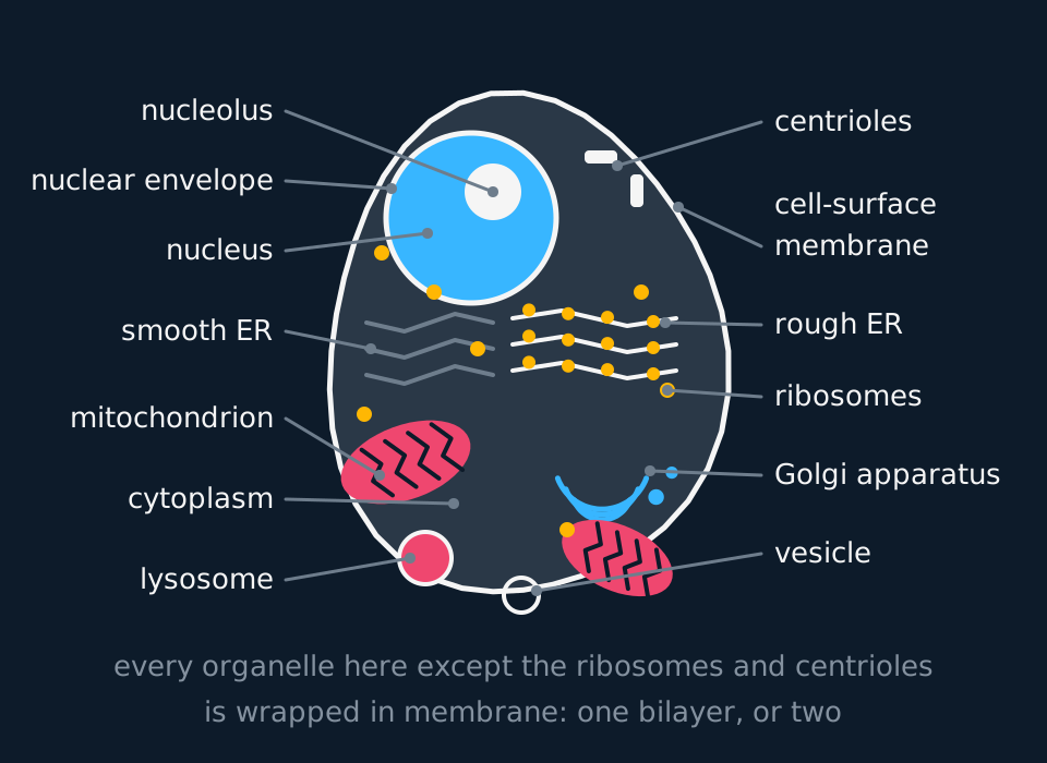
Describe the drawing
An animal cell drawn in section. A large round nucleus with a dense nucleolus inside it sits towards the top, wrapped in a double nuclear envelope. Flattened sacs of rough endoplasmic reticulum studded with ribosomes run beside it, with smooth endoplasmic reticulum free of ribosomes on the other side. A stack of Golgi sacs with vesicles budding off lies below, together with two mitochondria showing folded inner membranes, a lysosome, free ribosomes scattered through the cytoplasm, a pair of centrioles, and the cell-surface membrane enclosing everything. There is no cell wall, chloroplast or vacuole.
Membrane number is a useful structural distinction. The nucleus, mitochondria and chloroplasts have double membranes; the Golgi apparatus, lysosomes, vesicles and endoplasmic reticulum are bounded by a single membrane; ribosomes have no surrounding membrane. In an exam answer, link each structural feature to its function, for example: "Cristae increase the inner-membrane surface available for the electron transport chain and ATP synthase."
- Organelle
- A structure inside a cell with a distinct function, usually but not always surrounded by its own membrane.
- Ultrastructure
- The detail of a cell's internal structure as revealed by an electron microscope.
- Cytoplasm
- Everything inside the cell-surface membrane except the nucleus: the cytosol and the organelles suspended in it.
The nucleus, and two kinds of endoplasmic reticulum
The nucleus is wrapped in two membranes, and the pair together is the nuclear envelope. It is punched through with nuclear pores, a few thousand of them in a typical cell, each around 40 to 100 nm across. Those pores are the reason the arrangement works: they are gateways with transport machinery rather than plain holes, exporting processed messenger RNA selectively while the chromosomes stay inside the envelope, so transcription occurs in the nucleus and translation in the cytoplasm.
Inside, the DNA is wound round histone proteins into chromatin. One patch is denser than the rest and takes up stain more heavily: the nucleolus, where ribosomal RNA is made and the two ribosomal subunits are assembled. A cell with a large nucleolus is building a lot of ribosomes, which usually means it is about to make a lot of protein.
The outer membrane of the nuclear envelope does not stop at the nucleus. It runs straight on into the endoplasmic reticulum, a system of flattened sacs called cisternae folded through the cytoplasm. There are two versions, and the defining structural difference between them is what is stuck to the outside.
Rough endoplasmic reticulum is studded with ribosomes. A protein made on one of those ribosomes is threaded into the sac as the chain grows, so it enters the membrane system at the point of synthesis rather than crossing a membrane later. Proteins for secretion, for lysosomes and for the cell-surface membrane are all made here. A plasma cell pouring out antibody is almost solid rough ER.
Smooth endoplasmic reticulum has no ribosomes on it and a completely different job list: it synthesises lipids, phospholipids and steroids. Liver cells use theirs to break down drugs and alcohol, which is why a liver cell has an unusual amount. In a muscle fibre a specialised version stores calcium ions and releases them to trigger contraction. Abundant smooth endoplasmic reticulum therefore indicates lipid synthesis or, in the liver, drug metabolism.
- Nuclear envelope
- The double membrane surrounding the nucleus, continuous with the endoplasmic reticulum and perforated by nuclear pores.
- Nucleolus
- A dense region within the nucleus where ribosomal RNA is made and ribosomes are assembled.
- Rough endoplasmic reticulum
- Flattened membrane sacs with ribosomes attached, which make and transport proteins for export or for membranes.
- Smooth endoplasmic reticulum
- Membrane sacs without ribosomes, which synthesise lipids and steroids and store calcium ions.
The secretory pathway
Proteins for secretion, lysosomes or cell-surface membranes are synthesised by ribosomes on the rough endoplasmic reticulum. Transport vesicles carry them to the Golgi apparatus, where they are modified and sorted. Secretory vesicles then carry selected proteins to the cell-surface membrane for exocytosis.
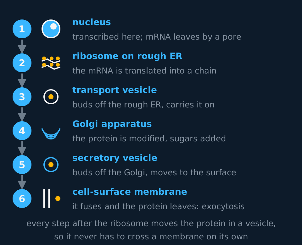
Describe the drawing
A numbered vertical route with six stations joined by downward arrows. Station one is the nucleus, where messenger RNA is made and leaves through a pore. Station two is a ribosome on the rough endoplasmic reticulum, where the protein is assembled. Station three is a transport vesicle budding off the rough endoplasmic reticulum. Station four is the Golgi apparatus, drawn as a stack of sacs, where the protein is modified. Station five is a secretory vesicle. Station six is the cell-surface membrane, drawn as two lines, where the vesicle fuses and the protein is released by exocytosis.
The Golgi apparatus is a stack of flattened sacs, usually four to eight of them, curved and with vesicles budding off one face. Vesicles arrive at one side carrying newly made protein, the contents move through the stack, and finished product leaves the other side. What happens in between is modification: sugars are trimmed and extended to finish glycoproteins (the first sugars are attached back in the ER), chains are cut to length, phosphate groups are attached, and each product is sorted and addressed to its destination. The stack allows a sequence of steps to happen in order, which a single compartment could not do.
Most folding begins in the rough endoplasmic reticulum. The Golgi modifies and sorts proteins; it is not the main site of protein folding. A secreted polypeptide folds as it is made, with chaperone proteins holding it while it does, and reaches the Golgi essentially in shape.
Lysosomes bud from the Golgi as well. The enzymes inside them are proteins, made on rough ER, packaged like any other export and then retained rather than secreted.
Reading a cell from its organelles
An electron micrograph shows a cell with a large nucleolus, cytoplasm packed with rough endoplasmic reticulum, several Golgi stacks, many mitochondria and a crowd of vesicles pressed against one face of the cell-surface membrane. Suggest what this cell does, and justify each part of your answer.
Show the working
It secretes protein, and at a high rate. Take the features one at a time.
The large nucleolus means ribosomes are being assembled in quantity, and the abundant rough ER means those ribosomes are making protein for export rather than for use in the cytoplasm.
Several Golgi stacks mean a lot of protein is being modified and packaged, and the vesicles gathered at one face are secretory vesicles waiting to fuse with the membrane.
Include the mitochondria. Secretion requires ATP for peptide-bond formation, for vesicle movement along the cytoskeleton and for fusion at the cell-surface membrane, so a cell secreting at this rate has a high rate of aerobic respiration.
The two organelles that handle energy
Mitochondria are usually between 1 and 10 µm long, about the size of a whole bacterium, which is not a coincidence. Each has two membranes. The outer one is smooth. The inner one is folded into cristae. The electron transport chain and ATP synthase are located in that inner membrane, so greater folding gives a larger surface area for them and a higher rate of ATP synthesis. The fluid inside is the matrix, where the Krebs cycle runs, and it contains enzymes, a small loop of DNA and 70S ribosomes of its own.
The number of mitochondria in a cell reflects how much ATP that cell needs. Muscle fibres, sperm cells and the epithelial cells lining the small intestine are all crowded with them; a mature red blood cell has none at all, and no nucleus either.
Chloroplasts, which plant cells have as well as mitochondria, are built on the same plan: a double envelope, a fluid interior called the stroma, and an internal membrane system. Here the membranes form flattened discs called thylakoids, stacked into piles called grana, and the chlorophyll sits in the thylakoid membranes. Stacking gives a large area of pigment-bearing membrane in a small volume, so a lot of light gets absorbed. The stroma holds the enzymes of the light-independent reactions, along with starch grains, a loop of DNA and, again, 70S ribosomes.
Those loops of DNA and small ribosomes are the evidence behind the endosymbiotic theory, that both organelles descend from free-living prokaryotes taken into an early eukaryotic cell. The theory itself is not required by any of the five specifications this site maps; the 70S ribosomes inside these organelles are examinable, because they qualify any simple statement that 70S ribosomes are found only in prokaryotes.
| Mitochondrion | Chloroplast | |
|---|---|---|
| Membranes | Two: outer smooth, inner folded | Two, plus a separate thylakoid system |
| Internal membrane | Cristae | Thylakoids stacked into grana |
| Fluid interior | Matrix, Krebs cycle | Stroma, light-independent reactions |
| Pigment | None | Chlorophyll, in the thylakoid membranes |
| Own DNA and ribosomes | Yes, 70S | Yes, 70S |
| Found in | Almost all eukaryotic cells | Plants and algae only |
Ribosomes, lysosomes and the rest of the small print
Ribosomes are the exception among the organelles described so far: they have no membrane at all. Each is built from ribosomal RNA and protein in two subunits that clamp round a strand of mRNA to translate it. In the cytoplasm of a eukaryotic cell they are 80S and about 25 nm across. In prokaryotes they are 70S and smaller. The ribosomes inside mitochondria and chloroplasts are prokaryote-like too, which A-level files under 70S: close to true for chloroplasts, while mammalian mitochondrial ribosomes have been remodelled over evolutionary time and sediment nearer 55S. The S is a Svedberg unit, which measures how fast a particle settles in a centrifuge rather than its mass, which is why a 60S and a 40S subunit make an 80S ribosome instead of a 100S one.
Lysosomes are spherical sacs, roughly 0.1 to 1 µm across, each holding up to fifty kinds of hydrolytic enzyme behind a single membrane. They digest worn-out organelles, break down material taken in by phagocytosis, and in a white blood cell they empty their contents onto engulfed bacteria. The single membrane is what makes this safe. Loose in the cytoplasm those enzymes would hydrolyse the cell that made them, so they are confined to a compartment and released only at the site of digestion.
Centrioles are pairs of hollow microtubule cylinders that organise the spindle during nuclear division. They are absent from the cells of flowering plants.
- Ribosome
- A structure of ribosomal RNA and protein, in two subunits, with no membrane, which is the site of translation. 80S in eukaryotic cytoplasm, 70S in prokaryotes and in mitochondria and chloroplasts.
- Lysosome
- A vesicle containing hydrolytic enzymes, bounded by a single membrane, which digests worn-out organelles and engulfed material.
- Crista
- A fold of the inner mitochondrial membrane, giving a large surface area for the electron transport chain.
The cytoskeleton
Organelles are not suspended in featureless cytosol. The cytoplasm is threaded through with a network of protein fibres called the cytoskeleton, which gives the cell its shape, holds each organelle in position, and moves material, and in some cases whole cells, from place to place. Three kinds of fibre make it up, distinguished by their diameter and their protein.
Microfilaments are the thinnest, about 7 nm, built from the globular protein actin. They gather in a mesh just under the cell-surface membrane, where they let the cell change shape: a phagocyte pushing out extensions around a bacterium is rearranging its actin, and so is an animal cell in cytokinesis, where a contractile ring of microfilaments pinches the cytoplasm in two.
Intermediate filaments, around 10 nm, are the mechanical ones: tough rope-like fibres, keratins among them, that spread tension through the cell and anchor the nucleus and other organelles in position. A cell pulled about by its neighbours, as skin cells constantly are, survives because these filaments carry the load.
Microtubules are the widest at about 25 nm: hollow cylinders of the protein tubulin, constantly built and taken apart, radiating from an organising region near the nucleus that in animal cells contains the centrioles. They provide tracks for directed transport. Motor proteins, with kinesin and dynein the named examples, attach to a vesicle or an organelle and move step by step along a microtubule, each step requiring ATP. Transport and secretory vesicles are moved along microtubules by motor proteins rather than by diffusion. The spindle that moves chromosomes during nuclear division is microtubules again, organised in an animal cell by the centrioles, and in a plant cell, which has none, by the poles themselves.
- Cytoskeleton
- The network of protein fibres (microfilaments, intermediate filaments and microtubules) that gives a cell its shape, anchors its organelles and provides tracks for movement.
- Microtubule
- A hollow cylinder of tubulin about 25 nm wide, along which motor proteins move vesicles and organelles, and which builds the spindle, cilia and flagella.
- Motor protein
- A protein such as kinesin or dynein that uses ATP to walk along a cytoskeletal fibre, carrying a vesicle or organelle with it.
Microtubules also build the cell's moving appendages. A cilium and a eukaryotic flagellum have the same internal plan: nine pairs of microtubules arranged in a ring around two central ones, the 9 + 2 arrangement, with dynein arms between the pairs. When the dynein walks, neighbouring pairs slide against each other, and because the pairs are anchored the sliding becomes bending. A sperm flagellum and the cilia of a ciliated epithelial cell use the same internal structure at different lengths. The bacterial flagellum is a different structure: a rotating protein filament that shares only the name. Microvilli are different again: finger-like folds of the cell-surface membrane, held out by microfilaments, that increase the surface area for absorption, as on the epithelial cells of the small intestine.
Structures found in plant cells and not in animal cells
Every organelle so far appears in a plant cell too. Plant cells respire, so they have mitochondria; they make and export protein, so they have rough ER and Golgi. Four structures are added, and one is taken away.
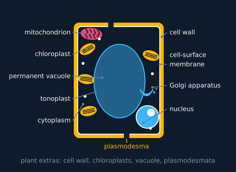
Describe the drawing
A plant cell drawn in section as a box with rounded corners. A thick cellulose cell wall forms the outer band, pierced by narrow plasmodesmata, with the cell-surface membrane pressed against its inside. A large permanent vacuole bounded by a tonoplast fills the centre and pushes the cytoplasm into a thin layer round the edge. Four chloroplasts with stacked grana, a mitochondrion with cristae, a Golgi stack, free ribosomes and a nucleus with a nucleolus lie in that cytoplasm layer.
The cell wall is cellulose, not membrane, and that matters twice over. It is freely permeable, so it is no barrier to water or dissolved substances and cannot control what enters the cell. The cell-surface membrane still does that. And it is strong in tension, so when water enters by osmosis the wall pushes back and the cell becomes turgid rather than bursting. Turgid cells packed against each other are what holds a non-woody plant up.
The permanent vacuole is a large sac of cell sap behind a single membrane called the tonoplast. It stores water, sugars, ions and sometimes pigments, and by filling with water it keeps the cell pressed against its wall. Animal cells have vacuoles too, but small and temporary ones.
Chloroplasts are the third addition. Plasmodesmata are the fourth: narrow channels through the walls of neighbouring cells, lined with membrane, through which cytoplasm is continuous from one cell to the next. They are how a plant moves substances and signals between cells without crossing a wall.
| Structure | Animal cell | Plant cell |
|---|---|---|
| Cell wall | Absent | Present, cellulose |
| Chloroplasts | Absent | Present in cells exposed to light |
| Permanent vacuole | Absent (small temporary ones only) | Present, large and central |
| Plasmodesmata | Absent | Present |
| Amyloplasts | Absent | Present in starch-storing tissue |
| Middle lamella | Absent | Present between neighbouring walls |
| Centrioles | Present | Absent in flowering plants |
| Nucleus, mitochondria, ER, Golgi, ribosomes | Present | Present |
| Carbohydrate store | Glycogen | Starch |
TRY IT: Explaining a difference rather than listing one
A palisade mesophyll cell from a leaf and a cell from the lining of your cheek are both eukaryotic. Give two structures found in the palisade cell and not in the cheek cell, and for each one explain what difference it makes to the way the cell lives.
Check your answer
Chloroplasts and a permanent vacuole are the safest pair; the cell wall and plasmodesmata would also do.
Chloroplasts contain chlorophyll in stacked thylakoid membranes, so the palisade cell can absorb light and make its own organic molecules by photosynthesis. The cheek cell cannot, so it depends entirely on molecules brought to it in the blood.
The permanent vacuole fills with water and presses the cytoplasm against the cellulose wall, so the palisade cell becomes turgid and helps support the leaf. A cheek cell in the same water would swell and burst, because it has no wall to push back.
Each answer names a structure and then states its consequence for the cell. State both.
Amyloplasts, pits and the middle lamella
Pearson Edexcel Biology A lists three more plant structures than the four above: amyloplasts, pits and the middle lamella. All three can be identified in an electron micrograph of the right tissue.
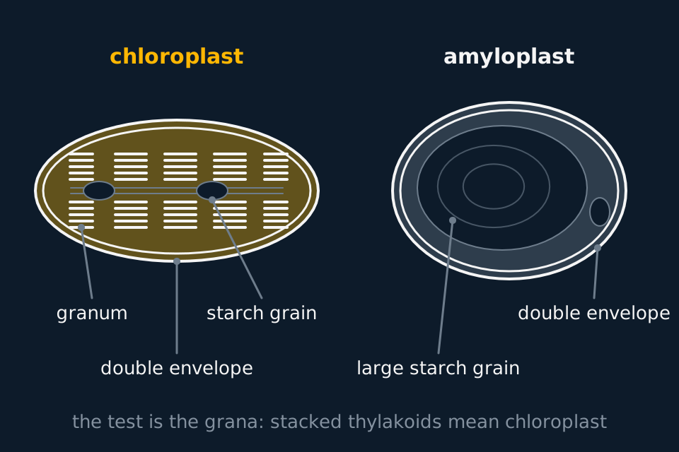
Describe the drawing
Two plastids drawn side by side as they appear in a transmission electron micrograph. On the left a lens-shaped chloroplast is bounded by two envelope membranes and holds several grana, each a stack of flattened thylakoids seen as dark parallel lines, joined by longer single membranes, with two small unshaded starch grains lying between the stacks. On the right an amyloplast of similar size is also bounded by two envelope membranes but has no thylakoids at all; one large unshaded starch grain with faint growth rings fills most of its interior, with a smaller grain beside it and a thin layer of stroma round both.
An amyloplast is a plastid that stores starch. Plastids are the family of organelles to which the chloroplast belongs, all with a double-membrane envelope, their own circular DNA and 70S ribosomes, and all developing from small undifferentiated precursors called proplastids. An amyloplast has no chlorophyll and almost no internal membrane. Its stroma holds the enzymes that build starch from glucose, made from sucrose delivered by the phloem, and one or several large starch grains take up most of its volume. Amyloplasts are found in storage tissue that receives no light, such as the tuber of a potato and the endosperm of a cereal grain, which is where the starch you eat was laid down.
A chloroplast stores starch too, but only for a short time. Starch made in the light is kept as small grains in the stroma and hydrolysed again at night, when the sugars are exported to the rest of the plant. An amyloplast is a long-term store. Amyloplasts in the cells of a root cap have a second job: their dense starch grains settle to the lower side of the cell, and that settling is one of the ways a root detects the direction of gravity. The two kinds of plastid can also convert one into the other. A potato tuber left in the light turns green at the surface as its amyloplasts develop thylakoids and chlorophyll.
The middle lamella is the layer that cements neighbouring plant cells together. It is the first layer laid down when a dividing plant cell forms a new wall, so it sits in the centre of every shared wall, with the cellulose wall of one cell on one side of it and the wall of the next cell on the other. It is made mainly of pectins, polysaccharides held together by calcium ions as calcium pectate. Dissolving it separates the cells. Ripening fruit softens partly because enzymes break down the pectins of the middle lamella, and the hydrochloric acid used in a root-tip squash works the same way, hydrolysing the middle lamella so the cells spread apart when pressed.
A pit is a thin region of a cell wall. Many plant cells, xylem among them, lay down a thick secondary wall inside the first, and a pit is a gap in that secondary wall where only the thin primary wall and the middle lamella are left. Pits in neighbouring cells usually line up, forming a pit pair, so that water and dissolved substances cross from one cell to the next through a very thin barrier. In xylem vessels, whose walls are thickened with lignin, pits are how water moves sideways between vessels. In living cells the plasmodesmata are often clustered in these thin regions.
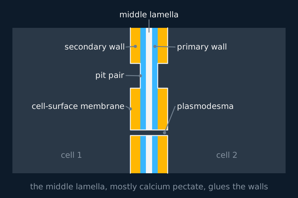
Describe the drawing
The wall between two neighbouring plant cells drawn in section at electron microscope scale, with the cytoplasm of one cell on the left and the other on the right. Crossing from left to right, the wall is a thick secondary wall, a thin primary wall, a dark middle lamella in the centre, then a primary wall and a secondary wall belonging to the second cell. Near the top the secondary walls of both cells stop opposite each other, leaving only the primary walls and middle lamella between them: a pit pair, with each cell-surface membrane following the dip. Lower down a narrow plasmodesma runs straight through every layer, lined by membrane continuous from one cell to the other.
None of these three has an equivalent in an animal cell. Animal cells store carbohydrate as glycogen granules lying free in the cytoplasm, not inside an organelle, and they are held together by junction proteins and the extracellular matrix, not by a cementing layer between walls they do not have.
- Amyloplast
- A colourless plastid with a double-membrane envelope that synthesises and stores starch, found in storage tissues such as potato tubers and in root cap cells.
- Middle lamella
- The layer, mainly calcium pectate, between the walls of neighbouring plant cells, which cements the cells together.
- Pit
- A thin region of a plant cell wall where no secondary wall has been laid down, usually matched by a pit in the neighbouring cell, allowing water and solutes to pass between them.
- Plastid
- Any of the family of plant organelles, including chloroplasts and amyloplasts, that have a double-membrane envelope and their own DNA and develop from proplastids.
Recognising plant organelles in an electron micrograph
Pearson Edexcel Biology A asks for every plant structure in this lesson to be identified from an electron micrograph. A transmission electron micrograph is a black-and-white image of a thin section, so identification depends on shape, size, the number of membranes and what lies inside them, not on colour.
| Structure | What to look for in a TEM image |
|---|---|
| Cell wall | A thick, evenly grey band outside the cell-surface membrane, shared between neighbouring cells |
| Middle lamella | A thin line down the centre of a shared wall, often darker than the wall on either side |
| Pit | A place where the wall suddenly narrows, usually opposite a matching narrowing in the next cell |
| Plasmodesma | A fine channel running straight through the wall, with membrane continuous from one cell to the next |
| Vacuole and tonoplast | A large pale region, almost empty of detail, edged by a single membrane |
| Chloroplast | Lens-shaped, two envelope membranes, parallel dark stacks of thylakoids (grana), sometimes small pale starch grains between them |
| Amyloplast | Two envelope membranes, no thylakoid stacks, almost filled by one or more large pale starch grains |
| Mitochondrion | Usually smaller, two membranes, the inner one folded into cristae, no starch |
Amyloplast and chloroplast are the pair most often confused, because both are plastids with a double envelope and both can contain starch. Decide on the internal membranes. Grana, seen as dark stripes in stacks, mean a chloroplast however much starch is present. No grana and a starch grain occupying most of the organelle mean an amyloplast. The tissue the cell came from is supporting evidence only: a leaf mesophyll cell holds chloroplasts and a cell from a potato tuber holds amyloplasts.
Naming a plastid from a micrograph
A transmission electron micrograph of a cell from a potato tuber shows several oval organelles, each about 10 µm across. Each is bounded by two membranes and contains one large, almost structureless grain, with a thin layer of granular stroma round it. No internal membranes are visible. Identify the organelle and explain how you ruled out a chloroplast.
Show the working
The two membranes make it a plastid or a mitochondrion; a nucleus also has two membranes but would show nuclear pores and chromatin, and there is only one per cell. A mitochondrion would show its inner membrane folded into cristae, and a mitochondrion does not store starch, so it is a plastid.
A chloroplast would contain thylakoid membranes stacked into grana, which in the image would be groups of dark parallel lines. There are none.
The large, structureless grain is starch, and it fills most of the organelle. A plastid with no thylakoids and a large starch grain is an amyloplast.
The tissue agrees with that conclusion. A tuber grows underground and stores starch, so its cells need amyloplasts and have no use for chloroplasts. State the structural reason first and give the tissue as support.
In the exam
- Count the membranes and state the number. Nucleus and mitochondrion: two. Golgi sac, lysosome, vacuole, vesicle, both kinds of ER: one. Ribosome: none. State that a ribosome has no surrounding membrane.
- Pair every structure with a function: 'cristae give a large surface area for the enzymes of the electron transport chain', rather than 'the inner membrane is folded' on its own.
- For the secretory pathway, give the order and name the vesicles. Rough ER, transport vesicle, Golgi, secretory vesicle, cell-surface membrane, exocytosis.
- 80S is the eukaryotic cytoplasmic ribosome and 70S the prokaryotic one, but mitochondria and chloroplasts also contain 70S ribosomes, so include them when asked where 70S ribosomes occur in a plant cell.
- The cell wall does not control what enters the cell. It is fully permeable; the cell-surface membrane is the partially permeable barrier.
- To tell an amyloplast from a chloroplast, name the grana. Stacked thylakoids mean chloroplast; no thylakoids and a large starch grain mean amyloplast. Starch alone decides nothing, because both store it.
Check yourself
A cell taken from the pancreas secretes digestive enzymes. Describe the path taken by one of those enzyme molecules from the gene that codes for it to the moment it leaves the cell, naming every organelle involved and saying what each contributes.
Answer
The gene is transcribed in the nucleus. Messenger RNA is made against the DNA template and, once processed, is exported through a nuclear pore, whose machinery passes mRNA out while the chromosomes stay put.
In the cytoplasm the mRNA binds to a ribosome attached to the rough endoplasmic reticulum. Translation assembles the polypeptide, and the chain is fed into the cisterna as it grows, so the enzyme is inside the membrane system from the moment it exists.
A transport vesicle pinches off the rough ER and carries the enzyme to the Golgi apparatus, where it is modified, commonly by having sugar groups added, then sorted and packaged. It folded in the ER as it was made, so what the Golgi does is finish it and address it, not shape it.
A secretory vesicle buds from the other face of the Golgi and moves to the cell-surface membrane, where the two membranes fuse and the enzyme is released outside. That last step is exocytosis, and it needs ATP, which is why a secretory cell like this one is also rich in mitochondria.
Questions
Question 14 marks
Compare the structure of a mitochondrion with the structure of a chloroplast.
Mark scheme
- B1 both are surrounded by two membranes, but a chloroplast has a separate internal thylakoid membrane system as well
- B1 the inner mitochondrial membrane is folded into cristae, whereas the chloroplast membranes form thylakoids stacked into grana
- B1 the fluid interior of a mitochondrion is the matrix, where the Krebs cycle runs, whereas that of a chloroplast is the stroma, holding the enzymes of the light-independent reactions
- B1 a chloroplast contains chlorophyll in its thylakoid membranes, whereas a mitochondrion contains no pigment
Question 24 marks
Explain why the enzymes of a lysosome must be kept behind a membrane, and explain how those enzymes reach the lysosome after they have been made.
Mark scheme
- B1 a lysosome holds hydrolytic enzymes, which digest worn-out organelles and material taken in by phagocytosis
- B1 loose in the cytoplasm those enzymes would hydrolyse the cell that made them, so they are released only where they are wanted
- B1 the enzymes are proteins, made on ribosomes attached to the rough endoplasmic reticulum
- B1 they are packaged at the Golgi apparatus, and a lysosome is simply a vesicle budded from it and kept rather than secreted
Question 34 marks
Describe the internal structure of a cilium, and describe how this structure produces bending.
Mark scheme
- B1 nine pairs of microtubules are arranged in a ring around two central microtubules, the 9 + 2 arrangement
- B1 dynein arms sit between each pair of microtubules and its neighbour
- B1 when the dynein arms use ATP to walk along the neighbouring pair, the two pairs slide against each other
- B1 because the pairs are anchored at the base, they cannot slide apart, and the sliding is converted into bending of the whole cilium
Question 44 marks
Compare a plant cell with an animal cell, referring to the cell wall, the permanent vacuole, plastids and centrioles.
Mark scheme
- B1 a plant cell has a cellulose cell wall outside its cell-surface membrane, whereas an animal cell has none
- B1 a plant cell has a large permanent vacuole bounded by a tonoplast, whereas an animal cell has at most small, temporary vacuoles
- B1 a plant cell exposed to light has chloroplasts, whereas an animal cell never does
- B1 an animal cell has centrioles that organise its spindle, whereas centrioles are absent from the cells of flowering plants
Question 54 marks
Describe the middle lamella and a pit in the walls of plant cells, giving the function of each.
Mark scheme
- B1 the middle lamella is the layer between the walls of two neighbouring cells, made mainly of pectins or calcium pectate
- B1 it cements, or holds, neighbouring cells together
- B1 a pit is a thin region of the wall where no secondary wall has been laid down, leaving only the primary wall and middle lamella, usually matched by a pit in the neighbouring cell
- B1 pits allow water and dissolved substances to pass between neighbouring cells, for example sideways between xylem vessels
Question 63 marks
Describe the structure of the Golgi apparatus, and describe what happens to a protein while it passes through it.
Mark scheme
- B1 a stack of flattened membrane sacs, usually four to eight, with vesicles budding from one face
- B1 vesicles carrying newly made protein arrive at one side and the contents move through the stack
- B1 the protein is modified, for example by having sugar groups added to make a glycoprotein, then sorted and packaged into a vesicle addressed to its destination
Question 73 marks
An electron micrograph of a cell from the adrenal gland shows very little rough endoplasmic reticulum but an unusually large amount of smooth endoplasmic reticulum. The adrenal gland releases steroid hormones. Suggest an explanation for these two observations.
Mark scheme
- B1 smooth endoplasmic reticulum is the site of lipid and steroid synthesis
- B1 a large area of smooth endoplasmic reticulum allows the cell to make steroid hormone in quantity
- B1 the cell exports very little protein, so few attached ribosomes and therefore little rough endoplasmic reticulum are needed
Question 83 marks
Describe the route taken by an extracellular enzyme from the ribosome where it is made to the outside of the cell, naming each structure it passes through in order.
Mark scheme
- B1 the protein is made on a ribosome attached to the rough endoplasmic reticulum and is threaded into the cisterna as the chain grows
- B1 a transport vesicle buds off the rough endoplasmic reticulum and carries the protein to the Golgi apparatus, where it is modified and packaged
- B1 a secretory vesicle buds off the Golgi, moves to the cell-surface membrane and fuses with it, releasing the enzyme outside the cell by exocytosis
Question 93 marks
Cells that secrete a lot of protein are crowded with mitochondria. Explain why a secretory cell needs them.
Mark scheme
- B1 secretion is active: forming each peptide bond, moving vesicles along the cytoskeleton and fusing them with the cell-surface membrane all require ATP
- B1 mitochondria are the site of aerobic respiration, which produces that ATP
- B1 the more protein the cell exports, the more ATP it spends, so a large number of mitochondria is needed to keep the supply level with the demand
Question 103 marks
Describe the structure of the nuclear envelope, and describe the role played by the nuclear pores.
Mark scheme
- B1 the nuclear envelope is a double membrane surrounding the nucleus, continuous with the rough endoplasmic reticulum, and perforated by nuclear pores
- B1 the pores are not plain holes but carry transport machinery of their own, so movement across them is controlled rather than free
- B1 they allow processed messenger RNA to leave the nucleus for the cytoplasm while the chromosomes stay inside, keeping transcription and translation in separate compartments
Question 113 marks
State what a ribosome is made of, state how many subunits it has, and state where 80S ribosomes are found in a eukaryotic cell.
Mark scheme
- B1 a ribosome is made of ribosomal RNA and protein
- B1 it consists of two subunits, which clamp round a strand of messenger RNA in order to translate it
- B1 80S ribosomes occur free in the cytoplasm and attached to the rough endoplasmic reticulum
Question 123 marks
Name the three types of protein fibre that make up the cytoskeleton, and name the protein each one is built from.
Mark scheme
- B1 microfilaments, built from the protein actin
- B1 intermediate filaments, built from tough rope-like proteins such as keratins
- B1 microtubules, built from the protein tubulin
Question 133 marks
Explain why the cellulose cell wall of a plant cell does not control which substances enter the cell, and explain what stops the cell bursting when water enters it by osmosis.
Mark scheme
- B1 the cell wall is freely permeable, so it is no barrier to water or dissolved substances; control of what enters is carried out by the cell-surface membrane, which lies inside the wall
- B1 the wall is strong in tension, so as water enters by osmosis and the cytoplasm presses outward, the wall resists and pushes back
- B1 the cell becomes turgid rather than bursting, because the wall's resistance stops the cell expanding any further once it is fully inflated
Question 143 marks
Explain the function of plasmodesmata, and explain why they are necessary given that a cellulose cell wall separates one plant cell from the next.
Mark scheme
- B1 plasmodesmata are narrow channels through the walls of neighbouring plant cells, lined with membrane, through which the cytoplasm is continuous from one cell to the next
- B1 a rigid cellulose wall would otherwise be a complete barrier between neighbouring cells, isolating each one's cytoplasm from its neighbour's
- B1 plasmodesmata let substances and signals pass directly from cell to cell without having to cross the wall or two separate cell-surface membranes
Question 153 marks
Compare the structure of an amyloplast with the structure of a chloroplast.
Mark scheme
- B1 both are plastids bounded by an envelope of two membranes
- B1 a chloroplast contains thylakoid membranes stacked into grana, whereas an amyloplast has no thylakoids or grana
- B1 a chloroplast contains chlorophyll and only small starch grains, whereas an amyloplast has no chlorophyll and is largely filled by one or more large starch grains
Question 163 marks
A cell from the cap of a root, seen with an electron microscope, contains several oval organelles. Each is bounded by two membranes, has no internal membranes, and is almost filled by large, pale, structureless grains. Identify these organelles and give two pieces of evidence from the description that rule out a chloroplast.
Mark scheme
- B1 they are amyloplasts, plastids that store starch
- B1 there are no internal membranes, so no thylakoids or grana, which a chloroplast always has
- B1 the cells come from the root cap, which receives no light, so chloroplasts would not be expected there
Question 172 marks
State one function of smooth endoplasmic reticulum, and state the one structural feature that distinguishes it from rough endoplasmic reticulum.
Mark scheme
- B1 synthesises lipids, phospholipids or steroids; storing calcium ions also accepted
- B1 smooth endoplasmic reticulum has no ribosomes attached to its surface, whereas rough endoplasmic reticulum does
Question 182 marks
Name the cytoskeletal fibre along which vesicles are carried through the cytoplasm, and name one of the motor proteins that does the carrying.
Mark scheme
- B1 a microtubule, the hollow tubulin cylinder that acts as the cell's rail network
- B1 kinesin or dynein, which walks along the microtubule using ATP
Question 192 marks
On an electron micrograph at a magnification of ×4500, an amyloplast from a potato tuber measures 36 mm long. Calculate its actual length in micrometres.
Mark scheme
- M1 actual size = image size divided by magnification, with 36 mm converted to 36 000 µm
- A1 36 000 ÷ 4500 = 8 µm
Question 202 marks
As a pear ripens, its flesh softens and its cells separate from one another more easily. Suggest an explanation.
Mark scheme
- B1 enzymes break down the pectins, or calcium pectate, of the middle lamella
- B1 the middle lamella holds neighbouring cells together, so once it is broken down the cells come apart and the tissue is softer
Worth remembering
- Two membranes: nucleus, mitochondrion, chloroplast. One: ER, Golgi, lysosome, vesicle, vacuole. None: ribosome.
- Rough ER makes protein for export; smooth ER makes lipids.
- Rough ER, transport vesicle, Golgi, secretory vesicle, membrane, out.
- 80S in eukaryotic cytoplasm; 70S in prokaryotes and inside mitochondria and chloroplasts.
- Wall, chloroplasts, permanent vacuole and plasmodesmata are the plant additions; centrioles are the animal one.
- Microfilaments change a cell's shape, intermediate filaments carry its loads, and microtubules are the tracks that motor proteins walk along, spending ATP as they go.
- Amyloplast: a plastid with a double envelope, no thylakoids and a large starch grain. Middle lamella: calcium pectate cementing two walls. Pit: a gap in the secondary wall.
CHECK YOUR PROGRESS
Rate how confident you are with each objective for this lesson. Ratings are saved in this browser, on this device, unless you sign in.
- Describe the ultrastructure of an animal cell and of a plant cell, and say which structures belong to which.
- Link the structure of the nucleus, mitochondrion, chloroplast, Golgi apparatus, lysosome and both kinds of endoplasmic reticulum to what each does.
- Put the organelles of the secretory pathway in the order a protein meets them.
- State where 80S and 70S ribosomes are found, and what a ribosome is made of.
- Name the structures a plant cell has and an animal cell does not, and give a function for each.
- Name the three fibre types of the cytoskeleton with a role for each, and describe how motor proteins, cilia and flagella produce movement.
- Describe amyloplasts, pits and the middle lamella, and tell an amyloplast from a chloroplast in an electron micrograph.
Open the full revision checklist to see every objective in the curriculum in one place.
Practise this lesson
WORKBOOK
The written questions from this topic, on paper with room to work, and a separate book of mark schemes. The question that is set on a diagram stays on this page, where the diagram can be drawn. Free to use; please do not redistribute or sell.
Used later in Translation and protein synthesis · The cell cycle and mitosis · Pathogens and non-specific defences · Chloroplast structure and the light-dependent reactions