Biology › Nervous coordination, receptors and muscles › Muscles and movement: the sliding-filament model
Muscles and movement: the sliding-filament model
Skeletal muscle fibres contain myofibrils made of repeating sarcomeres. During contraction, actin and myosin filaments slide past one another without changing length. Sarcomeres shorten, so the myofibrils, fibres and whole muscle can shorten. Calcium exposes myosin-binding sites on actin, and ATP is required for cross-bridge detachment and re-cocking of the myosin head.
Before this Synaptic transmission and the neuromuscular junction · Anaerobic respiration and the respiratory quotient
COMMON MISCONCEPTION
Muscles shorten because the filaments in them shorten.
During contraction, actin and myosin filaments slide past one another without changing length. Sarcomeres shorten, bringing Z lines closer together; muscle fibres and the whole muscle can therefore shorten. The A band remains constant while the I band and H zone narrow.
What you should be able to do
- Describe the structure of skeletal muscle from whole muscle down to the sarcomere.
- Explain how an impulse at the neuromuscular junction leads to calcium release inside the fibre.
- Describe the sliding filament mechanism, including the roles of calcium, tropomyosin, ATP and the myosin head.
- State what happens to the A band, I band and H zone during contraction, and why.
- Compare slow and fast twitch fibres, and explain how ATP is supplied to a working muscle.
From muscle to sarcomere
Skeletal muscles move the body by pulling on bones. A muscle is attached to bone by tendons, which are tough and inelastic, so the whole of its pull is passed to the bone, and bones meet at joints held together by ligaments. A muscle can pull but cannot push, so muscles work in antagonistic pairs across a joint, acting against an incompressible skeleton: the flexor contracts to bend the joint and the extensor contracts to straighten it, as the biceps and triceps do at the elbow.
Skeletal muscle is built in nested layers, and the levels are named in order from whole muscle down to sarcomere.
A muscle is a bundle of muscle fibres. Each fibre is a single cell, but an unusual one: it is up to several centimetres long, it is multinucleate, its cell surface membrane is called the sarcolemma, its cytoplasm the sarcoplasm, and it is packed with mitochondria. Folds of the sarcolemma run deep into the fibre as transverse tubules, and a specialised endoplasmic reticulum, the sarcoplasmic reticulum, wraps around the myofibrils and stores calcium ions.
Inside each fibre lie hundreds of myofibrils, and each myofibril is a chain of sarcomeres joined end to end at Z lines. The sarcomere is the functional unit of contraction, and the mechanism below operates within it.
Two kinds of filament fill it. Thick filaments are made of myosin, whose molecules have long tails bundled together and globular heads projecting outwards. Thin filaments are made of actin, wound with a fibrous protein called tropomyosin and studded with troponin. The thin filaments are anchored to the Z lines; the thick ones sit in the middle, held together at the M line.
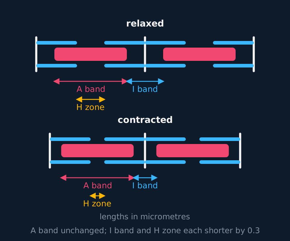
Describe the drawing
Two rows of sarcomeres, relaxed above and contracted below, each drawn between three Z lines. Thick filaments sit in the middle of each sarcomere and thin filaments reach in from every Z line. Brackets under each row mark the A band, which is the same width in both rows, the I band around the shared Z line and the H zone at the centre of the thick filaments, both of which are visibly narrower in the contracted row.
- Sarcomere
- The functional unit of a myofibril, running from one Z line to the next.
- A band
- The dark band, the full length of the thick filaments, including any overlap with thin filaments.
- I band
- The light band, containing thin filaments only, spanning a Z line.
- H zone
- The lighter region at the centre of the A band, containing thick filaments only.
- Sarcoplasmic reticulum
- The membrane system inside a muscle fibre that stores and releases calcium ions.
The banding pattern during contraction
The changing banding pattern is the evidence for the sliding filament mechanism, and each change can be derived from what each band contains.
The A band is the length of the thick filaments. During contraction the thick filaments neither shorten nor move relative to the centre of the sarcomere, so the A band does not change. A constant A band is the direct evidence that the filaments slide rather than shorten.
The I band contains only thin filaments. As the thin filaments are pulled in towards the centre, more of their length lies inside the A band, so the region containing thin filaments alone gets shorter.
The H zone contains only thick filaments, the part of the A band that the thin filaments have not yet reached. As they slide further in, that gap closes, so the H zone gets shorter too, and in a strong contraction it can disappear entirely.
The sarcomere as a whole shortens by the same amount the I band loses, which is also the amount the H zone loses, because the Z lines are dragged towards each other. A muscle shortens because millions of sarcomeres in series each shorten by a fraction of a micrometre.
| Region | What is in it | During contraction |
|---|---|---|
| A band | Thick filaments, plus overlapping thin | Unchanged |
| I band | Thin filaments only | Shorter |
| H zone | Thick filaments only | Shorter |
| Sarcomere | Z line to Z line | Shorter |
| Thick and thin filaments | Myosin, actin | Unchanged in length |
How the sliding is driven
The mechanism is a cycle, and the order of its steps carries the explanation. It begins at the neuromuscular junction.
Acetylcholine released there depolarises the sarcolemma, and the depolarisation spreads along it and down the transverse tubules into the depths of the fibre. That triggers the sarcoplasmic reticulum to release calcium ions into the sarcoplasm.
Calcium binds to troponin, which changes shape and pulls the tropomyosin aside, exposing the myosin binding sites on the actin. Until this moment the binding sites were covered and no cross-bridge could form, which is why a resting muscle does not contract despite having everything else it needs.

Describe the drawing
An animation of the myosin head cycle in five stages, drawn as a thin actin filament above a thick myosin filament carrying two heads, with a dashed mark left at the thin filament's starting position. Stage one: tropomyosin lies over the binding sites and the cocked heads cannot attach. Stage two: calcium binds troponin, tropomyosin is pulled aside and the sites are exposed. Stage three: a head attaches to the thin filament and the cross-bridge is formed, with ADP and phosphate still bound. Stage four: the head bends in the power stroke, ADP and phosphate leave, and the thin filament is dragged about ten nanometres past the dashed mark while the thick filament stays where it is. Stage five: ATP binds, the head lets go and hydrolysis re-cocks it, with the thin filament left in its new position.
A myosin head, carrying ADP and phosphate from a previous round, attaches to the exposed binding site and forms a cross-bridge. The head then bends in the power stroke, releasing ADP and phosphate and pulling the thin filament about 10 nm towards the centre of the sarcomere. An ATP molecule then binds to the head, which causes it to detach from the actin. Hydrolysis of that ATP by the head's own ATPase activity returns the head to its upright position, ready to attach further along the filament.
Hundreds of heads on each thick filament do this out of step with one another, so at any instant some are attached while others are detached and the filament is never released. As long as calcium remains and ATP is available, the cycle repeats and the filaments keep sliding.
Relaxation also requires ATP. When impulses stop, calcium ions are actively pumped back into the sarcoplasmic reticulum, which costs ATP; calcium leaves troponin, tropomyosin slides back over the binding sites, and cross-bridges can no longer form. The muscle is then lengthened by its antagonistic partner or by gravity, because a muscle can pull and cannot push.
Rigor mortis makes the role of ATP unusually clear. After death, respiration stops and ATP runs out. Without ATP the myosin heads cannot detach from actin, and without ATP calcium cannot be pumped back, so the cross-bridges stay formed and the muscles are locked rigid until the proteins themselves begin to break down.
Fibre types and the supply of ATP
Skeletal muscles contain a mixture of fibre types, and the proportions differ between muscles and between individuals. Every row of the comparison below follows from one difference: the main route by which each fibre type makes ATP.
| Slow twitch | Fast twitch | |
|---|---|---|
| Contraction | Slower, sustained | Rapid, powerful, short-lived |
| Main respiration | Aerobic | Anaerobic |
| Mitochondria | Many | Few |
| Myoglobin | High: dark red fibres | Low: paler fibres |
| Capillary supply | Dense | Sparse |
| Glycogen store | Lower | High |
| Fatigue | Resistant | Fatigues quickly |
| Found in | Postural muscles, calf muscles of distance runners | Biceps, leg muscles of sprinters |
Slow twitch fibres are built around a continuous aerobic supply: many mitochondria to make ATP, dense capillaries to bring oxygen and glucose, and myoglobin to store oxygen inside the fibre. Fast twitch fibres are built for a short burst instead, relying on anaerobic glycolysis and a large glycogen store, and they tire quickly. Accumulating lactate is the traditional exam marker of that fatigue, though the fatigue itself has several causes, phosphate build-up and ion imbalances among them, rather than lactate acting as a simple acid poison.
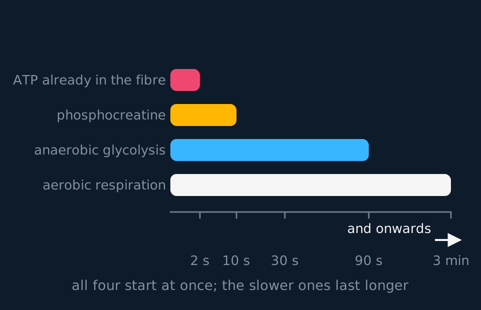
Describe the drawing
A horizontal bar chart of four sources of ATP for a muscle working hard, on a time axis marked at two seconds, ten seconds, thirty seconds, ninety seconds and three minutes. The ATP already in the fibre lasts about two seconds, phosphocreatine regenerates ATP for about ten, anaerobic glycolysis carries on to about ninety seconds, and aerobic respiration runs on past the right-hand edge of the chart. All four bars start at zero, because every source is in use from the first second.
The ATP a muscle contains at rest supports only a second or two of hard work. Three supplies extend it. Phosphocreatine, stored in the sarcoplasm, transfers its phosphate group directly to ADP, regenerating ATP rapidly and anaerobically for only a short period, of the order of ten seconds at maximal effort. Anaerobic glycolysis, with the pyruvate reduced to lactate, can support high power output for longer but yields only two ATP per glucose and contributes to fatigue. Aerobic respiration supplies ATP more slowly and requires oxygen delivery, but yields around 30 ATP per glucose and can sustain activity for much longer, subject to oxygen delivery, substrate availability, temperature and fatigue.
ATP supply during a 400 m race
A 400 m runner finishes in 48 seconds. Explain which sources of ATP supply the muscles during the race, and why the runner is breathing hard for several minutes afterwards.
Show the working
The first second or two runs on ATP already present in the fibres. Phosphocreatine then regenerates ATP for roughly the next ten seconds, covering the start and the first part of the back straight.
For most of the remaining 40 seconds the dominant supply is anaerobic glycolysis, because the demand exceeds what oxygen delivery can support. This yields ATP rapidly but produces lactate, which accumulates and contributes to the muscles failing near the finish.
Afterwards the runner continues to breathe hard because oxygen is needed to oxidise the lactate that has built up and to resynthesise phosphocreatine and ATP stores. Both processes are aerobic and both continue long after the muscles have stopped contracting.
TRY IT: Explaining an electron micrograph measurement
A student measures a sarcomere in a relaxed muscle as 2.4 micrometres long, with an A band of 1.6 micrometres. In a contracted sample from the same muscle the sarcomere measures 2.1 micrometres. Predict the width of the A band in the contracted sample and explain your answer, then state what has happened to the I band.
Check your answer
The A band is still 1.6 micrometres. The A band is the length of the thick filaments, and the thick filaments do not change length during contraction; they are not pulled shorter, and they do not move relative to the centre of the sarcomere.
The sarcomere has shortened by 2.4 − 2.1 = 0.3 micrometres, and all of that has come out of the light regions. The I band contains thin filaments only, and the thin filaments have slid further in between the thick ones, so the I band is 0.3 micrometres narrower.
The H zone will have narrowed by the same 0.3 micrometres, because it is the part of the A band that the thin filaments have not yet reached and they have now reached further into it.
If your prediction for the A band was 1.4 or 1.3 micrometres, you assumed the filaments shortened along with the sarcomere. Measurements like this one are the historical evidence that they do not, which is why the model is called the sliding filament theory.
In the exam
- State that the A band does not change, and that the I band and the H zone both get shorter.
- State that the filaments slide past one another. The filaments themselves do not shorten, and actin does not contract.
- Calcium binds to troponin, not to tropomyosin. Troponin changes shape and moves tropomyosin off the binding site: two proteins with two different roles.
- ATP is used at three points: for the myosin head to detach, for hydrolysis to re-cock it, and for pumping calcium back into the sarcoplasmic reticulum during relaxation. Include the third.
- For fibre-type comparisons, give a structural difference and the functional consequence together, as in 'more mitochondria, so more aerobic ATP, so resistant to fatigue', rather than a list of features.
- Phosphocreatine regenerates ATP from ADP directly and anaerobically. It is not a form of respiration and it does not produce lactate.
Check yourself
Explain how the arrival of an impulse at a neuromuscular junction leads to the shortening of a sarcomere, and state what happens to the width of the A band, the I band and the H zone as it does so.
Answer
Acetylcholine released at the neuromuscular junction binds to receptors on the sarcolemma and depolarises it. The depolarisation spreads along the sarcolemma and down the transverse tubules into the fibre, and causes the sarcoplasmic reticulum to release calcium ions into the sarcoplasm.
Calcium ions bind to troponin, changing its shape so that it moves tropomyosin away from the myosin binding sites on the actin. Myosin heads, already carrying ADP and phosphate, attach to those sites and form cross-bridges.
Each head then bends in a power stroke, releasing ADP and phosphate and pulling the thin filament about 10 nm towards the centre of the sarcomere. ATP binds to the head, which detaches, and hydrolysis of that ATP returns the head to its original position so it can attach further along. With heads working out of step, the thin filaments are drawn steadily inwards and the Z lines move closer together.
The A band is unchanged, because it is the length of the thick filaments and they neither shorten nor move. The I band and the H zone both get shorter, because the thin filaments now occupy more of the A band and leave less of it unoccupied. The sarcomere shortens by as much as each of those two regions narrows.
Questions
Question 15 marks
Describe how the release of calcium ions from the sarcoplasmic reticulum causes the thin filaments of a sarcomere to slide inwards.
Mark scheme
- B1 calcium ions bind to troponin, which changes shape
- B1 troponin pulls tropomyosin aside, exposing the myosin binding sites on the actin, which were covered before
- B1 a myosin head carrying ADP and phosphate attaches to an exposed binding site and forms a cross-bridge
- B1 the head bends in a power stroke, releasing the ADP and phosphate and pulling the thin filament about 10 nm towards the centre of the sarcomere
- A1 ATP binds to the head so that it detaches, and hydrolysis of that ATP returns it to its upright position to attach further along; heads work out of step so the filament is never released
Question 24 marks
Explain the three ways ATP is used by a contracting and relaxing muscle fibre, and explain why the muscles of a body stiffen after death.
Mark scheme
- B1 ATP binding to the myosin head causes it to detach from the actin at the end of a power stroke
- B1 hydrolysis of that ATP by the head's own ATPase activity returns the head to its upright position, ready to attach further along the thin filament
- B1 ATP is used to pump calcium ions back into the sarcoplasmic reticulum during relaxation, so tropomyosin can slide back over the binding sites
- A1 after death respiration stops and ATP runs out, so the heads cannot detach and calcium cannot be pumped back, and the cross-bridges stay formed until the proteins themselves break down
Question 34 marks
Compare slow twitch muscle fibres with fast twitch muscle fibres, linking each structural difference to the way the fibre works.
Mark scheme
- B1 slow twitch fibres contract more slowly but in a sustained way, whereas fast twitch fibres contract rapidly and powerfully for a short time
- B1 slow twitch fibres have many mitochondria and respire aerobically, whereas fast twitch fibres have few and rely on anaerobic glycolysis with a large glycogen store
- B1 slow twitch fibres have a dense capillary supply and much myoglobin, so oxygen is delivered and stored, whereas fast twitch fibres have a sparse supply and little myoglobin and are paler
- A1 slow twitch fibres therefore resist fatigue and dominate postural muscles and the calf muscles of distance runners, whereas fast twitch fibres accumulate lactate and tire quickly, and dominate the leg muscles of sprinters
Question 44 marks
A relaxed muscle fibre is 3.0 cm long and its myofibrils are made of sarcomeres each 2.4 micrometres long, joined end to end. Calculate the number of sarcomeres in one myofibril of this fibre, and calculate the length of the fibre in centimetres when every sarcomere has shortened to 2.0 micrometres.
Mark scheme
- M1 convert to the same unit: 3.0 cm is 30 000 micrometres
- M1 number of sarcomeres is the fibre length divided by the length of one sarcomere, so 30 000 ÷ 2.4
- A1 12 500 sarcomeres in series
- A1 12 500 × 2.0 = 25 000 micrometres, which is 2.5 cm
Question 54 marks
A sprinter's leg muscles fail after about 40 seconds of maximal effort. A student says this is because the muscles have used up all their ATP. Suggest why that explanation is wrong, and suggest what does limit the muscles over that time.
Mark scheme
- B1 the ATP already in the fibres lasts only about two seconds, so a muscle working for 40 seconds must be regenerating ATP throughout rather than running on a store of it
- B1 phosphocreatine in the sarcoplasm transfers its phosphate group directly to ADP to regenerate ATP anaerobically, but it is spent within about ten seconds
- B1 most of the rest of the effort is supplied by anaerobic glycolysis, because oxygen cannot be delivered fast enough for aerobic respiration to meet the demand
- A1 anaerobic glycolysis yields only two ATP per glucose and the lactate it produces accumulates, so what fails is the rate at which ATP can be resupplied rather than the quantity of ATP present
Question 64 marks
Explain how an action potential arriving at a neuromuscular junction leads to the release of calcium ions from the sarcoplasmic reticulum into the sarcoplasm.
Mark scheme
- B1 acetylcholine released at the neuromuscular junction binds to receptors on the sarcolemma and depolarises it
- B1 the depolarisation spreads across the sarcolemma and down the transverse tubules, which are folds of the sarcolemma running deep into the fibre
- B1 this carries the depolarisation to the sarcoplasmic reticulum, the specialised endoplasmic reticulum that surrounds the myofibrils and stores calcium ions
- B1 the depolarisation triggers the sarcoplasmic reticulum to release its stored calcium ions into the sarcoplasm
Question 74 marks
Compare thick filaments with thin filaments in a sarcomere, referring to what each is made of, where each is anchored, and what happens to their length during contraction.
Mark scheme
- B1 thick filaments are made of myosin, with tails bundled together and heads projecting outwards, whereas thin filaments are made of actin wound with tropomyosin and studded with troponin
- B1 thick filaments sit in the middle of the sarcomere, unattached to the Z lines, whereas thin filaments are anchored to the Z lines at each end
- B1 during contraction the thin filaments slide inward past the thick filaments, whereas the thick filaments stay in a fixed position relative to the centre of the sarcomere
- B1 neither type of filament changes in length during contraction; only the degree of overlap between them changes
Question 84 marks
A myofibril contains sarcomeres each 2.5 micrometres long when relaxed. During a contraction each sarcomere shortens by 15%. Calculate the length of one contracted sarcomere, and calculate the number of sarcomeres needed, joined end to end, to make a contracted myofibril 1.7 mm long.
Mark scheme
- M1 contracted sarcomere length = 2.5 × (1 − 0.15) = 2.5 × 0.85
- A1 2.1 micrometres to two significant figures, keeping the unrounded 2.125 for the next step
- M1 convert 1.7 mm to micrometres, 1700 µm, and divide by the contracted sarcomere length: 1700 ÷ 2.125
- A1 800 sarcomeres in the contracted myofibril
Question 94 marks
A drug blocks the calcium pumps in the sarcoplasmic reticulum, so calcium ions cannot be pumped back in after release. Suggest the effect of this drug on a muscle fibre's ability to relax after a contraction, and suggest what would eventually happen to its ATP concentration if contraction continued unchecked.
Mark scheme
- B1 with the pumps blocked, calcium ions would stay in the sarcoplasm rather than being returned to the sarcoplasmic reticulum, so calcium would remain bound to troponin
- B1 tropomyosin would not move back over the myosin binding sites on actin, so cross-bridges could keep forming and the fibre could not relax
- B1 the cross-bridge cycle would continue running for as long as ATP was available, with each cycle using ATP
- B1 ATP would be used up faster than in a normal resting muscle, and once it ran out the myosin heads could not detach from actin, so the muscle would become permanently contracted, similar to rigor mortis
Question 103 marks
State what happens to the width of the A band, the width of the I band and the width of the H zone when a sarcomere contracts.
Mark scheme
- B1 the A band is unchanged
- B1 the I band gets shorter
- B1 the H zone gets shorter, and in a strong contraction it can disappear altogether
Question 113 marks
Describe the structural organisation of a skeletal muscle fibre, from the whole fibre down to a single sarcomere.
Mark scheme
- B1 a muscle fibre is a single, multinucleate cell containing hundreds of myofibrils
- B1 each myofibril is a chain of sarcomeres joined end to end at Z lines
- B1 the sarcomere, running from one Z line to the next, is the functional unit of contraction, containing the thick and thin filaments
Question 123 marks
Explain why the A band does not change in width during contraction, while the I band and the H zone both get shorter.
Mark scheme
- B1 the A band is the length of the thick filaments, which neither shorten nor move relative to the centre of the sarcomere during contraction, so its width stays the same
- B1 the I band contains only thin filaments, and as they slide further into the A band, less of their length lies outside it, so the I band narrows
- B1 the H zone is the part of the A band not yet reached by thin filaments; as they slide further in, this unreached region shrinks, so the H zone narrows too
Question 133 marks
Explain why phosphocreatine can regenerate ATP more rapidly than aerobic respiration can, but only for a short time.
Mark scheme
- B1 phosphocreatine regenerates ATP by transferring its phosphate group directly to ADP in a single step, whereas aerobic respiration requires a longer sequence of reactions including the delivery of oxygen, so phosphocreatine responds faster
- B1 the store of phosphocreatine in the sarcoplasm is small, so it is used up within about ten seconds of maximal effort
- B1 aerobic respiration continues for as long as oxygen and substrate keep being delivered, so it sustains ATP production for much longer once it is underway
Question 142 marks
Name the protein that makes up the thick filaments of a sarcomere, and name the two proteins found on the thin filaments in addition to actin.
Mark scheme
- B1 myosin makes up the thick filaments
- B1 tropomyosin and troponin are found on the thin filaments, in addition to actin
Question 152 marks
State what the term multinucleate means as applied to a muscle fibre, and state the name given to a muscle fibre's cell surface membrane.
Mark scheme
- B1 multinucleate means the cell contains many nuclei rather than just one
- B1 the cell surface membrane of a muscle fibre is called the sarcolemma
Worth remembering
- Muscle, fibre, myofibril, sarcomere, and the sarcomere runs from one Z line to the next.
- Calcium binds troponin, troponin moves tropomyosin, and the binding site is exposed.
- Attach, power stroke, ATP binds and the head detaches, hydrolysis re-cocks it. Repeat while calcium lasts.
- The A band never changes; the I band and H zone shorten by the same amount as the sarcomere.
- Stored ATP, phosphocreatine, anaerobic glycolysis and aerobic respiration supply ATP at decreasing rates and for increasing lengths of time.
CHECK YOUR PROGRESS
Rate how confident you are with each objective for this lesson. Ratings are saved in this browser, on this device, unless you sign in.
- Describe the structure of skeletal muscle from whole muscle down to the sarcomere.
- Explain how an impulse at the neuromuscular junction leads to calcium release inside the fibre.
- Describe the sliding filament mechanism, including the roles of calcium, tropomyosin, ATP and the myosin head.
- State what happens to the A band, I band and H zone during contraction, and why.
- Compare slow and fast twitch fibres, and explain how ATP is supplied to a working muscle.
Open the full revision checklist to see every objective in the curriculum in one place.
Practise this lesson
WORKBOOK
The questions from this topic, on paper with room to work, and a separate book of mark schemes. Free to use; please do not redistribute or sell.