Biology › Nervous coordination, receptors and muscles › The synapse: transmission, and why it runs one way
The synapse: transmission, and why it runs one way
A synapse separates two neurones by a cleft about 20 nm wide, which an action potential cannot cross. A neurotransmitter released from the presynaptic membrane and bound by receptors on the postsynaptic membrane carries the signal across. The sequence of transmission at a cholinergic synapse, the structural reason for unidirectionality, and spatial and temporal summation are set out here.
Before this The action potential and the refractory period · Active transport, co-transport and bulk transport
COMMON MISCONCEPTION
The impulse crosses the synapse.
The impulse arrives at the presynaptic membrane and stops there. A chemical is released, diffuses across a gap about 20 nm wide and binds on the far side, where, if enough arrives, a new action potential begins in a different cell. The message continues; the impulse does not.
What you should be able to do
- Label a cholinergic synapse and say what each part does.
- Describe transmission across it in the right order, from arriving impulse to enzyme clearing the cleft.
- Explain why transmission is unidirectional, using the position of vesicles and receptors.
- Distinguish spatial from temporal summation, and explain what an inhibitory synapse does.
- Compare a neuromuscular junction with a synapse between two neurones.
- Explain how a named drug produces its effect at a synapse.
- Explain how too little dopamine contributes to Parkinson's disease and too little serotonin to depression, and how L-Dopa, SSRIs and MDMA change transmission.
What is actually there
A synapse is the junction between two neurones, or between a neurone and an effector. The one worth knowing in detail is the cholinergic synapse, which uses acetylcholine as its neurotransmitter and appears at neuromuscular junctions and throughout the nervous system.
The axon of the presynaptic neurone ends in a swelling, the synaptic knob. It is packed with mitochondria and with vesicles of acetylcholine, and its membrane carries voltage-gated calcium channels. Across a synaptic cleft of about 20 nm sits the postsynaptic membrane, whose receptor proteins have binding sites complementary to acetylcholine and which are themselves ion channels. The ones here, called nicotinic receptors, open to let positive ions through, mostly sodium inwards, so 'sodium channel' is the working exam shorthand. (A second family of acetylcholine receptors elsewhere in the body is not a channel at all and signals more slowly.)
Two features of that arrangement account for the direction of transmission: the vesicles are only on the presynaptic side, and the receptors are only on the postsynaptic side.
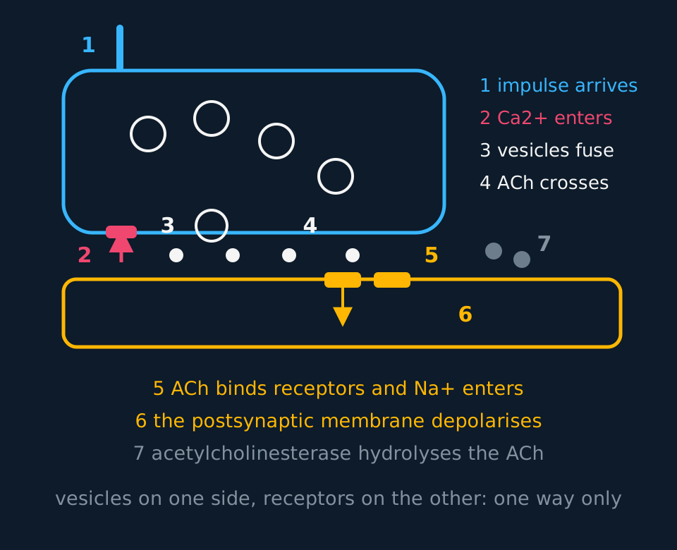
Describe the drawing
A cholinergic synapse in section. A presynaptic knob at the top left holds four vesicles with a fifth already fused to the membrane, an axon entering from above and a calcium channel marked on its lower membrane. Acetylcholine molecules are drawn as dots crossing the cleft to receptor blocks set in the postsynaptic membrane below, where an arrow shows sodium entering, and broken-down transmitter lies to the right. Seven numbered points on the drawing match a numbered list running from the impulse arriving to acetylcholinesterase hydrolysing the transmitter.
- Synapse
- The junction between the end of one neurone and another neurone or an effector cell, across which chemical transmission occurs.
- Neurotransmitter
- A chemical released from a presynaptic neurone that diffuses across the cleft and binds to receptors on the postsynaptic membrane.
- Cholinergic synapse
- A synapse that uses acetylcholine as its neurotransmitter.
- Excitatory postsynaptic potential
- A depolarisation of the postsynaptic membrane that makes an action potential more likely.
Sequence of transmission at a cholinergic synapse
Describe the events in causal order, from depolarisation of the presynaptic membrane to removal of acetylcholine.
The action potential arrives at the synaptic knob and depolarises its membrane. Voltage-gated calcium channels open, and calcium ions diffuse into the knob from the cleft, where their concentration is much higher.
The influx of calcium causes vesicles of acetylcholine to move to the presynaptic membrane and fuse with it, releasing their contents into the cleft by exocytosis. Acetylcholine then crosses the cleft by diffusion down its concentration gradient. No energy is required, and over 20 nm the crossing takes well under a millisecond.
On the far side, acetylcholine binds to receptor sites on the postsynaptic membrane. The receptors are sodium channels, and binding changes their shape so that they open. Sodium ions diffuse into the postsynaptic neurone and depolarise it, producing an excitatory postsynaptic potential. If enough receptors are occupied for the membrane to reach threshold, voltage-gated sodium channels open and a new action potential begins.
Then the cleft is cleared. Acetylcholinesterase, an enzyme in the cleft, hydrolyses acetylcholine into choline and ethanoic acid. The choline is reabsorbed into the presynaptic knob and recombined into acetylcholine, the acetyl group coming from acetyl-CoA and the energy from ATP made by those mitochondria, and the vesicles are refilled.
Hydrolysis of acetylcholine is necessary for repeated transmission. Without it, acetylcholine stays bound to the receptors and the channels stay open. The result is not continuous firing: a membrane held depolarised cannot reset its voltage-gated channels, so after an initial burst transmission fails, a state called depolarisation block. Several of the drugs described below act this way.
Putting a delay to work
Transmission across one synapse takes roughly 0.7 ms. A reflex pathway contains two synapses, and the total time from stimulus to response is 30 ms. What fraction of the delay is synaptic, and what does that imply about a pathway routed through the brain instead?
Show the working
Two synapses at 0.7 ms give about 1.4 ms of synaptic delay, which is 1.4 ÷ 30 ≈ 0.047, so under 5% of the total. Most of the 30 ms is conduction along the neurones.
A conscious response involves many neurones in the brain and therefore many more synapses; adding a hundred of them would add about 70 ms on its own, before any of the extra conduction distance is counted.
Each synapse also adds a junction at which signals can be combined, blocked or redirected, so the number of synapses in a pathway trades speed against that processing. A reflex arc contains as few as possible.
Why transmission is unidirectional
Neurones are connected into branching networks, and transmission through them runs in one direction only. The reason is structural.
Vesicles of neurotransmitter are found only in the presynaptic knob, so acetylcholine can only be released on that side. Receptors are found only on the postsynaptic membrane, so acetylcholine can only be detected on the other. An impulse arriving at the postsynaptic side of a synapse finds nothing to release and nothing on the far side to detect it.
Transmission therefore runs reliably from receptor to effector, and a network has defined inputs and outputs. Two separate mechanisms produce that directionality: the refractory period keeps an impulse travelling one way along an axon, and the distribution of vesicles and receptors keeps transmission one way between neurones.
Summation and inhibition
Between neurones, one impulse arriving at one synapse rarely triggers anything. Each vesicle releases a fixed quantity of acetylcholine, and a single excitatory postsynaptic potential is typically a few millivolts against the 15 mV needed to reach threshold from −70 mV. Postsynaptic potentials are summed, by two mechanisms.
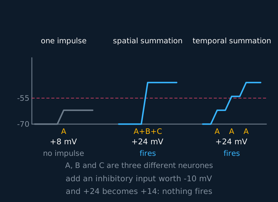
Describe the drawing
Three membrane-potential traces on shared axes, all starting from minus seventy millivolts with a dashed threshold line at minus fifty-five. The first rises one step of eight millivolts and stops short of the line. The second rises in one step of twenty-four millivolts as three different neurones fire at the same moment, and the third climbs in three steps of eight as one neurone fires three times in quick succession. Both cross the threshold, so both fire.
Spatial summation adds inputs from different presynaptic neurones arriving at the same postsynaptic cell at the same time. Several small depolarisations together take the membrane past threshold. This is the mechanism behind rod convergence in the retina, and it is how a postsynaptic neurone can respond to a pattern of inputs rather than to any one of them.
Temporal summation adds impulses from a single presynaptic neurone arriving in quick succession. The transmitter from the first has not yet been cleared when the second arrives, so the depolarisations build on one another. High-frequency firing therefore reaches threshold where the same number of impulses spread over a longer time would not, which is a further consequence of coding intensity as frequency.
Not every synapse is excitatory. At an inhibitory synapse the receptors open chloride channels, letting negative chloride ions in, or potassium channels, letting positive potassium ions out. Either way the postsynaptic membrane becomes more negative, or hyperpolarised, so a larger excitatory input is now needed to reach threshold. Inhibition sharpens signals, suppresses unwanted movements and limits the spread of excitation through a network.
A neuromuscular junction is the cholinergic synapse between a motor neurone and a skeletal muscle fibre. Transmission across it follows the same sequence, with some differences from a synapse between neurones. The postsynaptic membrane is the sarcolemma, folded at the motor end plate to hold many receptors, with acetylcholinesterase in the folds. The junction is always excitatory, never inhibitory. One impulse releases enough acetylcholine to take the fibre to threshold every time, so no summation is needed. The action potential it starts spreads along the muscle fibre and leads to contraction; it does not pass on to another neurone.
- Spatial summation
- Summation of inputs arriving at the same time from several presynaptic neurones.
- Temporal summation
- Summation of impulses arriving in quick succession from one presynaptic neurone.
- Inhibitory synapse
- A synapse whose transmitter hyperpolarises the postsynaptic membrane, making an action potential less likely.
- Neuromuscular junction
- The synapse between a motor neurone and a skeletal muscle fibre.
- Acetylcholinesterase
- The enzyme in the synaptic cleft that hydrolyses acetylcholine into choline and ethanoic acid.
Where drugs act
Most drugs that affect the nervous system act at a synapse, and they act at a small number of points in the transmission sequence. An unfamiliar substance can be reasoned about by identifying which step it alters.
| Drug | What it does | Effect |
|---|---|---|
| Nicotine | Binds to acetylcholine receptors and activates them | Postsynaptic neurone depolarised as though ACh had bound: more impulses |
| Organophosphate insecticides | Inhibit acetylcholinesterase | ACh is not broken down and receptors stay occupied; an initial burst of firing, then depolarisation block |
| Curare | Blocks acetylcholine receptors without activating them | ACh cannot bind, no depolarisation, muscles are paralysed |
| Cocaine | Blocks reuptake of dopamine from the cleft | Dopamine stays in the cleft and keeps binding, so the postsynaptic neurone fires repeatedly |
| Botulinum toxin | Prevents vesicles releasing acetylcholine | No transmitter released, no depolarisation, muscles cannot contract |
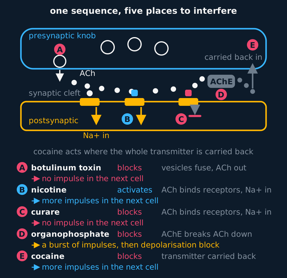
Describe the drawing
A synapse drawn in section with the presynaptic knob above, the cleft, and the postsynaptic membrane below, and five lettered points marked on it: A where a vesicle fuses and releases acetylcholine, B and C at two receptors in the postsynaptic membrane, one with a drug in it holding the channel open and sodium entering and the other with a drug in it and the channel shut, D at the enzyme in the cleft, and E at the arrow carrying the transmitter back into the knob. Beneath, five lines name the drug at each letter, the step it acts on and whether it blocks or activates that step, and give the result: botulinum toxin and curare leave no impulse in the next cell, nicotine and cocaine give more impulses, and the organophosphate gives a burst of impulses and then depolarisation block.
Nicotine and curare both bind to the acetylcholine receptor; nicotine activates it and curare does not, so one increases firing and the other abolishes it. The word to use is complementary: both molecules have shapes complementary to the binding site, and what differs is whether binding opens the channel.
Pearson Edexcel's Biology B specification names three substances. Nicotine is in the table. Cobra venom acts as curare does: its neurotoxins bind tightly to the acetylcholine receptors on the postsynaptic membrane at neuromuscular junctions without opening them, so acetylcholine cannot bind, no sodium ions enter, the muscle fibre is not depolarised and the muscle is paralysed. A bite can be fatal because the diaphragm and intercostal muscles are paralysed with the rest. Lidocaine acts before the synapse, on the axon: it blocks voltage-gated sodium channels, so action potentials are neither produced nor propagated, none arrives at the synaptic knob, and no transmitter is released. Its use as a local anaesthetic is described in the action potential lesson.
TRY IT: Reasoning about an unfamiliar poison
A nerve agent inhibits acetylcholinesterase at neuromuscular junctions. Explain why exposure causes twitching and cramps followed by paralysis, and suggest why it is eventually fatal.
Check your answer
Acetylcholinesterase normally hydrolyses acetylcholine in the cleft. Inhibited, the enzyme cannot break it down, so acetylcholine stays in the cleft and remains bound to the receptors on the postsynaptic membrane.
The receptor channels therefore stay open and the muscle fibre membrane is held depolarised. At first action potentials fire in a burst and the muscle twitches and cramps; but a membrane held depolarised cannot reset its voltage-gated channels, so transmission then fails, a state called depolarisation block, and the overstimulated muscle falls into flaccid paralysis.
It is fatal because the same junctions serve the muscles used in breathing: with the diaphragm and intercostal muscles paralysed, and the agent also acting in the brain and flooding the airways with secretions, ventilation stops. Real antidotes come as a pair: atropine blocks the acetylcholine receptors of glands and organs, a different receptor family from the neuromuscular junction, and an oxime such as pralidoxime reactivates the inhibited enzyme itself.
Brain chemicals, illness and new drugs
Dopamine and serotonin are neurotransmitters, each released by particular groups of neurones in the brain. Too little of either at the synapses where it is used contributes to illness: dopamine to Parkinson's disease, serotonin to depression. Knowing which transmitter is in short supply, and which step of transmission could be changed to make up for it, is where the development of a drug starts.
In Parkinson's disease, dopamine-secreting neurones in a region of the brainstem called the substantia nigra die. These neurones release dopamine in parts of the brain that control movement, and with less dopamine released there, fewer postsynaptic receptors are stimulated. The symptoms are a tremor at rest, stiff muscles, slow movement and difficulty starting a movement, and balance and speech can be affected as well. They usually appear only after many of the neurones have already been lost.
Giving dopamine itself does not work, because it cannot cross the blood-brain barrier. L-Dopa (levodopa), the molecule from which neurones make dopamine, can. Inside the brain an enzyme converts it to dopamine, so the surviving neurones release more and the symptoms are reduced. It is given with a second drug, such as carbidopa, that stops the conversion happening in the rest of the body, so more L-Dopa reaches the brain and side effects such as nausea are reduced. L-Dopa makes up for the shortage of dopamine but does not replace the neurones, so the disease continues to progress. Other drugs for Parkinson's disease act at other steps: dopamine agonists bind to dopamine receptors and activate them, and MAO-B inhibitors slow the breakdown of dopamine. Replacing the lost neurones with cells grown from stem cells is a different approach, discussed in the stem cells lesson.
Serotonin is released by neurones whose axons spread from the brainstem to many parts of the brain, including regions involved in mood, sleep and appetite. After it has bound to its postsynaptic receptors, a transport protein in the presynaptic membrane takes it back into the presynaptic neurone, where some is broken down by the enzyme monoamine oxidase (MAO). Depression has been linked to reduced serotonin transmission, and the most widely prescribed antidepressants were developed from that idea.
Selective serotonin reuptake inhibitors (SSRIs), such as fluoxetine, bind to the serotonin transport protein and block it. Serotonin stays in the cleft for longer and goes on binding to postsynaptic receptors, so transmission at those synapses increases. Older antidepressants act at a different step: MAO inhibitors slow the breakdown of serotonin in the presynaptic neurone, so more is available for release.
The link with depression is not a simple shortage. An SSRI raises serotonin in the cleft within hours, but mood usually takes several weeks to improve, and the drugs do not help every patient. Depression is thought to arise from several genetic and environmental factors, with altered serotonin transmission one contributor among them. An answer should say that an imbalance of serotonin contributes to depression, which is the claim the specification makes.
MDMA, the active substance in Ecstasy, acts on the same transport protein. It binds to it, blocking reuptake, and also makes it carry serotonin out of the presynaptic neurone into the cleft, so the concentration of serotonin in the cleft rises sharply. The postsynaptic neurones are strongly stimulated, which produces the raised mood the drug is taken for. Afterwards the presynaptic neurones' stores of serotonin are depleted, which is associated with low mood in the following days. MDMA can also raise body temperature dangerously and disturb the body's water balance, and these effects can be fatal.
| Drug | Transmitter | Step it changes | Effect at the synapse |
|---|---|---|---|
| L-Dopa | Dopamine | Supplies the molecule dopamine is made from | More dopamine made and released |
| SSRI, such as fluoxetine | Serotonin | Blocks the reuptake transport protein | Serotonin stays in the cleft for longer |
| MAO inhibitor | Serotonin and dopamine | Slows breakdown inside the presynaptic neurone | More transmitter available for release |
| MDMA | Serotonin | Blocks reuptake and reverses the transport protein | A large rise in serotonin, then depletion |
The search for new drugs starts the same way. Once the transmitter involved is known, each step in its life is a possible target: its synthesis, its release, its receptors, its reuptake and its breakdown. Candidate molecules can then be screened for their effect on that one step before any are tested in patients.
- Dopamine
- A neurotransmitter in the brain; too little where it controls movement contributes to Parkinson's disease.
- Serotonin
- A neurotransmitter in the brain involved in mood, sleep and appetite; an imbalance of it contributes to depression.
- L-Dopa
- The precursor of dopamine, which crosses the blood-brain barrier and is converted to dopamine in the brain; used to treat Parkinson's disease.
- SSRI
- Selective serotonin reuptake inhibitor: an antidepressant that blocks the reuptake of serotonin from the synaptic cleft.
- MDMA
- The active substance in Ecstasy, which blocks serotonin reuptake and causes serotonin to be released into the cleft.
In the exam
- Give the events in order: impulse arrives, calcium enters, vesicles fuse, acetylcholine diffuses, receptors bind, sodium enters, threshold, enzyme hydrolyses.
- Calcium ions enter the presynaptic knob and sodium ions enter the postsynaptic neurone. Name the correct ion on each side.
- Acetylcholine crosses by diffusion down a concentration gradient over 20 nm; it is not pumped or actively transported.
- For unidirectionality, state both halves: vesicles are only in the presynaptic knob, and receptors are only on the postsynaptic membrane.
- Spatial summation is several neurones at once; temporal summation is one neurone in quick succession. Identify which is shown in a given diagram.
- For a drug question, identify the step of the sequence affected and follow the consequences through to the postsynaptic membrane rather than stating that the synapse is blocked.
- For L-Dopa, say why dopamine itself cannot be given: it does not cross the blood-brain barrier, and L-Dopa does and is converted to dopamine in the brain.
- SSRIs and MDMA both act on the serotonin reuptake protein. SSRIs only block it; MDMA also makes it release serotonin into the cleft.
Check yourself
A postsynaptic neurone receives inputs from three presynaptic neurones. Each excitatory input depolarises it by 6 mV; one inhibitory input hyperpolarises it by 9 mV. The resting potential is −70 mV and threshold is −55 mV. Determine whether an action potential is produced when all three excitatory neurones fire together, and when the inhibitory neurone fires alongside them. Explain what the neurone is doing in each case.
Answer
Threshold requires a depolarisation of 15 mV, from −70 mV to −55 mV. Three excitatory inputs together give 3 × 6 = 18 mV, which takes the membrane to −52 mV. That is past threshold, so voltage-gated sodium channels open and an action potential is produced. This is spatial summation: no single input would have done it, since 6 mV alone reaches only −64 mV.
With the inhibitory input firing as well, the total is 18 − 9 = 9 mV of depolarisation, reaching −61 mV. That is short of threshold, so no action potential is produced.
The inhibitory synapse achieves this by opening chloride or potassium channels, making the inside of the postsynaptic neurone more negative and so raising the excitatory input needed to reach threshold.
Excitatory and inhibitory postsynaptic potentials arriving at the same time are summed, and an action potential is produced only if the total reaches threshold. A neurone in the brain may receive thousands of such inputs, summed in the same way.
Questions
Question 15 marks
Describe what happens at a cholinergic synapse from the moment an action potential arrives at the presynaptic knob to the moment the cleft is cleared again.
Mark scheme
- B1 the arriving action potential depolarises the membrane of the knob, so voltage-gated calcium channels open and calcium ions diffuse in
- B1 the influx of calcium causes vesicles of acetylcholine to move to the presynaptic membrane and fuse with it, releasing their contents by exocytosis
- B1 acetylcholine diffuses across the 20 nm cleft down its concentration gradient and binds to receptor sites on the postsynaptic membrane
- B1 the receptors are sodium channels, so binding opens them, sodium ions diffuse into the postsynaptic neurone and depolarise it, and a new action potential begins if threshold is reached
- A1 acetylcholinesterase in the cleft hydrolyses the acetylcholine, and the products are reabsorbed into the knob and recombined using ATP from its mitochondria
Question 24 marks
Explain how an inhibitory synapse reduces the chance that the postsynaptic neurone produces an action potential, and explain why inhibition is useful rather than a fault in the system.
Mark scheme
- B1 the receptors at an inhibitory synapse open chloride channels, letting negative chloride ions in, or potassium channels, letting positive potassium ions out
- B1 either movement makes the inside of the postsynaptic membrane more negative, so it is hyperpolarised rather than depolarised
- B1 a larger excitatory input is now needed to bring the membrane to the threshold of about −55 mV
- A1 this lets a postsynaptic neurone weigh positive against negative inputs, so signals are sharpened, unwanted movements are suppressed and the whole network does not fire at once
Question 34 marks
A toxin from a marine snail prevents the voltage-gated calcium channels in presynaptic knobs from opening. Suggest the effect of this toxin on transmission at a neuromuscular junction, and suggest why an injection of extra acetylcholine into the cleft would not restore normal control of the muscle.
Mark scheme
- B1 calcium ions could not diffuse into the synaptic knob when an action potential arrived
- B1 without the calcium influx the vesicles would not move to the presynaptic membrane and fuse with it, so no acetylcholine would be released into the cleft
- B1 no receptors on the postsynaptic membrane would be occupied, so no sodium channels would open, the muscle fibre membrane would not depolarise and the muscle would be paralysed
- A1 injected acetylcholine would bind and depolarise the fibre, but the amount present would no longer depend on the impulses arriving, so the contraction could not be graded or timed by the nervous system
Question 44 marks
An imbalance of serotonin in the brain is one of the factors that contributes to depression. Explain how a selective serotonin reuptake inhibitor (SSRI) can reduce the symptoms of depression.
Mark scheme
- B1 serotonin is released from the presynaptic neurone into the synaptic cleft and binds to receptors on the postsynaptic membrane
- B1 it is normally removed from the cleft by a transport protein that takes it back into the presynaptic neurone
- B1 an SSRI binds to this transport protein and blocks reuptake, so serotonin stays in the cleft for longer
- B1 serotonin goes on binding to the postsynaptic receptors, so transmission at serotonin synapses increases, which makes up for the reduced serotonin transmission linked to depression
Question 54 marks
Explain why people with Parkinson's disease have difficulty controlling movement, and explain why they are treated with L-Dopa rather than with dopamine.
Mark scheme
- B1 dopamine-secreting neurones in the brain that are involved in controlling movement die
- B1 less dopamine is released, so fewer receptors on the postsynaptic neurones are stimulated, giving tremor, stiffness and slow movement
- B1 dopamine cannot cross the blood-brain barrier, so dopamine given as a drug would not reach the brain
- B1 L-Dopa can cross it and is converted to dopamine in the brain, so the surviving neurones release more dopamine
Question 64 marks
Explain what determines the size of the excitatory postsynaptic potential produced by a single impulse, and explain why one impulse at one synapse does not usually trigger an action potential in the postsynaptic neurone.
Mark scheme
- B1 the size depends on how much acetylcholine is released, set by how many vesicles fuse, each holding a fixed quantity, and on how many receptor channels it opens; typically a few millivolts
- B1 reaching threshold from the resting potential of −70 mV requires a depolarisation of about 15 mV
- B1 a single small excitatory postsynaptic potential is far short of that on its own, so no action potential is triggered by one impulse at one synapse acting alone
- B1 threshold is reached only once several such potentials are combined, by spatial or temporal summation
Question 74 marks
Explain how nicotine and curare can both bind to the acetylcholine receptor on a postsynaptic membrane, yet produce opposite effects on transmission.
Mark scheme
- B1 both nicotine and curare have a shape complementary to the acetylcholine receptor, so both are able to bind to it
- B1 nicotine binds and activates the receptor, opening its channel as acetylcholine would, so the postsynaptic membrane is depolarised and more impulses are produced
- B1 curare binds the same receptor but does not activate it, so the channel stays closed
- B1 while curare occupies the receptor, acetylcholine cannot bind there either, so no depolarisation occurs and transmission is blocked
Question 84 marks
Compare an excitatory synapse with an inhibitory synapse, referring to the ions the postsynaptic channels let through and the effect each has on the chance of an action potential.
Mark scheme
- B1 at an excitatory synapse the postsynaptic channels let sodium ions in, whereas at an inhibitory synapse they let chloride ions in or potassium ions out
- B1 the excitatory synapse depolarises the postsynaptic membrane, making an action potential more likely, whereas the inhibitory synapse hyperpolarises it, making one less likely
- B1 both act by changing which ions cross the postsynaptic membrane, one moving it towards the threshold of about −55 mV and the other away from it
- B1 a postsynaptic neurone usually receives both kinds of input at once and sums them, so whether it fires depends on the balance between the two
Question 94 marks
Transmission across a single synapse takes about 0.5 ms. A pathway containing four synapses also involves 6 ms of conduction along its neurones. Calculate the total time from stimulus to response, and calculate the percentage of that time accounted for by the synapses.
Mark scheme
- M1 total synaptic delay = 4 × 0.5 = 2.0 ms
- A1 total time from stimulus to response = 2.0 + 6 = 8.0 ms
- M1 percentage = synaptic delay ÷ total time × 100 = 2 ÷ 8 × 100
- A1 25%, one quarter of the total
Question 103 marks
Explain why transmission across a synapse can happen in one direction only, and explain why this matters for a reflex arc.
Mark scheme
- B1 vesicles of acetylcholine are found only in the presynaptic knob, so the neurotransmitter can only be released on that side
- B1 receptors complementary to acetylcholine are found only on the postsynaptic membrane, so it can only be detected on the other side
- A1 an impulse arriving at the postsynaptic side would find nothing to release and nothing on the far side to detect it, so a reflex arc runs reliably from receptor to effector and never backwards
Question 113 marks
Compare spatial summation with temporal summation at a postsynaptic neurone.
Mark scheme
- B1 spatial summation adds inputs arriving at the same time from several different presynaptic neurones, whereas temporal summation adds impulses arriving in quick succession from a single presynaptic neurone
- B1 in spatial summation the depolarisations are produced at different points on the membrane at once, whereas in temporal summation the transmitter from the first impulse has not yet been hydrolysed when the second arrives
- B1 both reach threshold by adding small excitatory postsynaptic potentials together, and in neither case could threshold be reached by one impulse at one synapse acting alone
Question 123 marks
MDMA, the active substance in Ecstasy, binds to the serotonin reuptake proteins in presynaptic membranes. Explain how MDMA raises mood, and suggest why users often report low mood in the days that follow.
Mark scheme
- B1 MDMA blocks the reuptake of serotonin and also causes serotonin to be released from the presynaptic neurone into the cleft
- B1 the concentration of serotonin in the cleft rises, so more binds to postsynaptic receptors and the postsynaptic neurones are strongly stimulated
- B1 afterwards the presynaptic neurones' stores of serotonin are depleted, so less is released and serotonin transmission falls
Question 133 marks
Nicotine and cobra venom both bind to acetylcholine receptors. Compare their effects on transmission at a neuromuscular junction.
Mark scheme
- B1 both have shapes complementary to the binding site of the acetylcholine receptor on the postsynaptic membrane
- B1 nicotine mimics acetylcholine: it opens the receptor channels, sodium ions enter and the membrane is depolarised as though acetylcholine had bound
- B1 cobra venom binds without opening the channels and stops acetylcholine binding, so the muscle fibre is not depolarised and the muscle is paralysed
Question 143 marks
Name the presynaptic structure that contains vesicles of acetylcholine and voltage-gated calcium channels, name the ion that enters it when an action potential arrives, and name the process by which the vesicles release their contents into the cleft.
Mark scheme
- B1 the synaptic knob
- B1 calcium ions, entering down their gradient
- B1 exocytosis, the vesicles fusing with the presynaptic membrane
Question 153 marks
Cocaine blocks the protein that normally removes dopamine from a synaptic cleft by reuptake. Suggest what this does to the concentration of dopamine in the cleft after an impulse, and suggest what effect this has on how often the postsynaptic neurone fires.
Mark scheme
- B1 without reuptake, dopamine is not removed from the cleft as quickly as normal, so its concentration there stays higher for longer after each impulse
- B1 the transmitter remains bound to postsynaptic receptors for longer, or is able to re-bind after dissociating, so a single impulse produces a longer-lasting depolarising effect
- B1 the postsynaptic neurone therefore fires more often, or is brought to threshold more easily, than it would be with reuptake working normally
Question 163 marks
Organophosphate insecticides inhibit acetylcholinesterase. Explain why they cause an initial burst of increased firing at a synapse before transmission fails altogether.
Mark scheme
- B1 acetylcholine is not hydrolysed, so it builds up in the cleft and stays bound to the postsynaptic receptors
- B1 the receptor channels stay open and sodium ions keep entering, so the postsynaptic neurone fires repeatedly at first
- B1 a membrane held continuously depolarised cannot reset its voltage-gated sodium channels, which are needed to generate a new action potential, so transmission eventually fails, a state called depolarisation block
Question 173 marks
Describe the structure of a cholinergic synapse, naming a component on the presynaptic side, one in the cleft, and one on the postsynaptic side.
Mark scheme
- B1 on the presynaptic side, the synaptic knob contains vesicles of acetylcholine and voltage-gated calcium channels
- B1 the cleft, about 20 nm wide, contains the enzyme acetylcholinesterase, which hydrolyses the transmitter; acetylcholine diffusing across the cleft also scores
- B1 on the postsynaptic side, receptor proteins with binding sites complementary to acetylcholine act as sodium channels
Question 182 marks
Give the name of the neurotransmitter used at a cholinergic synapse, and give the name of the enzyme that removes it from the synaptic cleft.
Mark scheme
- B1 the neurotransmitter is acetylcholine
- B1 the enzyme is acetylcholinesterase, which hydrolyses it into choline and ethanoic acid
Question 192 marks
State the approximate width of the synaptic cleft, and state how acetylcholine crosses it.
Mark scheme
- B1 about 20 nm
- B1 by diffusion down its concentration gradient, requiring no energy
Worth remembering
- The impulse stops at the presynaptic membrane; a chemical crosses the gap and a new impulse starts.
- Calcium in, vesicles fuse, acetylcholine diffuses across, sodium in, threshold, enzyme clears the cleft.
- Vesicles on one side and receptors on the other is the whole reason transmission is one-way.
- Spatial summation is several neurones at once; temporal summation is one neurone in quick succession.
- Inhibitory synapses hyperpolarise the postsynaptic membrane, so more excitation is needed to fire it.
CHECK YOUR PROGRESS
Rate how confident you are with each objective for this lesson. Ratings are saved in this browser, on this device, unless you sign in.
- Label a cholinergic synapse and say what each part does.
- Describe transmission across it in the right order, from arriving impulse to enzyme clearing the cleft.
- Explain why transmission is unidirectional, using the position of vesicles and receptors.
- Distinguish spatial from temporal summation, and explain what an inhibitory synapse does.
- Compare a neuromuscular junction with a synapse between two neurones.
- Explain how a named drug produces its effect at a synapse.
- Explain how too little dopamine contributes to Parkinson's disease and too little serotonin to depression, and how L-Dopa, SSRIs and MDMA change transmission.
Open the full revision checklist to see every objective in the curriculum in one place.
Practise this lesson
WORKBOOK
The questions from this topic, on paper with room to work, and a separate book of mark schemes. Free to use; please do not redistribute or sell.
Used later in Brain imaging, development and learning