Biology › Nervous coordination, receptors and muscles › Brain imaging, development and learning
Brain imaging, development and learning
CT and MRI scans show the structure of the brain, while PET and fMRI show which regions are active. Hubel and Wiesel's experiments on kittens and monkeys showed that the visual cortex needs stimulation from both eyes during a critical period early in life. Habituation is learning to stop responding to a repeated, harmless stimulus, and the contributions of nature and nurture to the brain are investigated by several methods, each with its own weaknesses.
Before this The organisation of the nervous system and the regions of the brain · Synaptic transmission and the role of calcium ions · Haemoglobin and oxygen dissociation curves
COMMON MISCONCEPTION
A kitten whose eye was kept closed in its first months is blind in that eye because the eye itself failed to develop.
The eye was normal and its retina still responded to light. The loss was in the visual cortex: during the critical period the closed eye's synapses carried few impulses and were removed, and the open eye's connections took over.
What you should be able to do
- Describe what MRI, fMRI, PET and CT scans each detect, and say whether each shows the structure or the function of the brain.
- Explain what happens in the visual cortex during the critical period, using Hubel and Wiesel's experiments with kittens and monkeys.
- Explain why animal models have been used in research into brain development and function, and the limits of what they show.
- Discuss the use of animals in medical research from two ethical standpoints.
- Explain how habituation happens at a synapse, and describe how to investigate habituation to a stimulus.
- Evaluate the methods used to investigate nature and nurture in brain development: new-born babies, animal experiments, damaged brain areas, twin studies and cross-cultural studies.
Four ways of looking inside a living brain
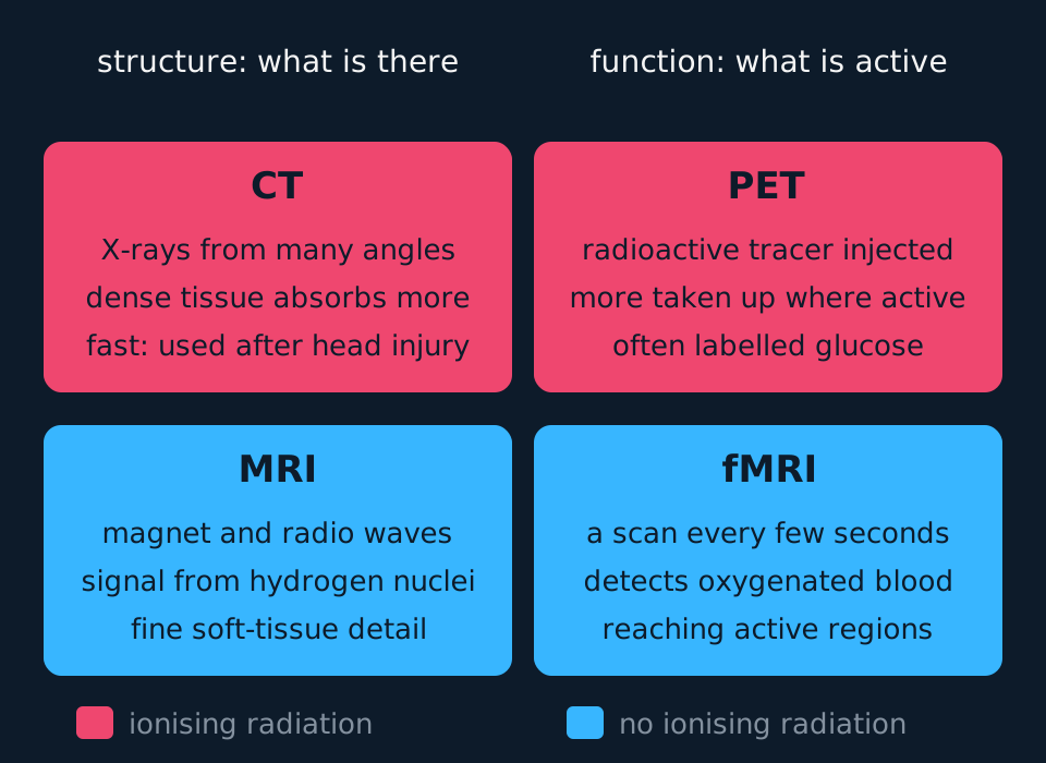
Four scanning methods image the brain of a living patient, and they divide into two kinds. A structural scan shows what is there: the shape and size of regions, a tumour, a bleed, an area of dead tissue. A functional scan shows which regions are active while the person does something, such as reading, moving a hand or recalling a word. Doctors use both kinds in medical diagnosis, and researchers use them to investigate how brain structure relates to function.
Describe the drawing
A two by two grid of cards comparing four brain scans. The left column, headed structure, holds CT, which builds an image from X-rays passed through the head at many angles, with dense tissue absorbing more, and below it MRI, which uses a strong magnet and radio waves and detects a signal from hydrogen nuclei, giving fine soft-tissue detail. The right column, headed function, holds PET, in which an injected radioactive tracer such as labelled glucose is taken up most by active regions, and below it fMRI, which repeats MRI scans every few seconds to detect oxygenated blood reaching active regions. CT and PET are coloured as using ionising radiation; MRI and fMRI are coloured as using none.
Computed tomography (CT) passes a thin beam of X-rays through the head from many angles as the X-ray source rotates round it, and detectors on the far side measure how much of the beam gets through. Dense tissue absorbs more X-rays than soft tissue, so bone appears white, fluid dark and brain tissue in shades of grey. A computer combines the measurements into cross-sectional images, or slices. A CT scan takes only minutes, which makes it the usual first scan after a head injury or a suspected stroke: it shows a bleed, and so tells a doctor whether a stroke is caused by bleeding or by a clot. It shows soft tissue in less detail than MRI, and the X-rays are ionising radiation, which carries a small risk of damaging DNA.
Magnetic resonance imaging (MRI) places the patient in a very strong magnetic field, which lines up the nuclei of hydrogen atoms in the water and fat of the tissues. Pulses of radio waves knock the nuclei out of alignment, and as they realign they give out radio signals of their own, which the scanner detects. Tissues with different water and fat contents give different signals, so MRI distinguishes grey matter from white matter and shows soft-tissue detail far finer than CT. It is used to find tumours, the damage left by a stroke, and the patches of lost myelin in multiple sclerosis. MRI uses no ionising radiation, but it is slower than CT, the patient must lie still in a narrow, noisy tube, and the magnet rules it out for some people with metal implants or pacemakers.
Functional magnetic resonance imaging (fMRI) uses the same scanner to take a fresh image every few seconds while the person carries out a task. Active neurones respire faster and use more oxygen, and within a few seconds the blood flow to that region increases, bringing more oxygenated blood than the tissue uses. Oxyhaemoglobin and deoxyhaemoglobin behave differently in a magnetic field, so the scanner detects the rise in oxygenated blood, and the computer marks the regions where it occurs. Comparing scans taken during a task with scans taken at rest shows which regions the task uses. fMRI is used to map language and movement areas before brain surgery, so that the surgeon can avoid them, and in research into memory and language. Because it follows blood flow rather than impulses, it lags a few seconds behind the activity it records.
Positron emission tomography (PET) follows a radioactive tracer injected into the blood. The commonest is a form of glucose labelled with a positron-emitting isotope of fluorine. Active brain cells take up more glucose, so more tracer collects where the brain is most active. When a positron meets an electron, gamma rays are given out, and a ring of detectors round the head picks them up and locates the tracer. PET shows reduced activity in particular regions in Alzheimer's disease, highly active tumour tissue, and, with tracers taken up by dopamine-releasing neurones, the loss of those neurones in Parkinson's disease. The patient receives a dose of ionising radiation, the image is less sharp than MRI's, and the short-lived isotope has to be made shortly before use, which makes PET expensive.
| CT | MRI | fMRI | PET | |
|---|---|---|---|---|
| What it detects | X-ray absorption by tissues | Radio signals from hydrogen nuclei in a magnetic field | The rise in oxygenated blood flowing to active regions | Gamma rays from a radioactive tracer taken up by active cells |
| Structure or function | Structure | Structure | Function | Function |
| Ionising radiation | Yes: X-rays | No | No | Yes: radioactive tracer |
| Typical use | Bleeds, fractures, the first scan after a head injury or stroke | Tumours, stroke damage, multiple sclerosis | Mapping regions used in a task, before surgery and in research | Alzheimer's disease, tumours, Parkinson's disease |
- Computed tomography (CT)
- A scan that builds cross-sectional images of the body from the absorption of X-rays passed through it from many angles.
- Magnetic resonance imaging (MRI)
- A scan that uses a strong magnetic field and radio waves to image soft tissue from the signals given out by hydrogen nuclei.
- Functional magnetic resonance imaging (fMRI)
- A series of MRI scans that detects increases in oxygenated blood flow, showing which brain regions are active during a task.
- Positron emission tomography (PET)
- A scan that locates a radioactive tracer, usually labelled glucose, showing where cells are most metabolically active.
The critical period in visual development
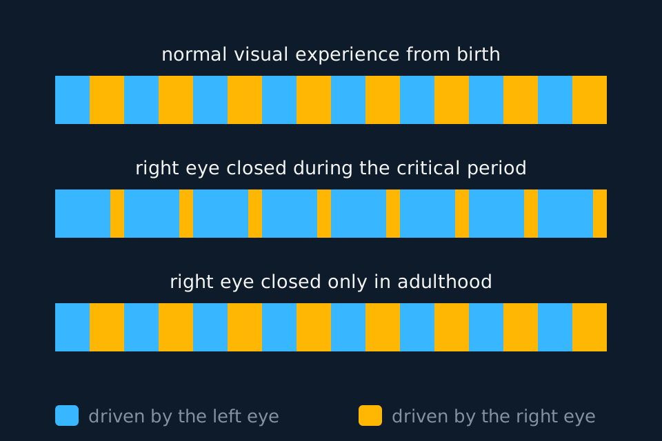
At birth the eyes of a kitten, a monkey or a human are almost fully formed, but the connections behind them in the brain are not finished. Axons carrying impulses from both eyes reach the visual cortex, at the back of the cerebrum, and at first their endings overlap, so many cortical neurones receive input from both eyes. Over the following weeks or months the inputs are sorted. Synapses that are used, because light falling on that eye sends impulses along them, are strengthened and kept; synapses that carry little activity are weakened and removed. The two eyes therefore compete for territory in the cortex, and normal experience leaves them with equal shares, in alternating bands called ocular dominance columns.
This sorting happens during a critical period: a window early in life when the visual cortex needs visual stimulation to develop its connections fully. If stimulation is missing during that window, the loss is permanent. If it is missing only later, the connections already made are kept.
The evidence comes from David Hubel and Torsten Wiesel, who recorded the impulses of single neurones in the visual cortex of cats and monkeys with fine microelectrodes, and shared a Nobel Prize for the work in 1981. In kitten experiments in the early 1960s they sewed one eyelid shut soon after birth and reopened it after about three months. The kitten then behaved as if blind in that eye. The eye itself was normal, and its retina still responded to light, but almost no neurones in the visual cortex responded to it: nearly all of them now responded to the eye that had stayed open. The same closure in an adult cat had no such effect. By varying when the eye was closed, they placed the critical period in cats roughly between the fourth week and the end of the third month after birth.
In monkeys, in the 1970s, they made the columns visible. A radioactive amino acid injected into one eye is carried along the neurones of the visual pathway to the cortex, and photographic emulsion spread over thin slices of the cortex shows that eye's territory as a set of stripes. In monkeys reared normally the stripes from the two eyes were of equal width. In monkeys that had one eye closed from soon after birth, the stripes of the open eye were wide and those of the closed eye narrow, because the open eye's active synapses had taken over cortex that the inactive ones had lost.
Describe the drawing
Three strips of visual cortex drawn as alternating blue and amber stripes, blue for regions driven by the left eye and amber for regions driven by the right. With normal visual experience from birth the stripes are of equal width. When the right eye was closed during the critical period, the amber stripes are narrow and the blue ones wide, because the open eye's connections took over territory. When the right eye was closed only in adulthood, the stripes are equal, as in the normal strip. The widths are schematic.
The same principle applies to children. A baby born with a cataract, a cloudy lens, receives no clear image in that eye, and unless the cataract is removed early the eye may have poor vision for life. A child with a squint, whose eyes point in different directions, may come to rely on one eye; covering the stronger eye with a patch forces the brain to use the weaker one while its connections can still be strengthened, which works best in early childhood.
- Critical period
- A window of time early in development when the nervous system needs a particular stimulus, such as light entering both eyes, for its connections to develop fully.
- Visual cortex
- The region at the back of the cerebrum that receives and processes impulses from the eyes.
- Ocular dominance columns
- Alternating bands of the visual cortex, each responding mainly to the left eye or mainly to the right eye.
Animal models, and the ethics of using them
An animal model is an animal studied in place of a human because some part of its biology is similar enough for the results to tell us about our own. Hubel and Wiesel's work could not have been done on people. It needed electrodes pushed into the cortex to record from single neurones, eyes deliberately deprived of light, and, in some experiments, the brain examined after death. Cats and monkeys were chosen for specific reasons. Both have forward-facing eyes and binocular vision as we do, with the two eyes' inputs meeting in a visual cortex organised much like ours, and the monkey's visual system is the closer of the two. Both develop faster than a human, so a critical period can be studied in months rather than years, and their rearing conditions can be controlled exactly.
Other questions need other models. Rats and mice are used to study learning and memory, and the sea slug Aplysia, with about 20 000 neurones, some large enough to be seen without a microscope, is where the cellular basis of habituation, below, was worked out. The limitation is the same in every case: a model is a different species. Its brain differs from ours in size and organisation, its critical periods run on a different timetable, and a result that holds in a kitten has to be checked in humans, usually by indirect evidence such as the outcome of cataract surgery at different ages, before it can be applied to them.
Whether such research should be done at all is an ethical question, and each of the two standpoints below can be used to argue for it or against it.
The utilitarian standpoint judges an action by its consequences: the right action produces the greatest overall good, or the least overall harm, for everyone affected. A utilitarian weighs the suffering of the animals against the benefits of the research. Hubel and Wiesel's kittens suffered permanent loss of vision in one eye; against that stand the understanding of how the visual system develops and the case it gave for treating children's cataracts and squints early. On that reckoning the experiments can be justified. The same framework can condemn a study whose benefits are small or uncertain, or whose results could have been obtained without animals, and many utilitarians count an animal's pain as carrying the same weight as the same pain in a human.
A rights-based standpoint holds that some actions are wrong whatever their consequences, because they break a duty or violate a right. If animals have a right not to be harmed, or not to be used merely as a means to someone else's ends, then experiments that harm them are wrong however many people benefit, and no weighing of benefits can make them acceptable. Others argue from the same framework that rights belong to beings capable of reasoning and of accepting duties in return, so animals do not hold them, though people still have a duty not to be cruel to them. On that view, carefully regulated research is permissible.
In the UK, research on animals is regulated by the Animals (Scientific Procedures) Act 1986, which protects living vertebrates and cephalopods such as octopuses; snails and Aplysia fall outside it. The establishment, the project and every person carrying out procedures each need a licence, and a project licence is granted only after its expected harms have been weighed against its likely benefits, a weighing of consequences of the kind a utilitarian makes. The Act also requires the three Rs to be applied: replacement of animals with other methods, such as cell cultures, computer models or scans of human volunteers; reduction of the number of animals used, through good experimental design; and refinement of procedures and housing to keep suffering to a minimum.
TRY IT: Arguing one case from two standpoints
A research group proposes to test a new drug for Parkinson's disease on monkeys whose dopamine-releasing neurones have been damaged, before any trial in humans. Give one argument a utilitarian might make for the study and one a utilitarian might make against it, then state how a rights-based standpoint could reach a different conclusion from both.
Check your answer
For, on utilitarian grounds: a small number of monkeys suffer, while a drug that works could relieve the symptoms of a very large number of patients, and testing in monkeys first protects the volunteers in the first human trial from an unsafe drug.
Against, on utilitarian grounds: if the drug's chance of success is low, if the results may not transfer from monkeys to humans, or if cell cultures or scans of patients could answer the question, the harm to the monkeys brings little benefit.
A rights-based standpoint does not weigh the two sides at all. If monkeys have a right not to be harmed, the study is wrong however large the benefit. If only beings that can accept duties hold rights, the study is not ruled out, but the researchers still have a duty to avoid unnecessary suffering.
- Animal model
- A species studied in place of humans because part of its biology is similar enough for the results to apply to humans.
- Utilitarianism
- The ethical standpoint that the right action is the one with the best overall consequences, the greatest good or the least harm for all affected.
- Rights-based ethics
- The ethical standpoint that some actions are wrong whatever their consequences, because they violate a right or break a duty.
- Three Rs
- Replacement, reduction and refinement: the principles for minimising the use and suffering of animals in research.
Habituation: learning to stop responding
Habituation is a reduced response to a stimulus that is repeated and turns out to be neither harmful nor rewarding. It is a simple form of learning, because the change in behaviour comes from experience, and it is found in animals from sea slugs to humans. A snail touched gently on the head pulls its tentacles in, but touched repeatedly it pulls them in less and brings them out again sooner; people stop noticing the noise of traffic outside a window. The animal stops spending time and energy on a stimulus that does not matter and can respond to ones that do, such as food or a predator.
Habituation is specific to the stimulus. A habituated snail still withdraws in full from a different stimulus, or from a much stronger touch, which shows that its muscles are not fatigued and that its receptors still work. It is also distinct from sensory adaptation, in which a receptor exposed to a constant stimulus produces a smaller generator potential and fewer impulses. In habituation the sensory neurone goes on sending impulses as before; the change is inside the nervous system, at a synapse.
The mechanism was worked out in Aplysia. Touching its siphon makes it withdraw its gill, through a short pathway in which sensory neurones from the siphon synapse with motor neurones to the gill muscles. When the touch is repeated:
| Step | What happens |
|---|---|
| 1 | Each touch still sends impulses along the sensory neurone to its presynaptic terminal. |
| 2 | With repeated stimulation, the calcium ion channels in the presynaptic membrane become less responsive, so fewer open when an impulse arrives. |
| 3 | Fewer calcium ions diffuse into the presynaptic terminal, so fewer vesicles fuse with the membrane and less neurotransmitter is released into the synaptic cleft. |
| 4 | Fewer transmitter molecules bind to receptors on the motor neurone, so its depolarisation is smaller and reaches threshold less often. |
| 5 | Fewer impulses travel to the gill muscles, and the withdrawal is weaker or does not happen. |
Core Practical 18 investigates habituation to a stimulus, usually in snails. A snail is placed on a clean, damp surface and left until it has emerged fully with its tentacles extended. It is then touched gently between the tentacles with a damp cotton bud, and the time from the touch until the tentacles are fully extended again is recorded. The touch is repeated at a fixed interval after each re-emergence, ten times or more. The touch must be applied to the same place, with the same pressure and by the same person, and temperature, light and vibration kept constant, because each of these affects the snail's response. Repeating the investigation with several snails shows whether the trend is repeatable. Snails should be handled gently, kept moist and returned unharmed to where they were collected, hands should be washed after handling them, and the school's risk assessment followed.
Reading a set of habituation results
A student touched one snail ten times and recorded the time for its tentacles to re-emerge after each touch. These results are constructed to show the pattern: 48, 41, 33, 27, 22, 15, 12, 8, 6 and 4 seconds. Describe the trend, explain it, and suggest how the student could test whether the trend is significant.
Show the working
Trend. The time to re-emerge falls with every touch, from 48 seconds after the first to 4 seconds after the tenth. The falls are larger over the first five touches than over the last five, as the time approaches zero.
Explanation. The snail is habituating. The touch is repeated and does it no harm, so at the synapses in the withdrawal pathway fewer calcium ion channels open, less transmitter is released, and fewer impulses reach the muscles that pull the tentacles in. The withdrawal is smaller and shorter each time.
Statistics. Touch number and re-emergence time are two variables measured on the same snail, so a correlation test such as Spearman's rank is appropriate. Ten pairs of values ranked in exactly opposite orders give a coefficient of −1. Its size, 1, is larger than the critical value of 0.648 for ten pairs at the 5% significance level, so the negative correlation is significant.
- Habituation
- A reduced response to a repeated stimulus that is neither harmful nor rewarding; a simple form of learning.
- Sensory adaptation
- A fall in the response of a receptor to a constant stimulus, so that fewer impulses are sent along the sensory neurone.
Nature and nurture in brain development
Nature means the contribution of genes to how the brain develops, and nurture the contribution of the environment and experience, including conditions in the womb. The critical period shows that the two cannot be fully separated: the genes build a visual cortex ready to be wired, and experience completes the wiring. Five kinds of evidence are used to judge how much each contributes to a particular ability, and each has weaknesses of its own.
The abilities of new-born babies. A new-born has had little chance to learn from the world, so an ability present at birth is likely to be innate. Within hours of birth babies turn their eyes and head further to follow a simple face-like pattern than the same features scrambled, which suggests that some preference for faces is built in. Babies cannot say what they perceive, so their abilities have to be inferred from behaviour such as how long they look at something, or how fast they suck. Learning also begins before birth: new-borns respond differently to their mother's voice, which they heard in the womb. An ability that appears months later may come from maturation of the brain or from experience gained in those months, and testing at that age cannot tell the two apart.
Animal experiments. Animals can be deprived of a particular experience in a way that would never be allowed with children. Hubel and Wiesel's deprivation experiments showed that the visual cortex needs visual experience to develop, which is evidence for nurture. They also found that in very young kittens that had not yet seen anything, many cortical neurones already responded to particular features, which is evidence that the basic wiring is laid down by nature. In a later experiment by Colin Blakemore and Grahame Cooper, kittens reared seeing only vertical stripes had few cortical neurones responding to horizontal lines and behaved as though they could not see horizontal edges, so experience also shapes what the cortex responds to. The weaknesses are the ones that apply to any animal model: the species differs from ours, and the ethics of depriving an animal limit what can be done.
Studies of individuals with damaged brain areas. When an injury, stroke or tumour damages one region and a particular ability is lost, the region is linked to that ability. In the 1860s Paul Broca studied patients who understood speech but could produce very little of it, and found damage to the same area of the left frontal lobe after death. That a function is localised to a region is consistent with nature having specified it. But children often recover lost abilities better than adults with similar damage, because other regions take over the function, and that capacity for rewiring shows the brain is shaped by use as well. The weaknesses are that damage is rarely confined to one region, the person's abilities before the damage are seldom known exactly, and each case is different, so samples are small.
Twin studies. Monozygotic (identical) twins share all their alleles, while dizygotic (non-identical) twins share on average half the alleles that vary between people. If monozygotic twins are more alike in an ability than dizygotic twins raised in similar homes, genes are likely to contribute to it. Monozygotic twins separated soon after birth and reared apart are especially useful, because any similarity between them is less likely to come from a shared home. The weaknesses are that monozygotic twins may be treated more alike than dizygotic twins, all twins share a womb, separated twins are often placed in similar families, and the number of twins reared apart is small.
Cross-cultural studies. These compare people brought up in different cultures. An ability found in the same form in every culture studied is likely to have a large genetic contribution; one that differs between cultures is likely to depend on experience. Studies in the 1960s found that people brought up in surroundings with few straight edges and right-angled corners were less easily taken in by the Müller-Lyer illusion, in which two lines of equal length look unequal because of the arrowheads at their ends, suggesting that the way we judge depth from lines is learned. The weaknesses are that populations differ in their genes as well as their culture, that a test devised in one culture may be unfamiliar or misunderstood in another, and that many other environmental factors differ at the same time.
| Method | What it can show | A main weakness |
|---|---|---|
| New-born babies | Abilities present before much learning is possible, so likely innate | Abilities inferred from behaviour; learning starts in the womb |
| Animal experiments | The effect of removing one kind of experience during development | A different species; ethics limit what can be done |
| Damaged brain areas | Which region an ability depends on, and how far other regions can take over | Damage is rarely confined to one region; small, varied samples |
| Twin studies | Whether greater genetic similarity gives greater similarity in an ability | Identical twins may share more similar environments too |
| Cross-cultural studies | Whether an ability is the same in different upbringings | Populations differ genetically as well as culturally |
- Nature
- The contribution of an organism's genes to a characteristic, such as an ability of the brain.
- Nurture
- The contribution of the environment and experience, including conditions in the womb, to a characteristic.
- Cross-cultural study
- A study comparing people brought up in different cultures, to see whether an ability depends on upbringing.
In the exam
- Say whether a scan shows structure or function before describing it. CT and MRI show structure; fMRI and PET show which regions are active.
- fMRI detects the increase in oxygenated blood reaching an active region. It does not detect impulses or neurotransmitter directly, so do not write that it does.
- In Hubel and Wiesel's kittens the deprived eye was normal. Place the loss in the visual cortex, and say that the inactive synapses were lost while the active ones from the open eye took over.
- An ethics answer from two standpoints must name both frameworks and use each one's own reasoning: consequences weighed for the utilitarian, rights and duties that hold whatever the consequences for the rights-based view.
- For the mechanism of habituation, go in order: fewer calcium ion channels open, less calcium enters, less neurotransmitter is released, the postsynaptic neurone is depolarised less, fewer impulses reach the effector.
- When evaluating a nature and nurture method, give a weakness that belongs to that method, such as learning in the womb for new-born studies, rather than a general remark about sample size.
Check yourself
A three-month-old kitten has had its right eye covered since birth. When the cover is removed, the right eye's lens and retina are found to be normal, but the kitten cannot see with that eye. Explain this observation, and explain why covering the right eye of an adult cat for the same length of time would not have this effect.
Answer
At birth, axons carrying impulses from both eyes connect with neurones in the visual cortex, and many cortical neurones receive input from both eyes.
During the critical period the connections are sorted by use. Synapses from the open left eye were active, because light reaching that eye sent impulses along them, so they were strengthened and kept.
Synapses from the covered right eye carried almost no impulses, so they were weakened and removed, and the left eye's connections took over the cortical territory. Few cortical neurones now respond to the right eye, so the impulses it sends produce little or no vision, even though the eye works.
In an adult cat the critical period is over. The connections from both eyes were made and stabilised while both eyes were in use, and they are kept even when one eye is covered, so vision returns when the cover is removed.
Questions
Question 16 marks
Discuss the use of animals in medical research from two ethical standpoints.
Mark scheme
- B1 a utilitarian standpoint judges the research by its consequences: it is right if it produces the greatest overall good or the least overall harm
- B1 a utilitarian may support it when the suffering of a limited number of animals is outweighed by the benefits, such as treatments for large numbers of patients or new understanding of disease
- B1 a utilitarian may oppose it when the benefits are small or uncertain, when the results may not apply to humans, or when the same results could be obtained without animals
- B1 a rights-based standpoint holds that some actions are wrong whatever their consequences, because they violate a right or break a duty
- B1 if animals have a right not to be harmed or used as a means to an end, the research is wrong however great the benefits
- B1 alternatively, if rights belong only to beings that can reason and accept duties, animals have none and regulated research is permissible, though there is still a duty to avoid cruelty, as in the three Rs
Question 25 marks
When the siphon of the sea slug Aplysia is touched, it withdraws its gill, but after many repeated touches it withdraws the gill only weakly. Explain how this habituation happens at the synapse between the sensory neurone and the motor neurone.
Mark scheme
- B1 the sensory neurone still sends impulses to its presynaptic terminal with each touch
- B1 with repeated stimulation the calcium ion channels in the presynaptic membrane become less responsive, so fewer open
- B1 fewer calcium ions enter the presynaptic terminal, so fewer vesicles fuse with the membrane and less neurotransmitter is released
- B1 less neurotransmitter binds to receptors on the motor neurone, so its depolarisation is smaller and reaches threshold less often
- B1 so fewer impulses reach the gill muscles and the withdrawal is weaker
Question 35 marks
Describe how you could investigate habituation to touch in a snail, including the variables you would control.
Mark scheme
- B1 place the snail on a clean, damp surface and wait until it has emerged fully with its tentacles extended
- B1 touch it gently between the tentacles with a damp cotton bud and time how long the tentacles take to extend fully again
- B1 repeat the touch at a fixed interval after each re-emergence, ten times or more, recording the time each time
- B1 control the place touched, the pressure of the touch and the person applying it, and keep temperature, light and vibration constant
- B1 repeat with several snails to check the trend is repeatable, and handle them gently, keep them moist and return them unharmed
Question 44 marks
Explain how functional magnetic resonance imaging (fMRI) is used to find which regions of the brain are active while a person reads aloud.
Mark scheme
- B1 neurones in an active region respire faster and use more oxygen
- B1 blood flow to the active region increases, bringing more oxygenated blood
- B1 oxyhaemoglobin and deoxyhaemoglobin behave differently in the magnetic field, so the scanner detects the rise in oxygenated blood
- B1 scans are taken every few seconds during the reading and compared with scans at rest, and the regions where oxygenated blood has increased are the ones the task uses
Question 54 marks
Compare computed tomography (CT) with magnetic resonance imaging (MRI) as ways of imaging the structure of the brain.
Mark scheme
- B1 CT detects the absorption of X-rays by tissues, whereas MRI detects radio signals from hydrogen nuclei in a strong magnetic field
- B1 both produce cross-sectional images of brain structure rather than of activity
- B1 MRI shows soft tissue in finer detail, such as grey against white matter, whereas CT gives poorer soft-tissue resolution
- B1 CT exposes the patient to ionising radiation but is quicker, whereas MRI uses no ionising radiation but is slower and unsuitable for some people with metal implants
Question 64 marks
Use the idea of a critical period to explain the difference between the results for animal B and animal C in the table. The figures are constructed to show the pattern of Hubel and Wiesel's results.
| Animal | Responding to the left eye only / % | Responding to both eyes / % | Responding to the right eye only / % |
|---|---|---|---|
| A: kitten, both eyes open | 20 | 60 | 20 |
| B: kitten, right eye closed for its first three months | 90 | 8 | 2 |
| C: adult cat, right eye closed for three months | 22 | 58 | 20 |
Mark scheme
- B1 kitten B's eye was closed during the critical period, when the connections from the eyes to the visual cortex are still being sorted by use
- B1 synapses from the closed right eye carried few impulses, so they were weakened and lost
- B1 synapses from the open left eye were active, so they were strengthened and took over the cortical neurones, which is why 90% respond to the left eye only
- B1 in the adult cat C the critical period was over, so the connections made while both eyes were in use were kept even though one eye received no light
Question 74 marks
Within hours of birth, babies turn their eyes and head further to follow a face-like pattern than to follow the same features scrambled. Evaluate the use of this evidence to conclude that the preference for faces is determined by nature.
Mark scheme
- B1 new-born babies have had very little experience of the world, so a preference present at birth is likely to be innate, which supports the conclusion
- B1 learning begins in the womb, so some experience has already occurred before birth
- B1 the preference is inferred from eye and head movements, because babies cannot report what they perceive, so the interpretation may be wrong
- B1 a judgement: the evidence supports a large genetic contribution to a basic preference, but cannot show that later face recognition owes nothing to experience
Question 84 marks
Explain how studies of people with damaged brain areas are used to investigate brain function and development, and give two limitations of these studies.
Mark scheme
- B1 when damage to one region causes a particular ability to be lost, that region is linked to the ability, as Broca linked speech production to an area of the left frontal lobe
- B1 recovery of an ability after damage, which is often greater in children than in adults, shows other regions can take over, so development is shaped by experience as well as genes
- B1 one limitation: the damage is rarely confined to one region, so the lost ability may depend on another region that was also damaged
- B1 another limitation: abilities before the damage are seldom known exactly, and every case differs, so samples are small
Question 93 marks
Describe how a positron emission tomography (PET) scan using labelled glucose shows which regions of the brain are most active.
Mark scheme
- B1 glucose labelled with a positron-emitting isotope is injected into the blood
- B1 the most active brain cells take up the most glucose, so the tracer collects in the most active regions
- B1 the radiation produced when the positrons meet electrons is detected by detectors round the head, and a computer locates where it came from to build the image
Question 103 marks
The table shows the percentage of neurones in the visual cortex that responded to light shone into each eye, in an experiment of the kind Hubel and Wiesel carried out. The figures are constructed to show the pattern of their results. Describe what the data show about the effect of closing the right eye.
| Animal | Responding to the left eye only / % | Responding to both eyes / % | Responding to the right eye only / % |
|---|---|---|---|
| A: kitten, both eyes open | 20 | 60 | 20 |
| B: kitten, right eye closed for its first three months | 90 | 8 | 2 |
| C: adult cat, right eye closed for three months | 22 | 58 | 20 |
Mark scheme
- B1 closing the right eye in the kitten (B) reduced the neurones responding to the right eye from 20% to 2%, and those responding to both eyes from 60% to 8%
- B1 in the kitten the neurones responding only to the open left eye rose from 20% to 90%
- B1 closing the right eye in the adult cat (C) had almost no effect: the percentages are close to those of the normal kitten (A)
Question 113 marks
Explain why kittens and monkeys were used as animal models in research into the development of human vision.
Mark scheme
- B1 they have forward-facing eyes and binocular vision, with a visual cortex organised much like the human one, so results are likely to apply to humans
- B1 the experiments required procedures that would not be allowed on humans, such as depriving an eye of light and recording from single neurones in the cortex with electrodes
- B1 they develop faster than humans, so the critical period can be studied in months, and their rearing conditions can be controlled exactly
Question 123 marks
A snail has habituated to gentle touches on its head, but when it is touched much more firmly it withdraws its tentacles fully. Explain what this observation shows about habituation.
Mark scheme
- B1 the muscles that withdraw the tentacles are not fatigued, because they can still produce a full response
- B1 the receptors and sensory neurones still work, so the reduced response is not sensory adaptation
- B1 the change is specific to the repeated, harmless stimulus and lies in the nervous system, at the synapses of the withdrawal pathway
Question 133 marks
People brought up in surroundings with few straight edges and right-angled corners were found to be less easily taken in by the Müller-Lyer illusion. Suggest why this finding does not prove that the way we judge depth from lines is learned.
Mark scheme
- B1 the two populations differ genetically as well as in their surroundings, so the difference could be partly genetic
- B1 the test was devised in one culture and may be understood or answered differently in another
- B1 many other environmental factors also differ between the two groups, such as schooling and experience of pictures, so the cause cannot be pinned to the surroundings alone
Question 142 marks
Of CT, MRI, fMRI and PET, state which two expose the patient to ionising radiation, and the source of the radiation in each.
Mark scheme
- B1 CT, which uses X-rays passed through the head
- B1 PET, which uses a radioactive tracer injected into the blood
Question 152 marks
A student touched a snail between its tentacles ten times and recorded the time for its tentacles to re-emerge after each touch. The results are constructed. Calculate the percentage decrease in the re-emergence time between the first touch and the tenth.
| Touch number | 1 | 2 | 3 | 4 | 5 | 6 | 7 | 8 | 9 | 10 |
|---|---|---|---|---|---|---|---|---|---|---|
| Time for tentacles to re-emerge / s | 48 | 41 | 33 | 27 | 22 | 15 | 12 | 8 | 6 | 4 |
Mark scheme
- M1 (48 − 4) ÷ 48 × 100
- A1 a decrease of 91.7% (accept 92%)
Worth remembering
- CT and MRI show structure; PET and fMRI show function.
- CT uses X-rays and PET a radioactive tracer; MRI and fMRI use a magnet and radio waves, with no ionising radiation.
- In the critical period, active synapses from the eyes are kept and inactive ones are lost, permanently.
- Habituation: repeated harmless stimulus, fewer calcium channels open, less transmitter, weaker response.
- Nature and nurture are studied through new-born babies, animal experiments, damaged brain areas, twins and other cultures, and each has its own weakness.
CHECK YOUR PROGRESS
Rate how confident you are with each objective for this lesson. Ratings are saved in this browser, on this device, unless you sign in.
- Describe what MRI, fMRI, PET and CT scans each detect, and say whether each shows the structure or the function of the brain.
- Explain what happens in the visual cortex during the critical period, using Hubel and Wiesel's experiments with kittens and monkeys.
- Explain why animal models have been used in research into brain development and function, and the limits of what they show.
- Discuss the use of animals in medical research from two ethical standpoints.
- Explain how habituation happens at a synapse, and describe how to investigate habituation to a stimulus.
- Evaluate the methods used to investigate nature and nurture in brain development: new-born babies, animal experiments, damaged brain areas, twin studies and cross-cultural studies.
Open the full revision checklist to see every objective in the curriculum in one place.
Practise this lesson
WORKBOOK
The written questions from this topic, on paper with room to work, and a separate book of mark schemes. The 3 questions that are set on a diagram stay on this page, where the diagram can be drawn. Free to use; please do not redistribute or sell.