Biology › Practicals › Required practical 9: respiration rate in yeast
Required practical 9: respiration rate in yeast
Yeast respiring glucose removes hydrogen from it, and methylene blue will take that hydrogen. Blue when oxidised and colourless when reduced, the dye fades from a tube of respiring yeast, and the time it takes measures how fast the cells are respiring. Repeat at several temperatures and the rates trace the curve of the enzymes doing the work.
The capability this develops
This guide is one investigation under capability P11: Measuring respiration and photosynthesis rates, which sets out the techniques the investigation belongs to, the calculations it carries and the errors that cost marks in all of them. The notes behind the biology are in Respiration and cellular energy.
Where this sits on the specifications
This library maps a qualification to its own specification one numbered heading at a time, against a document whose sha256 is recorded, and publishes the result as a route map. These are the headings whose practical work this investigation serves, on three of the five qualifications mapped so far. Each links to the route map the row belongs to.
| Qualification | Heading |
|---|---|
| OCR H020/H420 | 5.2.2 Respiration |
| Cambridge International 9700 | 12.2 Respiration |
| Pearson Edexcel Biology B 8BI0/9BI0 | 5.5 iv Anaerobic respiration |
Some boards number their practical activities, and those lists were read here from the same specifications as the headings above. This investigation is numbered on them as:
- AQA 7401/7402 required practical 9
- OCR Biology A H020/H420 PAG 12
- Pearson Edexcel Biology A (Salters-Nuffield) 8BN0/9BN0 core practical 16
The other qualifications above are matched by specification heading only, because none of them numbers an activity that is this investigation. Use your awarding organisation’s current specification and its practical handbook as the authority on which activities your course requires and how they are assessed.
The principle
A respirometer measures a gas. A second standard method measures the removal of hydrogen from a substrate instead. The enzymes catalysing that removal are the dehydrogenases, and their hydrogen normally goes to NAD. A redox indicator will take it instead. Methylene blue is blue when oxidised and colourless when reduced, so a suspension of respiring yeast with methylene blue in it goes from blue to cream, and the time that takes is the measurement.
Every tube holds the same amount of dye, so every tube times the same amount of reduction. A shorter time is a faster rate, and the rate is taken as 1 ÷ time. The dye stays reduced only while no oxygen reaches it, which is why the tube is stoppered and left still.
The dehydrogenases are enzymes, so the rate follows the enzyme curve: it rises with temperature to an optimum and falls steeply above it as the enzymes denature. Boiled yeast is the control that shows the colour change needs living cells.
- Dehydrogenase
- An enzyme that catalyses the removal of hydrogen from a substrate, passing it to a carrier such as NAD.
- Redox indicator
- A dye that changes colour when it is reduced or oxidised, such as methylene blue, which is blue when oxidised and colourless when reduced.
Apparatus
- Dried yeast, and glucose solution of known concentration
- Methylene blue solution, 0.05%
- Test tubes with bungs, a rack, and graduated pipettes or syringes for 10 cm³, 2 cm³ and 1 cm³
- Water baths at 20, 30, 40, 50 and 60 °C, or one bath reset between runs, and a thermometer
- A boiling water bath for the boiled-yeast control
- A white tile or card to read the colour against, and a stopwatch
- Eye protection
Safety
Work to your school’s risk assessment. Methylene blue stains skin and clothing, so wear eye protection and wash off any splash. Water baths scald: lift tubes with a holder, and keep cables and plugs clear of the water. Boil the control yeast in a water bath, not over a flame. Check the CLEAPSS Hazcard for methylene blue before starting.
Variables
Independent variable
Temperature, set by the water bath and checked with a thermometer: 20, 30, 40, 50 and 60 °C.
Dependent variable
The time for the blue colour to go from the body of the liquid, in seconds, converted to a rate as 1 ÷ time, in s⁻¹.
Control variables
- The yeast. The same volume of one suspension, stirred before each volume is drawn, so every tube has about the same number of cells.
- The substrate. The same volume of glucose solution at the same concentration.
- The dye. The same volume of the same methylene blue, since more dye takes longer to reduce and is toxic to the cells.
- pH. A buffer, because fermentation acidifies the suspension as it runs.
- Oxygen. Every tube stoppered after mixing and not shaken, so that no dye is re-oxidised.
- The endpoint. Judged against the same white background and the same reference tube every time.
Method
Preparing the yeast
- Make up a yeast suspension by stirring dried yeast into warm water with glucose in it.
- Leave it for fifteen or twenty minutes, so the cells are active before the experiment starts.
- Boil 15 cm³ of it in a water bath for 5 minutes for the control, and let it cool.
Setting up each tube
- Into a test tube put a measured volume of that suspension, say 10 cm³, and a measured volume of glucose solution of known concentration as the substrate, say 2 cm³.
- Stand the tube in a water bath and leave it long enough to reach the bath's temperature.
- Set up the boiled-yeast tube the same way at 40 °C, and a reference tube of yeast that has already lost its colour, to judge the endpoint against.
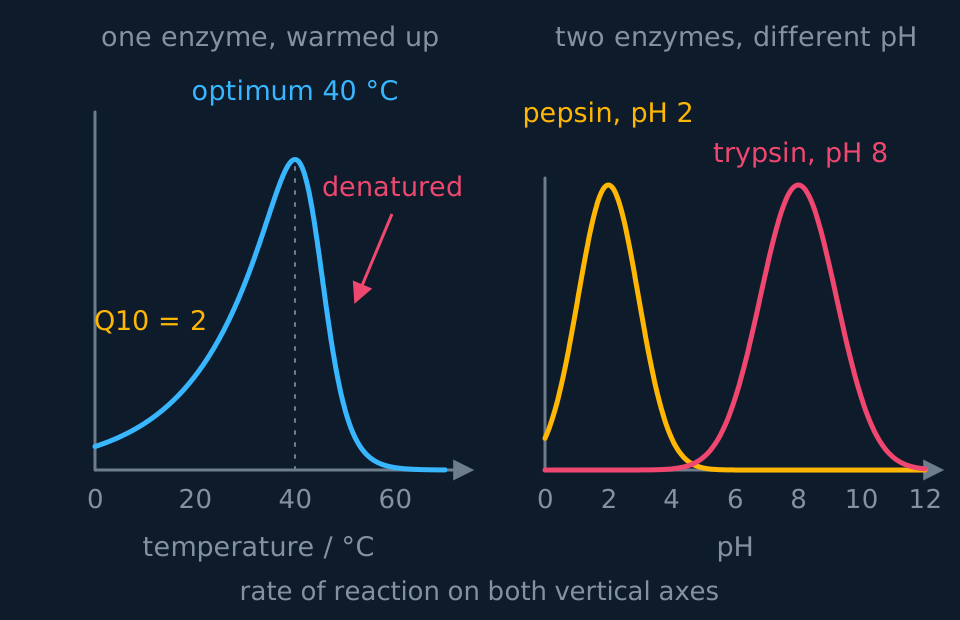
Describe the drawing
Two graphs side by side. On the left, rate against temperature for one enzyme: the curve doubles every ten degrees up to a peak at forty degrees Celsius, then drops away far more steeply than it rose and reaches zero by about sixty. On the right, rate against pH for two enzymes: pepsin gives a narrow peak at pH two and trypsin a narrow peak at pH eight, each falling to nothing a few units either side.
Timing the colour change
- Then add a small measured volume of dilute methylene blue (1 cm³ of a 0.05 per cent solution is typical).
- Stopper the tube, invert it once to mix, and start the clock.
- Leave the tube still in the bath. Shaking it lets air back in, and oxygen turns the reduced dye blue again.
- Stop the clock when the blue colour has gone from the body of the liquid.
- Repeat three times at each temperature, in a random order of temperatures, and take the mean.
Sample data
These times are idealised. They are the times the lesson works, each standing for the mean of three tubes, and real times scatter. Nothing here is a record of an experiment.
| Temperature / °C | Time to decolourise t / s | Rate 1 ÷ t / 10⁻³ s⁻¹ |
|---|---|---|
| 20 | 610 | 1.6 |
| 30 | 300 | 3.3 |
| 40 | 190 | 5.3 |
| 50 | 340 | 2.9 |
| 60 | still blue at 900 | below 1.1 |
Take the 40 °C row. The rate is 1 ÷ 190 s = 0.0053 s⁻¹, which is 5.3 × 10⁻³ s⁻¹.
Convert to rates first: 0.0016, 0.0033, 0.0053 and 0.0029 s⁻¹ for the first four, and effectively zero at 60 °C.
The 60 °C tube sets a limit, not a value. Still blue at 900 s, its rate is below 1 ÷ 900 s = 1.1 × 10⁻³ s⁻¹, and that is what the table records.
The boiled-yeast tube at 40 °C stayed blue for the whole run.
Reading the graph
Rate is the reciprocal of the time. A tube that decolourises in 60 s is respiring twice as fast as one that takes 120 s, so plotting time against temperature gives a curve with a minimum where the biology has a maximum. Plot 1 ÷ time, in units of s⁻¹, and the graph has the shape the explanation needs.
The rise from 20 to 40 °C is the usual kinetic argument. More kinetic energy means more frequent successful collisions between the dehydrogenases and their substrates, so hydrogen is removed faster and the indicator is reduced faster.
From 20 °C to 30 °C the rate doubles, from 1.6 to 3.3 × 10⁻³ s⁻¹. From 30 °C to 40 °C it rises by a smaller factor, to 5.3, and the highest rate measured is at 40 °C. The optimum lies somewhere between 30 °C and 50 °C, and these five temperatures cannot place it more closely.
The fall above 40 °C is denaturation. Hydrogen bonds holding the tertiary structure break, the active sites change shape, and by 60 °C enough of the dehydrogenases have been destroyed that no measurable reduction happens in fifteen minutes.
The boiled tube licenses that conclusion. It shows the dye is not being reduced by anything in the medium or by the heat itself, so the colour change in the other tubes was the yeast's enzymes and nothing else.
Other variables. The same tubes at one temperature test glucose concentration, where the rate rises and levels off as the enzymes approach saturation, or the sugar itself: yeast decolourises fastest on glucose, more slowly on sucrose and maltose, and hardly at all on lactose or starch, which it has no enzymes to hydrolyse.
Evaluation
Each of these is a real source of error in this investigation, and the middle column is the part an answer earns marks for: what it does to the result, and in which direction.
| Source of error | What it does to the result | What to do about it |
|---|---|---|
| The endpoint judged by eye | The blue fades rather than switching off, so two people stop the clock at different times. The scatter is in both directions, and it is a larger share of the short times. | A colorimeter with a red filter removes that judgement altogether, and turns the run into a series of absorbance readings in place of a single time. |
| Air reaching the reduced dye | Oxygen re-oxidises the dye and the blue comes back at the surface, so the endpoint is reached later and the rate comes out too low. | Stopper the tube or float a thin layer of oil on the liquid, and do not shake it once timing has begun. |
| Yeast settling in the suspension | Tubes filled later carry more or fewer cells than tubes filled earlier, which moves their rates up or down at random. | Stir the suspension immediately before every volume is drawn. |
| Dye added at room temperature | The cold dye lowers the temperature of the mixture for the first part of the run, most of all at the hottest baths, so those rates come out too low. | Stand the methylene blue in the same bath before it is added. |
| The yeast changing during the session | The suspension goes on respiring and using its glucose while it waits, so tubes set up late in the session differ from early ones for a reason that is not temperature. | Randomise the order of temperatures, and repeat the first one at the end to see how much the yeast has changed. |
| Too much methylene blue | More dye takes longer to reduce, and the dye is toxic to the cells, so the rate is lowered by the measurement itself. | Use the same small volume, 1 cm³ of 0.05%, in every tube. |
Questions
Question 1 (3 marks)
Explain why methylene blue loses its colour in a tube of respiring yeast.
Check your answer
dehydrogenase enzymes remove hydrogen from the glucose (or respiratory substrates);
the methylene blue accepts the hydrogen (in place of NAD) and is reduced;
reduced methylene blue is colourless.
Question 2 (2 marks)
A tube decolourised in 190 s. Calculate the rate of the reaction and give its unit.
Check your answer
rate = 1 ÷ 190;
= 0.0053 s⁻¹ (5.3 × 10⁻³ s⁻¹).
Question 3 (3 marks)
Explain why a tube of boiled yeast is a better control than a tube of water.
Check your answer
boiled yeast differs from the other tubes only in having no active enzymes, because boiling denatures the dehydrogenases;
water has no cells, so it looks different and the endpoint would be judged differently;
if the boiled tube stays blue, the colour change in the others was caused by the yeast's enzymes and not by anything else in the medium.
Question 4 (2 marks)
Explain why the tubes are stoppered and not shaken during the timing.
Check your answer
oxygen from the air re-oxidises the reduced methylene blue;
turning it blue again, so the endpoint would be delayed and the rate would be too low.
Question 5 (3 marks)
Describe and explain the effect of increasing the temperature from 40 °C to 60 °C on the rate of respiration.
Check your answer
the rate falls, from 5.3 × 10⁻³ s⁻¹ at 40 °C to almost zero at 60 °C;
hydrogen bonds holding the tertiary structure of the dehydrogenases break and the active sites change shape;
fewer enzyme–substrate complexes form, so hydrogen is removed more slowly.
Coverage maps are available for AQA 7401/7402, OCR Biology A H020/H420, Cambridge International 9700, Pearson Edexcel Biology A (Salters-Nuffield) 8BN0/9BN0 and Pearson Edexcel Biology B 8BI0/9BI0. Use the relevant route map to check what InkBiology covers, and your awarding organisation’s current specification as the final authority on examinable content.