Biology › Cells, microscopy and biological organisation › Cell specialisation and biological organisation
Cell specialisation and biological organisation
Most nucleated somatic cells contain the same genome, yet a neurone a metre long and a red blood cell with no nucleus are built completely differently, because different genes are expressed in each. As a cell differentiates, the range of cell types it can still become narrows.
Before this Cell ultrastructure and the organelles · DNA, genes and the idea of transcription
COMMON MISCONCEPTION
A neurone kept the nerve genes and lost the rest, and that is how it differs from a red blood cell.
Nothing is discarded. Almost every nucleated cell holds a complete genome, so a cell in your retina carries the insulin gene and never uses it. What differs between cell types is which genes are transcribed.
What you should be able to do
- Explain how cells with identical DNA come to differ, in terms of gene expression.
- Define totipotent, pluripotent and multipotent, and give a source of each.
- Describe the levels of organisation from cell to organ system, with examples.
- Relate the structure of squamous epithelium, ciliated epithelium, cartilage, xylem and phloem to their functions.
- Explain why differentiated cells cannot usually be converted back into other cell types.
- Explain how xylem vessels and sieve tubes both arise from one meristem, the procambium in a young stem and the vascular cambium in a thickening one.
- State the three parts of cell theory and explain why it is a unifying concept in biology.
Same genome, different cell
A fertilised egg divides into two, then four, then eight. The cells at that point are interchangeable, and each carries the whole genome. An adult body contains over two hundred recognisably different cell types, and most nucleated somatic cells still contain the same genome. Differentiation is the process leading from the first state to the second.
It works by switching genes on and off. In any given cell only a fraction of the genome is being transcribed at any moment: the genes that cell needs. Which ones those are is set by signals the cell receives: from hormones, from growth factors, from its neighbours and from its position in the developing embryo. Once the pattern is established it tends to be maintained through subsequent divisions, so a liver cell's daughters are liver cells.
The consequences appear in the ultrastructure. A cell specialised for protein secretion develops extensive rough ER and Golgi. A muscle fibre fills with protein filaments and mitochondria. A mature red blood cell goes further than any of them and loses its nucleus altogether, along with its mitochondria, leaving room for about 270 million molecules of haemoglobin, and no way of dividing or repairing itself, which is why it lasts only about 120 days.
A sperm cell makes the same trade in a different direction: almost no cytoplasm, a head packed with a haploid nucleus, a cap of digestive enzymes called the acrosome to get through the egg's outer layers, and a mid-piece packed with mitochondria to supply ATP for the tail. Each of those features is the visible result of a particular pattern of gene expression.
- Differentiation
- The process by which a cell develops the structures and characteristics needed to carry out a particular function, through some genes being expressed and others not.
- Specialised cell
- A cell whose structure is adapted to one particular function.
How much a stem cell can still become
A stem cell is an undifferentiated cell that can keep dividing and can develop into other cell types. Both halves matter, and the first is what keeps the second possible: stem cells divide in a way that replaces themselves as fast as they are spent, so the supply is not used up. The textbook picture is a single division giving one daughter that differentiates and one that stays a stem cell, and that does happen. It is not the only arrangement. In plenty of tissues both daughters differentiate, or both stay, and the balance is struck across the population rather than inside every division.
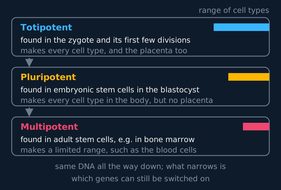
Stem cells are classified by the range of cell types they can still produce, and that range narrows as development proceeds. The three terms below are examinable, and totipotency differs from pluripotency in one respect only.
Describe the drawing
Three stacked cards, one for each kind of stem cell, joined by downward arrows. The top card is totipotent, found in the zygote and its first few divisions, able to make every cell type and the placenta. The middle card is pluripotent, found in embryonic stem cells of the blastocyst, able to make every cell type in the body but not the placenta. The bottom card is multipotent, found in adult stem cells such as those in bone marrow, able to make only a limited range. A coloured bar on each card shrinks from top to bottom.
- Totipotent
- Able to develop into any cell type in the organism and into the extra-embryonic tissues such as the placenta. Found in the zygote and the cells of the first few divisions.
- Pluripotent
- Able to develop into any cell type in the body, but not into the placenta. Found in embryonic stem cells in the blastocyst.
- Multipotent
- Able to develop into a limited range of cell types. Found in adult stem cells, such as those in bone marrow which produce the blood cells.
Bone marrow is the standard multipotent example and a good one because the limits are visible. A haematopoietic stem cell in the marrow can produce red blood cells, all the kinds of white blood cell and platelets, but nothing else. It cannot produce a neurone, because the genes a neurone requires remain unexpressed and are not reactivated by any signal the marrow normally supplies.
This narrowing is the central difficulty in stem-cell medicine. Pluripotent cells could in principle replace almost any tissue, but obtaining them from embryos raises ethical objections that have shaped the law in most countries. Induced pluripotent stem cells offer a way round: adult cells, usually from skin, are treated with specific transcription factors that switch previously silent genes back on, and the cells revert to a pluripotent state. Because they come from the patient, tissue made from them carries a much lower risk of rejection, though not zero: reprogramming and time in culture can alter the cells enough to provoke an immune response. Induced pluripotent cells also demonstrate that differentiation is difficult to reverse rather than genuinely irreversible: the genes are retained rather than lost, and specific transcription factors can restore their expression.
Cell theory
Cell theory is the statement that ties the rest of this unit together. It has three parts. All living organisms are made of one or more cells. The cell is the fundamental unit of structure, function and organisation in every living organism. New cells arise only from the division of existing cells.
The first two parts were set out by Matthias Schleiden for plants in 1838 and Theodor Schwann for animals in 1839, after improved light microscopes showed the same basic unit in plant tissues and in animal tissues such as cartilage. The third was established in the 1850s and is summed up in the phrase Rudolf Virchow made famous, omnis cellula e cellula, every cell from a cell. It ended the idea that cells could form spontaneously from non-living material, and it is why growth and repair are explained by cell division.
Cell theory is called a unifying concept because it applies to every organism, from a single bacterium to an oak tree, and because so much of biology can be explained at the level of the cell. Respiration, protein synthesis and the replication of DNA happen inside cells. A tissue is a group of similar cells, an organ a group of tissues, and the properties of each level depend on the cells it is built from. Disease, too, is understood as a failure of cells, which was Virchow's own application of the theory.
A few structures look like exceptions and are not. A striated muscle fibre contains many nuclei in one long cell, because it forms when many precursor cells fuse. A fungal hypha is often not divided into separate cells. A mature red blood cell has no nucleus, and a xylem vessel element is dead. Each of these is still made by cells and from cells, so the theory holds; what they show is that a cell can be modified a long way from the textbook diagram. Viruses are the test case in the other direction. A virus is not made of cells and cannot carry out any function outside one, which is the reason it is classed as non-living.
- Cell theory
- The unifying concept that all living organisms are made of one or more cells, that the cell is the fundamental unit of structure, function and organisation, and that new cells arise only from existing ones.
Cells, tissues, organs, systems
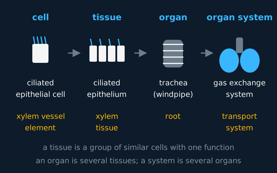
Multicellular organisms are organised in levels, and each level is built from the one below it. Learn the definitions in a form that can be written out precisely.
Describe the drawing
Four stages left to right joined by arrows, headed cell, tissue, organ and organ system. Above the labels are simple drawings: one ciliated cell, then four of them side by side as an epithelium, then a ringed tube, then that tube with two lungs attached. Beneath each stage two example chains are printed, an animal one reading ciliated epithelial cell, ciliated epithelium, trachea, gas exchange system, and a plant one reading xylem vessel element, xylem tissue, root, transport system.
- Tissue
- A group of similar cells working together to perform a particular function.
- Organ
- A structure made of several different tissues working together to perform a particular function.
- Organ system
- A group of organs working together to perform an overall life function.
The line that gets blurred is between tissue and organ, so use the number of tissue types to decide. Ciliated epithelium is one layer of similar cells doing one job, so it is a tissue. (The definition says similar cells rather than identical ones, which matters here: the goblet cells that make the mucus sit in the same layer.) The trachea contains that epithelium plus cartilage, smooth muscle, connective tissue and blood vessels, all working together to keep an airway open and clean, so it is an organ. The gas exchange system is the trachea plus the bronchi, bronchioles, lungs and diaphragm.
Two epithelia are the standard structure-and-function examples.
Squamous epithelium is a single layer of very flat cells, so thin that the nucleus makes a visible bulge. It lines the alveoli and forms the walls of capillaries, and the reason is diffusion distance: a thin barrier means a short path for oxygen and carbon dioxide, and the shorter the path, the faster they diffuse across it.
Ciliated epithelium is a layer of column-shaped cells each carrying hundreds of cilia that beat in a coordinated wave. It lines the trachea, bronchi and oviduct. In the airway it works with the goblet cells scattered among it: the goblet cells secrete mucus that traps dust and bacteria, and the cilia sweep the loaded mucus up towards the throat to be swallowed. Smoking damages the cilia, so mucus is no longer cleared from the airway and accumulates, and coughing becomes the only means of removing it.
Epithelia are not the only kind of animal tissue, and cartilage is the connective tissue worth knowing as the contrast. In an epithelium the cells sit shoulder to shoulder and the tissue is nearly all cell. Cartilage is nearly all matrix: its cells, called chondrocytes, sit isolated in small spaces named lacunae, surrounded by the firm, elastic extracellular material of collagen fibres and glycoproteins that they secreted around themselves. The matrix is the tissue's working part, smooth enough to glide and stiff enough to hold a shape while still absorbing compression.
That is why cartilage turns up where the body needs a surface or a support that must not collapse: capping the ends of bones at joints, shaping the ear and the nose, and stiffening the wall of the trachea, where the C-shaped rings of it are one of the several tissues that make the trachea an organ rather than a single tissue. Cartilage contains no blood vessels, so its chondrocytes are supplied by diffusion through the matrix alone, and that slow supply is why a damaged cartilage surface repairs so poorly compared with the bone beneath it.
- Cartilage
- A connective tissue in which chondrocytes, lying in lacunae, are surrounded by a firm extracellular matrix of collagen that they secrete; it has no blood vessels and supports structures such as the trachea, joints, ear and nose.
Muscle is the third kind of animal tissue to know, after epithelium and cartilage. Its cells are packed with protein filaments that slide past one another, so each cell can shorten. In the airway wall it is smooth muscle: spindle-shaped cells that contract together, without conscious control, to narrow the lumen.
Xylem and phloem
Plants are organised the same way. Xylem and phloem are tissues; a leaf, a root and a stem are organs; together they make a transport system. In both tissues the conducting cells lose organelles as they differentiate, and which organelles are lost determines how each tissue works.
Describe the drawing
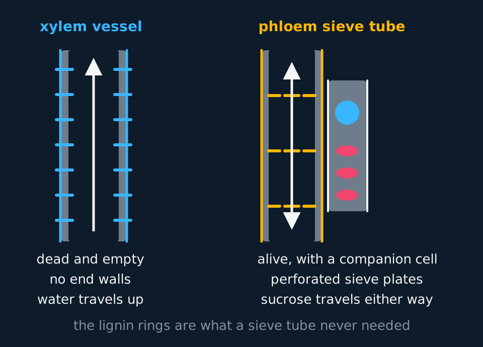
Two plant transport tubes drawn side by side in longitudinal section. On the left a xylem vessel: a wide open tube with no end walls anywhere along it, walls thickened with rings of lignin, nothing at all inside, and a single arrow running straight up it. On the right a phloem sieve tube: a narrower tube crossed at intervals by perforated sieve plates, with a companion cell alongside containing a nucleus and several mitochondria, and a double-headed arrow showing that sap can travel either way.
Xylem carries water and dissolved mineral ions from root to leaf, and in a transpiring plant the flow runs one way: upwards. A xylem vessel is a file of cells whose end walls have broken down completely, leaving one continuous hollow tube, sometimes metres long. The cells die in the process: the cytoplasm, nucleus and organelles are gone, so there is nothing to obstruct the flow. The side walls are thickened with lignin, laid down in rings and spirals, which waterproofs them and stops the tube collapsing inwards when the water inside is under tension. Small unlignified gaps called pits let water pass sideways into neighbouring cells.
Phloem carries sucrose and amino acids from where they are made or stored to wherever they are needed: up the plant in spring, down it in summer, either way at any time. That flexibility rules out the xylem solution. A sieve tube element keeps its plasma membrane and a thin layer of cytoplasm, but loses its nucleus, ribosomes and most other organelles to clear a path down the middle. Its end walls survive as sieve plates, perforated with pores that the contents flow through.
A cell with no nucleus and no ribosomes cannot synthesise its own proteins. Each sieve tube element therefore has a companion cell alongside it, connected through plasmodesmata, with a nucleus, dense cytoplasm and a great many mitochondria. It makes the proteins the sieve tube needs and supplies the ATP for actively loading sucrose into the tube. Take the companion cell away and the sieve tube stops working, because there is nothing left to replace its proteins or to pay for the loading.
| Xylem vessel | Phloem sieve tube | |
|---|---|---|
| Living? | No, dead and empty | Yes, but without a nucleus |
| End walls | Broken down completely | Remain as perforated sieve plates |
| Wall | Thickened with lignin | Cellulose, unthickened |
| Transports | Water and mineral ions | Sucrose and amino acids |
| Direction | Upwards only | Up or down |
| Partner cell | None | Companion cell, rich in mitochondria |
The two tissues contrast at every row of that table, but they share an origin. In a stem, xylem vessels and sieve tubes are produced by the same dividing population of unspecialised cells.
That population is a meristem, the plant equivalent of a multipotent animal stem-cell population. A meristem is a tissue of small, thin-walled, unspecialised cells that go on dividing while the cells around them have stopped, and the vascular tissue of a stem comes from two of them in succession. In a young shoot the strands of procambium running back from the apical meristem divide and their products differentiate in two directions: the ones left on the inner face of the strand thicken their walls with lignin, break down their end walls, lose their contents and become the first xylem vessels, while the ones left on the outer face keep their plasma membrane and cytoplasm, perforate their end walls into sieve plates and become the first sieve tubes. The two daughters did not differ at the moment of division. They differed in position, and therefore in the signals each received, which is the same mechanism of differentiation described for animal cells above.
In a dicot stem that goes on to thicken, a strip of that meristem is kept for life as the vascular cambium, the layer of dividing cells the plan-diagram convention shows between the xylem and the phloem of every bundle. It divides the way a stem cell divides: one daughter stays cambium, so the supply is never spent, and the other differentiates according to which side of the cambium it was left on. Inwards it becomes secondary xylem, outwards secondary phloem. Years of that adds up, and the result is visible from outside the plant: wood is accumulated secondary xylem, and the living inner layer of bark is secondary phloem. Both were made by the same cells.
The cambium can be classified in the same terms as an animal stem cell. A haematopoietic stem cell is multipotent and can make any blood cell and nothing else; the cambium is multipotent in the same sense, restricted to the vascular fates and their supporting parenchyma. The difference is that an animal keeps very few such populations and keeps them hidden, whereas a plant keeps meristems at every growing tip and around every thickening stem, for as long as it lives. That is why a cutting can grow a whole new plant and a severed finger cannot.
- Meristem
- A plant tissue of unspecialised cells that continue to divide, from which specialised cells such as xylem vessels and sieve tube elements differentiate.
- Vascular cambium
- The meristem layer lying between the xylem and the phloem of a vascular bundle, whose divisions add secondary xylem on its inner side and secondary phloem on its outer side.
TRY IT: Reading a function off a structure
A student examines a stained section of a plant stem and finds a tube with no cell contents at all, whose walls carry thick spiral bands that stain red for lignin. Identify the tissue and explain three ways its structure suits its function.
Check your answer
It is xylem, specifically a xylem vessel.
The absence of cell contents means the tube is hollow from end to end, with no cytoplasm, nucleus or end walls in the way, so water moves through it with very little resistance and the column stays unbroken.
The lignin waterproofs the thickened parts of the wall, so water is not lost sideways through them as it travels, and it also stiffens the tube so it does not collapse under the tension produced when water is pulled up from the leaves.
The lignin is arranged in spirals and rings rather than continuously, so the wall can still extend as the stem grows, and unlignified pits remain through which water can move sideways into neighbouring cells.
The same lignin gives the stem much of its mechanical strength, which is why wood is mostly old xylem.
In the exam
- Say 'genes are expressed', not 'genes are present'. State that most nucleated somatic cells contain the same genome but transcribe different parts of it.
- The difference between totipotent and pluripotent is the extra-embryonic tissue. Name the placenta explicitly when distinguishing the two.
- Use the tissue count to sort tissue from organ. One cell type doing one job is a tissue; several tissues working together is an organ. State how many tissues, and name one or two.
- State the consequence of every structural feature: 'squamous epithelium is thin, so the diffusion distance is short', rather than 'squamous epithelium is thin' on its own.
- For xylem, state: no end walls, no cell contents, lignified walls. For phloem: sieve plates with pores, cytoplasm retained, no nucleus, companion cell with mitochondria.
- Xylem and phloem arise from the same meristem, not from separate ones. Name the cambium, state that it divides, and state that the daughter left on the inner side becomes xylem while the one left outside becomes phloem.
- When asked for cell theory, give all three parts, and use the specification's words for the second: the cell is the fundamental unit of structure, function and organisation.
Check yourself
Cells lining the trachea and cells lining the alveoli both come from the same fertilised egg and carry the same DNA, yet one is a tall cell covered in cilia and the other is so flat it is barely visible in section. Explain how this difference arises and why each shape suits where the cell is.
Answer
Both cells hold a complete copy of the genome. The difference comes from gene expression: in each cell only the genes needed for that cell type are transcribed, and the rest stay switched off. Which genes those are was determined during development by the signals each cell received, including its position in the growing airway, and the pattern is then inherited by its daughter cells.
The ciliated cell in the trachea has expressed the genes for the proteins that build cilia and for the mitochondria that power them. Its job is to keep the airway clean: goblet cells among the epithelium secrete mucus that traps dust and bacteria, and the coordinated beating of the cilia sweeps that mucus up to the throat, away from the lungs.
The squamous cell in the alveolus has become extremely flattened, and its function is to form a short diffusion path. Oxygen and carbon dioxide diffuse across the alveolar wall, and rate of diffusion falls as the distance rises, so a wall one flattened cell thick keeps the path as short as possible.
Neither cell could do the other's job, and neither can change into the other, because differentiation is very hard to reverse once the pattern of expression is fixed.
Questions
Question 15 marks
Discuss the arguments for and against using embryonic stem cells rather than induced pluripotent stem cells in medical treatment.
Mark scheme
- B1 both are pluripotent and so can in principle be used to produce almost any tissue type, offering the same range of possible treatments
- B1 obtaining embryonic stem cells destroys the early embryo it came from, which raises an ethical objection that some people hold to be a serious moral cost
- B1 induced pluripotent cells are made by reprogramming adult cells, commonly from skin, so they avoid the need to use an embryo at all
- B1 induced pluripotent cells can be taken from the patient themselves, giving a much lower risk of immune rejection than tissue derived from a donor embryo
- B1 reprogramming and time spent in culture can alter induced pluripotent cells, so they carry their own, different, safety uncertainties that have to be weighed against the ethical cost of using embryonic cells
Question 24 marks
A stem cell in the bone marrow and a ciliated epithelial cell from the trachea carry exactly the same DNA. Explain how the two cells come to have such different structures.
Mark scheme
- B1 both cells hold a complete copy of the same genome, so the genes present in the two cells are the same
- B1 only a fraction of those genes is transcribed in any one cell, so different proteins are made and different structures develop
- B1 which genes are expressed is set by the signals a cell receives, such as hormones, growth factors, its neighbours and its position in the embryo
- B1 once established the pattern of expression is maintained through subsequent divisions, so a cell's daughters stay the same type
Question 34 marks
A phloem sieve tube element has lost its nucleus and almost all of its organelles, yet it is still alive. Explain how this suits it to its function, and explain why it cannot work without a companion cell.
Mark scheme
- B1 losing the nucleus and most organelles clears the centre of the tube, so sap flows through with little obstruction
- B1 the end walls survive as sieve plates perforated with pores, through which the contents pass from one element to the next
- B1 with no nucleus and no ribosomes the element cannot make its own proteins, so the companion cell makes them and passes them through plasmodesmata
- B1 the companion cell has dense cytoplasm and many mitochondria, and supplies the ATP for loading sucrose actively into the sieve tube
Question 44 marks
A xylem vessel and a phloem sieve tube in the same vascular bundle have almost nothing in common when they are mature. Explain how both can be produced by a single population of dividing cells.
Mark scheme
- B1 the vascular cambium, or the procambium in a young stem, is a meristem of unspecialised cells that go on dividing
- B1 one daughter of a division stays as cambium, so the dividing population is not used up, and the other differentiates
- B1 a daughter left on the inner side of the cambium differentiates into xylem and one left on the outer side into phloem, so position decides the fate
- B1 both daughters carry the same genome, and the signals each receives in its position switch on a different set of genes
Question 54 marks
Describe the structure of cartilage, and describe two examples of where it is found in the body.
Mark scheme
- B1 chondrocytes, the cells that secrete the tissue, lie isolated in small spaces called lacunae
- B1 they are surrounded by a firm, elastic extracellular matrix of collagen fibres and glycoproteins that they secreted
- B1 cartilage contains no blood vessels, so its chondrocytes are supplied only by diffusion through the matrix
- B1 any two examples of where it is found: capping the ends of bones at a joint, shaping the ear or nose, or the cartilage rings of the trachea
Question 64 marks
Compare the structure of a xylem vessel with the structure of a phloem sieve tube element, referring to whether each is living, its wall and its end walls.
Mark scheme
- B1 a xylem vessel is dead with no cell contents, whereas a sieve tube element is alive, though it has lost its nucleus
- B1 a xylem vessel wall is thickened with lignin, whereas a sieve tube element wall remains unthickened cellulose
- B1 a xylem vessel's end walls have broken down completely, whereas a sieve tube element's end walls survive as perforated sieve plates
- B1 xylem transports water and mineral ions upward only, whereas phloem transports sucrose and amino acids in either direction
Question 74 marks
A student says that a striated muscle fibre, which contains many nuclei, and a virus, which contains none, both show that cell theory is wrong. Evaluate this claim.
Mark scheme
- B1 a muscle fibre forms when many cells fuse, so it is still made from cells and by cells
- B1 it is the unit of structure and function of muscle tissue, so it is a modified cell and fits the theory
- B1 a virus is not made of cells and cannot carry out any function outside a host cell, so it is classed as non-living and cell theory, which concerns living organisms, does not apply to it
- B1 judgement: neither case contradicts cell theory; one is a modified cell and the other is outside the theory's scope
Question 83 marks
Compare a totipotent cell, a pluripotent cell and a multipotent cell, giving one place each type is found.
Mark scheme
- B1 a totipotent cell can produce any cell type in the organism and the extra-embryonic tissues such as the placenta, and is found in the zygote and the first few divisions
- B1 a pluripotent cell can produce any cell type in the body but not the placenta, and is found in the embryonic stem cells of the blastocyst
- B1 a multipotent cell can produce only a limited range of cell types, and is found in adult tissues such as bone marrow
Question 93 marks
Long-term smokers secrete more mucus than usual and the cilia on their airway epithelium are damaged. Suggest why such a smoker develops a persistent cough and gets more chest infections.
Mark scheme
- B1 cilia normally beat in a coordinated wave that sweeps mucus up towards the throat to be swallowed
- B1 with the cilia damaged the mucus is not moved, so it collects in the airways and coughing is the only way left to shift it
- B1 that mucus holds trapped dust and bacteria, which now stay in the airway and can multiply there, so infections are more likely
Question 103 marks
Explain how the structure of squamous epithelium suits it to lining the alveoli, referring to diffusion.
Mark scheme
- B1 squamous epithelium is a single layer of extremely flat, thin cells
- B1 this gives a short diffusion path across the alveolar wall
- B1 the shorter the path, the faster oxygen and carbon dioxide diffuse across it, so gas exchange is rapid
Question 113 marks
A student is given three structures to classify: ciliated epithelium from the trachea, the trachea itself, and the gas exchange system. Identify which is a tissue, which is an organ and which is an organ system, giving a reason for each identification.
Mark scheme
- B1 ciliated epithelium is a tissue, because it is one type of similar cells performing one function
- B1 the trachea is an organ, because it is built from several different tissues, such as epithelium, cartilage and smooth muscle, working together
- B1 the gas exchange system is an organ system, because it is a group of organs, including the trachea, bronchi, lungs and diaphragm, working together for an overall function
Question 123 marks
Explain how the loss of its nucleus and mitochondria suits a mature red blood cell to its function of carrying oxygen.
Mark scheme
- B1 losing the nucleus and mitochondria leaves more space inside the cell, filled instead with roughly 270 million molecules of haemoglobin, increasing the oxygen-carrying capacity of the cell
- B1 with no mitochondria the cell respires anaerobically, so it does not consume the oxygen it is carrying on its way to deliver it to other tissues
- B1 the trade-off is that the cell cannot divide or repair itself, which is why each one lasts only about 120 days
Question 133 marks
A surgeon proposes to repair a patient's damaged trachea using cartilage grown in the laboratory from a small sample of the patient's own cartilage. Suggest why cartilage is a realistic tissue to grow and transplant in this way, and suggest one reason repair might still be slow once it is in place.
Mark scheme
- B1 cartilage is mostly extracellular matrix secreted by a comparatively small number of chondrocytes, so a small number of cells can be grown to produce a useful mass of tissue
- B1 cartilage has no blood vessels of its own, so a piece grown outside the body does not need a blood supply to survive before or immediately after grafting, unlike a more vascular tissue
- B1 the chondrocytes in the graft are supplied only by diffusion through the matrix, which is slow, so nutrients reaching the repair site are limited and healing is correspondingly slow
Question 143 marks
State the three parts of cell theory.
Mark scheme
- B1 all living organisms are made of one or more cells
- B1 the cell is the fundamental unit of structure, function and organisation in living organisms
- B1 new cells arise only from the division of existing cells
Question 152 marks
State what is meant by a tissue, and state what is meant by an organ.
Mark scheme
- B1 a tissue is a group of similar cells working together to perform a particular function
- B1 an organ is several different tissues working together to perform a particular function
Question 162 marks
State what is meant by a meristem, and state where the vascular cambium lies in a vascular bundle of a dicot stem.
Mark scheme
- B1 a tissue of unspecialised cells that continue to divide, and from which specialised cells differentiate
- B1 as a layer between the xylem on the inner side of the bundle and the phloem on the outer side
Question 172 marks
State two ways, other than the loss of most of its cytoplasm, in which the structure of a sperm cell is adapted to its function.
Mark scheme
- B1 any one of: an acrosome, a cap of digestive enzymes used to digest through the outer layers of the egg; a mid-piece packed with many mitochondria, supplying ATP for the movement of the tail; or a long flagellum for swimming
- B1 any second, different adaptation from the same list
Question 182 marks
Explain why cell theory is described as a unifying concept in biology.
Mark scheme
- B1 it applies to every living organism, from bacteria to plants and animals
- B1 processes such as respiration, protein synthesis, growth and repair are explained at the level of the cell, and tissues and organs are built from cells
Worth remembering
- Most nucleated somatic cells contain the same genes; differentiation depends on which are expressed.
- Totipotent makes anything including the placenta; pluripotent anything in the body; multipotent a limited range.
- Cell, tissue, organ, organ system: each built from the one before it.
- Squamous epithelium is thin for a short diffusion path; ciliated epithelium sweeps mucus.
- Xylem is dead, hollow and lignified; phloem is alive, has sieve plates and needs a companion cell.
- Both come from one meristem: the cambium divides, inner daughters become xylem and outer daughters become phloem.
- Cell theory: all organisms are made of cells, the cell is the basic unit of structure, function and organisation, and cells come only from cells.
CHECK YOUR PROGRESS
Rate how confident you are with each objective for this lesson. Ratings are saved in this browser, on this device, unless you sign in.
- Explain how cells with identical DNA come to differ, in terms of gene expression.
- Define totipotent, pluripotent and multipotent, and give a source of each.
- Describe the levels of organisation from cell to organ system, with examples.
- Relate the structure of squamous epithelium, ciliated epithelium, cartilage, xylem and phloem to their functions.
- Explain why differentiated cells cannot usually be converted back into other cell types.
- Explain how xylem vessels and sieve tubes both arise from one meristem, the procambium in a young stem and the vascular cambium in a thickening one.
- State the three parts of cell theory and explain why it is a unifying concept in biology.
Open the full revision checklist to see every objective in the curriculum in one place.
Practise this lesson
WORKBOOK
The questions from this topic, on paper with room to work, and a separate book of mark schemes. Free to use; please do not redistribute or sell.
Used later in Stem cells and cell potency · Gas exchange in mammals: lungs and ventilation