Biology › Exchange surfaces and gas exchange › Gas exchange in mammals: lungs and ventilation
Gas exchange in mammals: lungs and ventilation
Human lungs contain a large alveolar surface for gas exchange. The airways are followed from trachea to alveoli, the tissues in each wall are related to their function, and ventilation is explained through changes in thoracic volume and pressure.
Before this Surface area to volume ratio and Fick's law · Specialisation and organisation: squamous and ciliated epithelium · Plan diagrams and tissue sections
COMMON MISCONCEPTION
Breathing in works by the lungs expanding and pulling air into themselves.
Lung tissue contains no skeletal muscle, and airway smooth muscle changes airway resistance rather than ventilating the lungs. Contraction of the diaphragm and the external intercostal muscles increases the volume of the thorax, so the pressure in the lungs falls below atmospheric and air moves in.
What you should be able to do
- Trace air from the trachea to an alveolus and name the tissues in the wall at each level.
- Recognise trachea, bronchus, bronchiole and alveolus in a section, and justify the identification from what is in the wall.
- Describe inspiration and expiration in the correct causal order: muscles, then volume, then pressure, then air.
- Explain why quiet expiration costs almost nothing while inspiration always costs energy.
- Calculate pulmonary ventilation rate, and read tidal volume, vital capacity and breathing rate off a spirometer trace.
- Read FEV₁ and FVC off a forced breath out, and use them to explain how pulmonary fibrosis, tuberculosis, asthma and emphysema affect ventilation and gas exchange.
- Evaluate the evidence linking smoking, pollution and dust to lung disease, separating correlation from cause, and explain how it led to laws such as the ban on smoking in enclosed public places.
- Use spirometer traces taken at rest and after exercise to find tidal volume, breathing rate, respiratory minute ventilation and oxygen consumption.
From trachea to alveolus, and what changes on the way
Air entering the nose is warmed, moistened and filtered, then passes down the trachea, a tube about 10 cm long held permanently open by rings of cartilage. The rings are C-shaped rather than complete, with the gap at the back where the oesophagus lies against the trachea, so a swallowed mouthful can bulge into that space instead of being crushed against a rigid tube.
The trachea divides into two bronchi, one to each lung, and each bronchus divides again and again into bronchioles of decreasing diameter. Around twenty-three rounds of branching separate the trachea from the smallest air spaces. The finest bronchioles open into clusters of alveoli, and that is where nearly all the gas exchange happens. Strictly, the last generations of bronchioles carry scattered alveoli in their own walls and exchange a little themselves; 'in the alveoli' remains the exam answer.
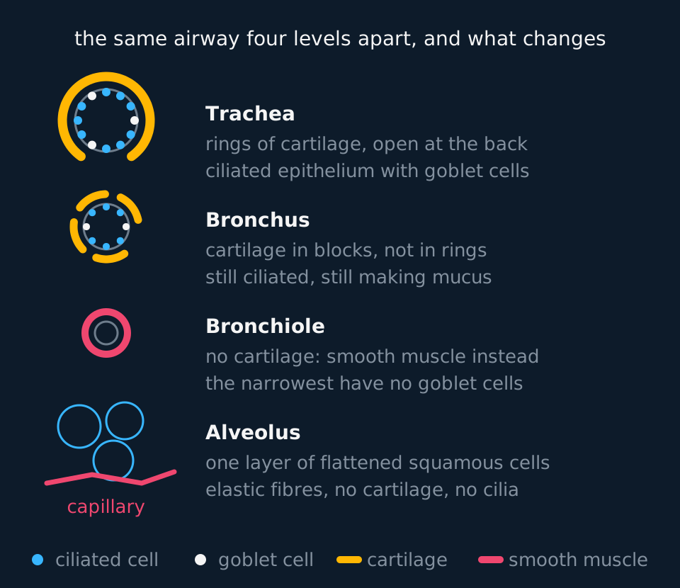
Describe the drawing
Four ring-shaped cross-sections drawn down the left of the figure. The trachea has one thick incomplete ring of cartilage with a lining of ciliated and goblet cells; the bronchus has four separate blocks of cartilage and the same lining; the bronchiole has a thick ring of smooth muscle and no cartilage; and the alveoli are three thin sacs lying against a capillary. A key beneath gives the colours for ciliated cells, goblet cells, cartilage and smooth muscle.
- Cartilage
- A firm, flexible supporting tissue. In the trachea it forms incomplete rings, and in the bronchi irregular blocks, holding the airway open against the pressure drop of inspiration.
- Ciliated epithelium
- An epithelium whose cells carry cilia that beat in a co-ordinated wave, sweeping mucus and trapped particles up towards the throat.
- Goblet cell
- A cell in the airway epithelium that secretes mucus, which traps dust, pollen and bacteria.
- Smooth muscle
- Involuntary muscle in the walls of hollow organs and vessels. In the airway wall its contraction narrows the lumen, and it is the tissue that contracts inappropriately during an asthma attack.
- Elastic fibres
- Fibres that stretch as the lungs inflate and recoil as the muscles relax, providing much of the force for quiet expiration.
- Squamous epithelium
- An epithelium of very flattened cells, giving a wall a fraction of a micrometre thick; the lining of an alveolus.
Cambridge International 9700 requires candidates to identify tissues in sections and micrographs of the gas-exchange system. Use cartilage distribution, smooth muscle and epithelial thickness to distinguish the trachea, bronchi, bronchioles and alveoli: rings of cartilage indicate trachea, irregular blocks of cartilage indicate bronchus, a conspicuous ring of smooth muscle without cartilage indicates bronchiole, and a single squamous layer against a capillary indicates alveolus. Beneath the epithelium of the trachea and bronchi lie mucous glands, which secrete mucus through ducts onto the lining; with the goblet cells, they supply the mucus that traps dust and pathogens for the cilia to sweep away.
Smooth muscle and elastic fibres are present along the length of the airway tree and act in opposition. Smooth muscle contracts to narrow an airway; elastic fibres stretch as it does and recoil to widen the airway when the muscle relaxes. Elastic fibres are not muscle, so they are not an antagonistic muscle pair.
The surface itself
Published estimates for an adult give of the order of 300 million alveoli and a combined surface area in the range of about 50 to 75 m², commonly quoted as roughly 70 m². Subdivision into alveoli provides that area within a thoracic cavity of a few litres, which raises the first term of Fick's law at little cost in volume.
Each alveolus is lined by a single layer of squamous epithelium and wrapped in a dense network of capillaries so narrow that red blood cells pass through in single file, squeezed against the capillary wall. Between the air and the haemoglobin lie only the alveolar epithelium, a basement membrane shared with the capillary, the capillary endothelium and a film of plasma.
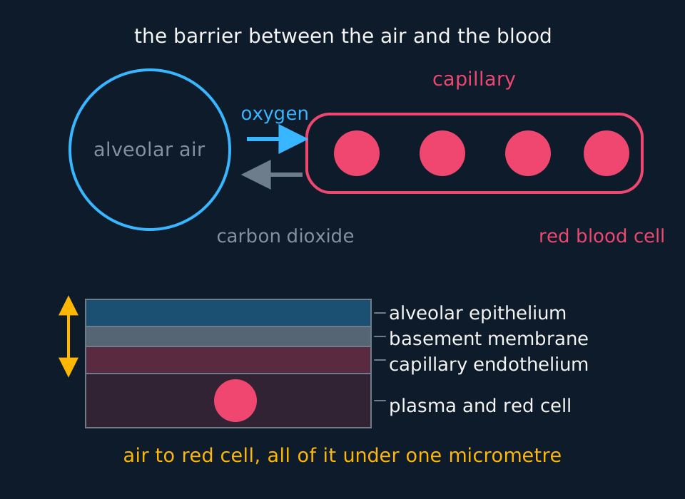
Describe the drawing
At the top, a round alveolus on the left with an arrow for oxygen crossing to a capillary on the right that carries four red blood cells, and an arrow for carbon dioxide crossing back. Below, the same barrier drawn as four stacked bands: alveolar epithelium, shared basement membrane, capillary endothelium, and the plasma holding a red cell.
The difference in concentration is maintained on both sides, which is the second term of Fick's law. Each breath replaces part of the alveolar air with fresh air, so the oxygen partial pressure in the alveolus stays around 13.3 kPa. Blood arriving from the pulmonary artery has an oxygen partial pressure of about 5.3 kPa. Each red cell loads within the first third or so of its passage along the capillary and is then carried away, while fresh deoxygenated blood keeps arriving behind it, so the gradient across the surface as a whole is never used up. Carbon dioxide crosses the other way down a much smaller gradient, roughly 6.1 kPa in the arriving blood against 5.3 kPa in the alveolus, and that suffices because carbon dioxide diffuses far more readily than oxygen.
Two further features of the surface matter. The alveolar lining is kept moist, because a gas has to dissolve before it can diffuse across a membrane. And the fluid contains a surfactant secreted by specialised cells in the alveolar wall, which lowers surface tension and stops the smallest alveoli collapsing shut between breaths. Premature babies are often short of it, which is why respiratory distress syndrome is a risk of early birth.
Elastic fibres in the alveolar walls stretch during inspiration and store energy while they do. Releasing it is what drives quiet expiration. In emphysema those fibres are destroyed and the alveolar walls break down into fewer, larger spaces, so the surface area falls and the recoil weakens at the same time. Both changes reduce gas exchange, and an account of emphysema should name both.
Ventilation: muscles, then volume, then pressure, then air
Lung tissue contains no skeletal muscle. Airway smooth muscle changes airway resistance but does not ventilate the lungs, and the muscles that do are in the wall of the sealed cavity the lungs sit in: the diaphragm underneath and the intercostal muscles between the ribs.
What ties lung to wall is the pleura: one membrane on the lung's surface, one lining the cavity, and between them a thin film of fluid at slightly below atmospheric pressure. The film lets the lungs slide against the wall but not pull away from it, so when the cavity expands the lungs are carried with it.

Describe the drawing
Breathing animated in four steps. Between breaths the ribcage and the domed diaphragm are still and no air moves. Then the external intercostal muscles contract, the ribs swing upwards and outwards and the diaphragm contracts and flattens downwards, enlarging the chest. With the chest larger the pressure inside falls and an arrow shows air flowing down the trachea into the lungs. Finally the muscles relax, the ribcage drops, the diaphragm domes upward again and the arrow reverses as air leaves.
| Inspiration | Quiet expiration | |
|---|---|---|
| Diaphragm | Contracts, flattens, moves down | Relaxes, returns to its domed shape |
| External intercostals | Contract, pulling ribs up and out | Relax |
| Internal intercostals | Relaxed | Relaxed in quiet breathing; contract in forced expiration |
| Volume of thorax | Increases | Decreases |
| Pressure in the lungs | Falls below atmospheric | Rises above atmospheric |
| Air | Moves in down the pressure gradient | Moves out down the pressure gradient |
| Energy | Always active: muscles are contracting | Largely passive: elastic recoil does the work |
The external and internal intercostals are an antagonistic pair. Externals contract for inspiration, internals for forced expiration, and at any moment one set is relaxed. In quiet breathing the internals do essentially nothing, because recoil of the stretched elastic tissue and the weight of the ribcage are enough. They come into play when you blow out hard, along with the abdominal muscles, which push the abdominal contents up against the diaphragm.
State the asymmetry explicitly. Inspiration always requires energy, because it always involves muscle contraction. Quiet expiration is largely passive: the diaphragm does not push upwards or contract upwards, since a muscle can only pull, and it returns to its domed shape when it stops contracting.
The pressure changes involved are small. Between breaths the pressure in the alveoli equals atmospheric pressure exactly, so no air moves. During quiet inspiration it falls by only a fraction of a kilopascal, which is ample: gases move quickly down even a shallow pressure gradient. On a trace of alveolar pressure against time, air enters wherever the trace lies below the atmospheric line and leaves wherever it lies above it.
Putting numbers on it
AQA names one calculation of its own in this topic, and it is the volume of air moved per minute. OCR A reaches the same quantity through a spirometer trace.
pulmonary ventilation rate = tidal volume × ventilation rateAlso called minute ventilation. Tidal volume in dm³, ventilation rate in breaths per minute, so the answer is in dm³ min⁻¹.
- Tidal volume
- The volume of air moved in or out in one normal breath at rest, about 0.5 dm³ in an adult.
- Ventilation rate
- The number of breaths taken per minute, about 12 to 15 at rest. Also called breathing rate.
- Vital capacity
- The largest volume that can be breathed out after the deepest possible breath in; roughly 4.6 dm³ in a young adult man and 3.1 dm³ in a young adult woman.
- Residual volume
- The volume left in the lungs after the hardest possible breath out, about 1.2 dm³. It cannot be measured with a spirometer, because it never leaves the lungs. Vital capacity plus residual volume is the total lung capacity.
From a trace to a rate
A spirometer trace shows twelve complete breaths in 48 seconds, each moving 0.45 dm³. Calculate the pulmonary ventilation rate. The same subject then exercises, and the tidal volume rises to 2.1 dm³ while the ventilation rate rises to 28 breaths per minute. Calculate the new rate and the factor by which it has changed.
Show the working
First convert to a rate per minute. Twelve breaths in 48 s is 12 × (60 ÷ 48) = 15 breaths per minute.
Pulmonary ventilation rate = 0.45 × 15 = 6.75 dm³ min⁻¹.
During exercise: 2.1 × 28 = 58.8 dm³ min⁻¹.
The factor is 58.8 ÷ 6.75 = 8.7. The breathing rate did not quite double, while the tidal volume rose more than fourfold, so the increase in depth contributes more than the increase in frequency.
The ventilation centre in the medulla oblongata makes these changes. During exercise the muscles release more carbon dioxide, more carbonic acid forms and the pH of the blood falls. Chemoreceptors in the carotid and aortic bodies and in the medulla detect the fall and send more impulses to the ventilation centre, which sends impulses more often to the diaphragm and the external intercostal muscles. They contract more strongly and more often, so breathing becomes deeper and faster, carbon dioxide is removed faster and the pH returns towards normal.
A spirometer records the volume of air in a closed circuit against time, and OCR A expects lung volumes and oxygen uptake to be read from one. Oxygen uptake is measured using the soda lime in the circuit: it absorbs the carbon dioxide the subject breathes out, so the total volume of gas in the apparatus falls steadily by exactly the volume of oxygen consumed. Draw a line through the midpoints of the oscillations, take its gradient, and that gradient is the rate of oxygen uptake. Without the soda lime the exhaled carbon dioxide would roughly replace the oxygen removed and the trace would stay level.
Oxygen uptake from the slope of the trace
On a spirometer trace with soda lime in the circuit, a line through the midpoints of the breaths falls from 3.10 dm³ to 2.60 dm³ over 120 seconds. Over the same two minutes the subject's pulmonary ventilation rate is 6.0 dm³ min⁻¹. Calculate the rate of oxygen uptake, and the percentage of the volume of air breathed that is taken up as oxygen.
Show the working
The fall in volume is 3.10 − 2.60 = 0.50 dm³, and 120 s is 2 minutes, so the rate of oxygen uptake is 0.50 ÷ 2 = 0.25 dm³ min⁻¹, or 250 cm³ min⁻¹.
Percentage taken up = 0.25 ÷ 6.0 × 100 = 4.2% of the volume breathed.
Air is about 21% oxygen, so only a fraction of the oxygen breathed in is taken up; the rest is breathed out again. During exercise the slope steepens as uptake rises, and tidal volume and breathing rate rise with it.
One further quantity qualifies these figures. About 150 cm³ of each breath never reaches an alveolus; it stays in the airways from the nose to the bronchioles, which are conducting tubes with no exchange surface. That volume is the dead space. It means a shallow, rapid breathing pattern moves less useful air than a slow deep one at the same pulmonary ventilation rate, because the dead space is paid for on every breath.
TRY IT: Explaining a histology slide
A student is shown a section of lung tissue containing a tube about 0.4 mm across. The wall contains a thick layer of smooth muscle and some elastic fibres, but no cartilage, and the lining cells carry no cilia. Identify the tube and justify the identification. Suggest why the absence of cartilage matters during an asthma attack.
Check your answer
It is a bronchiole. Cartilage is present in the trachea and bronchi and absent from bronchioles, so its absence rules the larger airways out, and the diameter and the thick smooth muscle layer both fit a bronchiole. The lack of cilia points to one of the smaller bronchioles close to the alveoli.
Without cartilage there is nothing rigid holding the tube open. When the smooth muscle contracts, as it does in an asthma attack, the lumen narrows sharply and the resistance to air flow rises, so less air reaches the alveoli per breath.
Identify the airway from the tissues in the wall rather than from its apparent size, because magnification varies between slides while the distribution of cartilage does not.
Lung disease and what it does to the data
AQA 7401/7402 asks students to interpret information about the effects of lung disease on gas exchange and ventilation, and to read lung-function data. The other boards do not include lung disease in their gas exchange topic, although every board teaches tuberculosis elsewhere as an infectious disease.
Each disease reduces the rate of gas exchange by acting on a term of Fick's law, or on ventilation, which keeps the concentration gradient up. The data show two broad patterns. In an obstructive disease the airways are narrowed, so air cannot be moved in and out quickly. In a restrictive disease the lungs cannot expand fully, so less air can be moved at all.
A clinic spirometer test separates the two. The patient breathes in as deeply as possible, then breathes out as hard and as fast as possible until no more air will leave, and two volumes are read from the forced breath out.
- Forced vital capacity (FVC)
- The total volume of air breathed out in one forced breath after the deepest possible breath in. In healthy lungs it is close to the vital capacity.
- Forced expiratory volume in one second (FEV₁)
- The volume of air breathed out in the first second of a forced breath out. It falls when the airways are narrowed, because air cannot leave them quickly.
- FEV₁ ÷ FVC ratio
- The fraction of the forced vital capacity breathed out in the first second. In a healthy adult it is usually above 0.7, often around 0.8; a value below 0.7 is the threshold commonly used to indicate airway obstruction.
- Obstructive lung disease
- A disease that narrows the airways and raises the resistance to air flow, so FEV₁ falls more than FVC and the ratio drops. Asthma and emphysema are examples.
- Restrictive lung disease
- A disease that stops the lungs expanding fully, so FVC and FEV₁ fall together and the ratio stays normal or rises. Pulmonary fibrosis is an example.
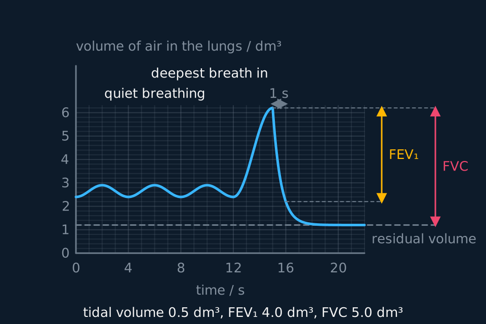
Describe the drawing
A spirometer trace plotted as the volume of air in the lungs, in cubic decimetres, against time in seconds, with constructed values for a healthy young adult. For twelve seconds the line rises and falls between 2.4 and 2.9, three quiet breaths of 0.5 cubic decimetres. It then climbs to 6.2 with the deepest possible breath in, and at fifteen seconds falls steeply as the subject breathes out as hard and fast as possible: 4.0 cubic decimetres leave in the first second, marked as FEV1, and the line levels off at 1.2, so the forced vital capacity is 5.0. A dashed line at 1.2 marks the residual volume.
Read every quantity as a difference. Tidal volume is the height of one quiet breath, here 2.9 − 2.4 = 0.5 dm³, and ventilation rate is 60 divided by the time for one breath, here 60 ÷ 4 = 15 breaths per minute. FEV₁ is the fall in the first second after the forced breath begins, 6.2 − 2.2 = 4.0 dm³. FVC is the whole fall from the top of the deepest breath to the level at which the trace flattens, 6.2 − 1.2 = 5.0 dm³. So this subject breathes out 4.0 ÷ 5.0 = 0.80 of the forced vital capacity in the first second.
| Disease | What happens in the lungs | Effect on ventilation and lung function | Effect on gas exchange |
|---|---|---|---|
| Pulmonary fibrosis | Scar tissue builds up in the alveolar walls, which become thick and stiff; in advanced disease some alveoli are lost | The lungs are harder to inflate, so tidal volume and FVC fall; FEV₁ falls with FVC, so the ratio stays normal or high; breathing becomes rapid and shallow | The thicker wall lengthens the diffusion pathway, and lost alveoli reduce the surface area |
| Tuberculosis | Mycobacterium tuberculosis infects the lungs and is walled into tubercles by the immune system; in active disease lung tissue is destroyed, and healing leaves scar tissue | Less of the lung can be ventilated, so FVC and tidal volume can fall; a persistent cough | Destroyed alveoli reduce the surface area, and scar tissue thickens the surface that remains |
| Asthma | A trigger such as pollen, dust-mite droppings or cold air inflames the airway lining; goblet cells secrete extra mucus and the smooth muscle of the bronchioles contracts | Narrowed airways raise the resistance, so FEV₁ falls far more than FVC and the ratio drops; the narrowing is largely reversed by a bronchodilator inhaler | Less fresh air reaches the alveoli, so the oxygen concentration gradient across the surface is smaller |
| Emphysema | Elastic fibres in the alveolar walls are digested and the walls between alveoli break down, leaving fewer, larger air spaces; most cases are caused by smoking | Without elastic recoil the small airways collapse during a forced breath out and air is trapped, so FEV₁ and the ratio fall; a bronchodilator makes little difference | The surface area falls, so the rate of diffusion falls; the breathing rate rises to compensate |
In asthma the lining of the airway reacts to something harmless. Mast cells in the lining release histamine and other chemicals, which cause inflammation, extra mucus and contraction of the bronchiole smooth muscle. In emphysema the damage starts with smoke: particles and tar inflame the alveoli, phagocytes gather there, and enzymes they release, such as elastase, digest the elastic fibres. Smoke also damages the cilia, so mucus and trapped particles are cleared more slowly.
Breathlessness in all four diseases has the same cause. Each breath delivers less oxygen to the blood and removes less carbon dioxide, so the ventilation centre raises the rate and depth of breathing. Where the lungs are stiff the depth cannot rise far, so the rate does, and because the dead space is paid on every breath, rapid shallow breathing delivers less air to the alveoli than its pulmonary ventilation rate suggests.
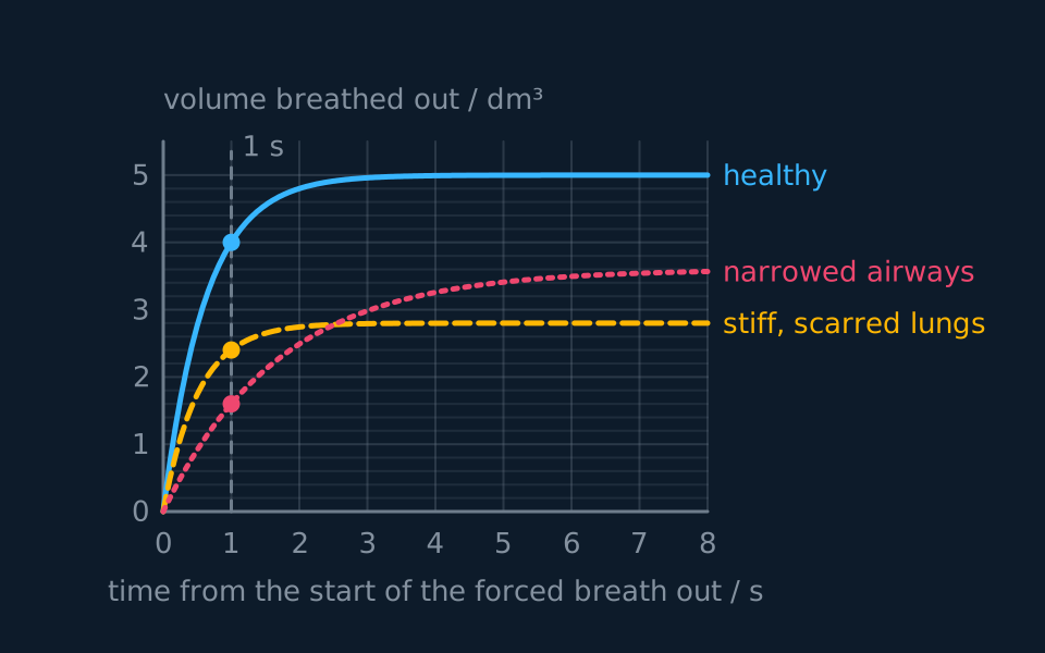
Describe the drawing
Three curves of volume breathed out against time over eight seconds of a forced breath out, with constructed values and a dashed line at one second. The healthy curve rises steeply to 4.0 cubic decimetres at one second and levels off at 5.0. The curve for stiff, scarred lungs rises almost as steeply but levels off early, at 2.4 by one second and 2.8 in all. The curve for narrowed airways rises slowly, reaching only 1.6 at one second and creeping up to about 3.6.
Reading FEV₁ and FVC, and naming the pattern
The figure shows a forced breath out for three adults. For each, read FEV₁ and FVC, calculate FEV₁ ÷ FVC, and say whether the pattern is obstructive or restrictive. Suggest which of asthma, emphysema and pulmonary fibrosis could produce each abnormal curve.
Show the working
Read FEV₁ where each curve crosses the one-second line and FVC where it flattens. Healthy: FEV₁ 4.0 dm³, FVC 5.0 dm³. Narrowed airways: FEV₁ 1.6 dm³, FVC 3.6 dm³. Stiff lungs: FEV₁ 2.4 dm³, FVC 2.8 dm³.
The ratios are 4.0 ÷ 5.0 = 0.80, 1.6 ÷ 3.6 = 0.44 and 2.4 ÷ 2.8 = 0.86.
The narrowed-airways ratio is well below 0.7. Air leaves slowly even though more than 3 dm³ can eventually be breathed out, which is an obstructive pattern. Asthma produces it when smooth muscle contraction and mucus narrow the bronchioles; emphysema produces it when the loss of elastic recoil lets the small airways collapse as the person blows out.
The stiff-lungs ratio is normal, but the FVC is little more than half the healthy value. Air leaves quickly and there is not much of it, which is a restrictive pattern, as in pulmonary fibrosis, where scar tissue stops the lungs expanding fully.
Both abnormal subjects have a low FEV₁, so FEV₁ alone does not identify the disease; the ratio does. To tell asthma from emphysema, repeat the test after a bronchodilator: in asthma FEV₁ rises substantially, in emphysema it changes little.
TRY IT: Rapid, shallow breathing
A person with pulmonary fibrosis breathes 22 times a minute with a tidal volume of 0.30 dm³. A healthy person breathes 12 times a minute with a tidal volume of 0.50 dm³. Both have a dead space of 0.15 dm³. Calculate the pulmonary ventilation rate and the volume reaching the alveoli each minute for each person, and comment on the result.
Check your answer
Pulmonary ventilation rate: 0.30 × 22 = 6.6 dm³ min⁻¹ for the person with fibrosis, and 0.50 × 12 = 6.0 dm³ min⁻¹ for the healthy person.
Volume reaching the alveoli: (0.30 − 0.15) × 22 = 3.3 dm³ min⁻¹, against (0.50 − 0.15) × 12 = 4.2 dm³ min⁻¹.
The person with fibrosis moves more air in total but delivers less of it to the alveoli, because half of each shallow breath stays in the dead space. With a thickened exchange surface as well, far less oxygen reaches the blood, which is why breathlessness is the main symptom.
Risk factors, evidence and the law
A risk factor is anything that raises the probability of developing a disease. For lung disease the main ones are smoking; air pollution, particularly the fine particles from traffic and burning fuel; dust and fibres breathed in at work, such as coal dust, silica and asbestos; and genetic make-up. AQA expects students to interpret data linking these factors to the incidence of lung disease, to recognise correlations and causal relationships, and to evaluate the way experimental data led to statutory restrictions on the sources of risk.
- Risk factor
- Anything that increases the probability that a person will develop a disease, such as smoking for lung cancer and emphysema.
- Incidence
- The number of new cases of a disease in a population over a stated period, usually given per 100 000 people per year so that populations of different sizes can be compared.
A correlation shows only that two variables change together. A third variable may drive both: if the smokers in a sample were also more likely to work in dusty jobs, part of the link between smoking and lung disease could come from the dust. Evidence for a causal link builds up when several kinds of finding agree, and the case against smoking is the standard example.
| Kind of evidence | What it showed for smoking |
|---|---|
| Case-control study | In 1950 Richard Doll and Austin Bradford Hill compared patients with lung cancer in London hospitals with patients admitted for other conditions; a far higher proportion of the lung cancer patients had smoked |
| Cohort study, giving the correct time order | The British Doctors Study recorded the smoking of more than 34,000 male doctors from 1951 and followed them for fifty years. Smokers died more often of lung cancer and chronic obstructive lung disease, and on average about ten years earlier than lifelong non-smokers |
| Dose-response | The risk rose with the number of cigarettes smoked a day |
| Reversal | Those who stopped smoking had a lower risk than those who carried on, and the earlier they stopped, the greater the benefit |
| Mechanism | Tar contains carcinogens; smoke damages cilia and inflames the alveoli, and enzymes from the phagocytes drawn in digest the elastic fibres |
Evidence that smokers harm themselves justified health warnings, taxes and limits on advertising. A ban on smoking in public places rests on a different claim: that smoke harms people who have not chosen to breathe it. That evidence came from studies of non-smokers exposed to other people's smoke, such as the non-smoking wives of smokers. In 1998 the Scientific Committee on Tobacco and Health, which advised the UK government, concluded that long-term exposure to other people's smoke raises a non-smoker's risk of lung cancer by roughly 20 to 30 per cent.
Bar and restaurant staff could not avoid the smoke at work, and protecting them was the main argument for the laws that followed. Smoking in enclosed public places and workplaces was banned in Scotland in March 2006, in Wales and Northern Ireland in April 2007 and in England in July 2007.
Other risk factors have followed the same route from data to law. The London smog of December 1952, after which about 4,000 more people died in the following weeks than would otherwise have been expected, mostly from heart and lung disease, led to the Clean Air Act 1956 and its smoke control areas. Evidence that asbestos fibres cause asbestosis, a form of pulmonary fibrosis, and cancers of the lung and pleura led the UK to ban the import and use of blue and brown asbestos in 1985 and of white asbestos, the last type still in use, in 1999.
To evaluate how data led to a restriction, weigh both sides. In favour of the conclusion: very large samples, consistent results across many studies and countries, a dose-response relationship, the correct time order, a fall in risk when the exposure stops, and a plausible mechanism. Against it: almost all the data are correlational, because nobody can be assigned at random to smoke or to breathe asbestos; smoking is usually self-reported; and confounding variables can be reduced by matching or statistical adjustment but never removed.
Data collected after a law need the same care. A fall in disease supports the law only if the trend beforehand is taken into account, since rates may already have been falling. Hospital admissions for childhood asthma in Scotland, for instance, had been rising before the 2006 law and fell after it.
Core practical 17: the effects of exercise, from spirometer traces
optional
Pearson Edexcel Biology A (9BN0) prints Core Practical 17 beside statement 7.9: Investigate the effects of exercise on tidal volume, breathing rate, respiratory minute ventilation and oxygen consumption using data from spirometer traces. Some schools record their own traces; others give students traces or data-logger files recorded earlier. The exam can ask about the method either way, so learn how the traces are made as well as how they are read.
The apparatus is a closed-circuit spirometer: a chamber with a lid that floats on water, or a sealed bellows, filled with medical-grade oxygen, with a canister of soda lime in the circuit to absorb the carbon dioxide breathed out. The subject breathes in and out of the chamber through a new or disinfected mouthpiece, wearing a nose clip. A kymograph or data logger records the movement of the lid, and so the volume of gas in the chamber, against time. Exercise is a step or an exercise bicycle, used at a rate set in advance.
| Step | What to do | Why |
|---|---|---|
| 1 | The teacher fills the spirometer with oxygen and checks that the soda lime is fresh | Spent soda lime lets carbon dioxide build up in the circuit |
| 2 | The subject sits and rests for five minutes, fits the nose clip, and breathes normally through the mouthpiece. Record for one minute once the breathing has settled | Gives the resting values; the first few breaths after connecting are often irregular |
| 3 | The subject disconnects and exercises for a fixed time at a fixed intensity, for example three minutes of stepping at a set rate | The same exercise for every run makes the results comparable |
| 4 | Reconnect at once, or after the same short delay every time, and record for another minute | Breathing starts to recover as soon as exercise stops, so the delay has to be the same |
| 5 | Repeat with other subjects, or on another day | One person's results do not show how people in general respond |
From each minute of trace, read four quantities. Tidal volume is the mean height of several breaths, from trough to peak, on the volume scale. Breathing rate is the number of complete breaths in one minute. Respiratory minute ventilation is tidal volume × breathing rate. Oxygen consumption is the fall per minute of a line drawn through the troughs of the trace, or through the midpoints of the breaths as above; because the soda lime removes the carbon dioxide, the gas lost from the chamber is the oxygen the subject has taken up.
The independent variable is exercise: resting against after exercise, or the intensity or length of the exercise. The dependent variables are the four quantities. Control the type, intensity and length of the exercise, the delay before recording, the length of each recording, and the subject's posture, and let the subject rest to the same baseline before each run.
Rest against exercise on one subject
These traces are invented to show the processing. At rest the trace shows 15 breaths in one minute with a mean height of 0.50 dm³, and a line through the troughs falls by 0.30 dm³ in that minute. Immediately after exercise it shows 24 breaths in one minute with a mean height of 1.20 dm³, and the line through the troughs falls by 0.90 dm³. Calculate the four quantities for each recording and describe the changes.
Show the working
At rest: tidal volume 0.50 dm³; breathing rate 15 min⁻¹; minute ventilation 0.50 × 15 = 7.5 dm³ min⁻¹; oxygen consumption 0.30 dm³ min⁻¹.
After exercise: tidal volume 1.20 dm³; breathing rate 24 min⁻¹; minute ventilation 1.20 × 24 = 28.8 dm³ min⁻¹; oxygen consumption 0.90 dm³ min⁻¹.
Minute ventilation rose by a factor of 28.8 ÷ 7.5 = 3.8, and oxygen consumption by a factor of 3.0. Tidal volume rose more than breathing rate did.
The muscles were respiring faster during exercise and stay at a raised rate for a while afterwards, so more oxygen is taken up. More carbon dioxide lowers the pH of the blood, the chemoreceptors detect it, and the ventilation centre makes breathing deeper and faster, as described earlier in this lesson.
| Source of error | Effect | Improvement |
|---|---|---|
| A leak at the mouthpiece or the nose clip | Gas escapes, so the trace falls faster and oxygen consumption is overestimated | Check the fit of the nose clip and the seal of the lips before recording |
| Spent soda lime | Carbon dioxide is not absorbed, so the trace falls less and oxygen consumption is underestimated | Use fresh soda lime |
| Recovery begins before recording starts | The values after exercise are too low and vary between runs | Reconnect as soon as the exercise stops, after the same short delay each time |
| The subject breathes unnaturally because of the apparatus | Resting values are not typical | Let breathing settle before recording, and ignore the first few breaths |
| One subject | The results may not apply to others | Use several subjects and compare the mean change |
Risk assessment. Only a healthy volunteer who has agreed to take part is a subject, and may stop at any time: anyone with asthma, epilepsy, a heart or lung condition, or who feels unwell, does not take part, and the school's risk assessment decides who may. The teacher fills the spirometer with medical-grade oxygen from the cylinder, and no naked flame is allowed nearby, since oxygen makes things burn far more fiercely. The soda lime must be fresh: spent soda lime lets carbon dioxide build up in the air the subject rebreathes, which causes breathlessness, headache and dizziness. Soda lime is corrosive: check the CLEAPSS Hazcard, and handle it only as the risk assessment allows. Each subject uses a new or disinfected mouthpiece, and the apparatus is disinfected between subjects as the school's procedure directs. Recordings are kept short, and the subject stops at once on feeling dizzy, light-headed or uncomfortable. The exercise is suited to the subject and supervised throughout.
- Respiratory minute ventilation
- The volume of air breathed in or out per minute: tidal volume × breathing rate, in dm³ min⁻¹. Also called pulmonary ventilation rate.
- Oxygen consumption
- The volume of oxygen the body takes up per minute. On a closed-circuit spirometer trace with soda lime present, it is the fall per minute of a line through the troughs.
In the exam
- Write the sequence in causal order: muscles contract, thorax volume increases, pressure in the lungs falls below atmospheric, air moves in. The links between the steps are the explanation.
- Say 'below atmospheric pressure', not 'negative pressure' and never 'a vacuum'. Air moves down a pressure gradient; nothing sucks it.
- Quiet expiration is passive. Name elastic recoil, and do not describe the diaphragm as contracting upwards.
- For pulmonary ventilation rate, check the units before multiplying. Tidal volume is often given in cm³ and the answer wanted in dm³ min⁻¹.
- When identifying an airway from a section, justify it from the wall: rings of cartilage, blocks of cartilage, smooth muscle without cartilage, or a single squamous layer. CAIE 9700 names this skill directly.
- For any question about the rate of gas exchange, come back to Fick's law. Alveoli give area, the squamous epithelium gives thinness, and ventilation plus blood flow give the difference in concentration.
- For lung-function data, compare FEV₁ with FVC rather than quoting either alone: obstruction lowers the ratio, while restriction lowers both volumes and leaves the ratio normal. AQA 7401/7402 examines this; the other boards do not.
- Where data link a risk factor to a disease, call the link a correlation, name a possible confounding variable, and say what further evidence would support a causal relationship.
- Read oxygen consumption from the fall of a line through the troughs of a spirometer trace, not from the height of a breath, and give it per minute.
Check yourself
Explain how the structure of an alveolus and the blood supply around it together maximise the rate of oxygen uptake. Refer to all three terms of Fick's law in your answer.
Answer
Surface area. There are about 300 million alveoli, giving a combined area of roughly 70 m². Area is on the top of the fraction, so a larger area means a proportionally faster rate.
Thickness. The alveolar wall is a single layer of flattened squamous epithelium and the capillary wall is a single layer of endothelium, sharing one basement membrane, so the whole barrier is under a micrometre. Thickness divides, so a short pathway means a fast rate. The capillaries are narrow enough to force red cells against the wall, which shortens the path further.
Difference in concentration. Ventilation keeps replacing the alveolar air, holding the alveolar oxygen partial pressure near 13.3 kPa, while blood flow keeps removing oxygenated blood and bringing in blood at around 5.3 kPa, so the gradient is maintained rather than used up.
The point that ties them together is that all three are maintained at once. Any one of them can fail on its own: scarring that thickens the wall, emphysema that destroys area, a blocked bronchiole that stops ventilation. Each of those reduces the rate by itself, which is why lung diseases with quite different causes produce the same breathlessness.
Questions
Question 15 marks
A spirometer trace shows 21 complete breaths in 84 seconds, each moving 0.55 dm³ of air. Calculate the pulmonary ventilation rate in dm³ min⁻¹. About 0.15 dm³ of every breath stays in the dead space; calculate the volume of air reaching the alveoli each minute.
Mark scheme
- M1 ventilation rate = 21 × (60 ÷ 84), converting the count to a number of breaths per minute
- A1 15 breaths per minute
- M1 pulmonary ventilation rate = tidal volume × ventilation rate = 0.55 × 15
- A1 8.3 dm³ min⁻¹ (8.25 unrounded), with the unit given
- A1 alveolar volume = (0.55 − 0.15) × 15 = 6.0 dm³ min⁻¹, because the dead space is paid for on every breath
Question 25 marks
The table shows the tissues found in the wall at three levels of the human airway. Compare the wall of the trachea with the walls of a small bronchiole and of an alveolus, and explain what each difference allows that part of the airway to do.
| Feature | Trachea | Small bronchiole | Alveolus |
|---|---|---|---|
| Cartilage | C-shaped rings | none | none |
| Lining | ciliated epithelium with goblet cells | cuboidal epithelium, goblet cells gone, cilia lost in the smallest | a single layer of squamous cells |
| Smooth muscle | present | a thick, conspicuous layer | none |
| Elastic fibres | present | present | present |
| Thickness of the wall | many layers of tissue | several layers of tissue | a fraction of a micrometre, against a capillary |
Mark scheme
- B1 the trachea has C-shaped rings of cartilage that hold it permanently open against the fall in pressure during inspiration, whereas neither the bronchiole nor the alveolus has any cartilage
- B1 the trachea is lined with ciliated epithelium and goblet cells, so mucus traps particles and the cilia sweep it up towards the throat, whereas the goblet cells and then the cilia are lost in the smallest bronchioles and the alveolus has neither
- B1 the bronchiole has a thick layer of smooth muscle and no cartilage, so contracting that muscle narrows the lumen and raises the resistance to air flow, while the alveolus has no smooth muscle
- B1 the alveolar wall is a single layer of squamous cells a fraction of a micrometre thick against a capillary, which gives the short diffusion path gas exchange needs, whereas the trachea's thick wall exchanges nothing, so the air in it is dead space
- B1 all three contain elastic fibres, which stretch during inspiration and recoil afterwards, driving quiet expiration and widening an airway again when its smooth muscle relaxes
Question 35 marks
A spirometer measures the volume of air a subject moves in and out of the lungs, with soda lime in the circuit absorbing the carbon dioxide they breathe out. Evaluate this apparatus as a way of measuring the total volume of air the lungs can hold and the subject's rate of oxygen consumption.
Mark scheme
- B1 every volume that actually moves is recorded directly and continuously, so tidal volume, vital capacity and ventilation rate all come from one record of the same session
- B1 for oxygen consumption it is well suited: soda lime removes the carbon dioxide breathed out, so the volume of gas in the circuit falls by exactly the volume of oxygen absorbed, and the gradient of that fall is the rate
- B1 against it: the residual volume never leaves the lungs however hard the subject breathes out, so a spirometer cannot measure it and cannot give total lung capacity, which needs a different method such as dilution of an inert gas
- B1 against it: the readings depend on the subject's effort and on there being no leak at the mouthpiece, and the apparatus must be sterile with fresh soda lime and the subject free of respiratory infection, or the value is both unreliable and unsafe to obtain
- A1 a judgement: it is the right instrument for the volumes that move and for oxygen uptake, but it answers the question only in part, because the one quantity it cannot reach is the total volume the lungs hold
Question 45 marks
A person with asthma was tested with a spirometer before and ten minutes after using a bronchodilator inhaler, which relaxes smooth muscle. The values are constructed for practice. Calculate the percentage increase in FEV1 after the inhaler, and explain why FEV1 changed much more than FVC.
| When tested | FEV1 / dm³ | FVC / dm³ |
|---|---|---|
| Before the inhaler | 1.8 | 3.9 |
| After the inhaler | 3.2 | 4.1 |
Mark scheme
- M1 percentage increase = (3.2 − 1.8) ÷ 1.8 × 100
- A1 78%, accepting 77.8%
- B1 the bronchodilator relaxes the smooth muscle in the walls of the bronchioles, so their lumen widens
- B1 the resistance to air flow falls, so air can be breathed out faster and more of it leaves in the first second
- B1 FVC depends on how far the lungs can fill and empty, which asthma barely changes, so given time almost the same volume leaves and FVC rises only slightly
Question 55 marks
Smoking in enclosed public places and workplaces was banned across the UK in 2006 and 2007. Evaluate the evidence used to justify this ban, using what you know about studies of smokers and of non-smokers exposed to other people's smoke.
Mark scheme
- B1 for: large studies, such as the British Doctors Study, consistently found more lung cancer and chronic obstructive lung disease in smokers, with risk rising with the number smoked and falling after stopping
- B1 for: studies of non-smokers living or working with smokers found a raised risk of lung cancer (about 20 to 30 per cent; the figure is not required), and the ban protects people such as bar staff who had not chosen that risk
- B1 for: there is a plausible mechanism, since tar contains carcinogens and smoke damages cilia and the elastic fibres of the alveoli
- B1 against: the data are correlational rather than from randomised experiments, so confounding variables such as occupation, diet or air pollution cannot be ruled out entirely, and exposure to other people's smoke is self-reported and hard to measure
- B1 a judgement: no single study proves cause, but the consistency of many studies, the dose-response relationship and the mechanism together make a causal link very likely, so the evidence justified the ban
Question 64 marks
Describe how air is drawn into the lungs during inspiration, giving the events in the order in which they happen.
Mark scheme
- B1 the external intercostal muscles contract, pulling the ribcage upwards and outwards
- B1 the diaphragm contracts, flattens and moves downwards
- B1 the volume of the thoracic cavity increases, and the lungs are stretched with it
- A1 the pressure inside the lungs falls below atmospheric pressure, so air moves in down the pressure gradient
Question 74 marks
Explain why inspiration always costs the body energy while quiet expiration costs almost none.
Mark scheme
- B1 inspiration depends on the diaphragm and external intercostal muscles contracting, and muscle contraction requires ATP
- B1 elastic fibres in the alveolar walls and airways are stretched as the lungs inflate, storing energy as they do so
- B1 in quiet expiration those muscles simply relax, and the stretched elastic tissue recoils, which needs no contraction of any muscle
- A1 the recoil reduces the volume of the thorax, so pressure in the lungs rises above atmospheric and air moves out
Question 84 marks
During vigorous exercise, expiration stops being largely passive. Explain how the muscles of the chest and abdomen drive air out of the lungs faster than elastic recoil alone can, giving the events in causal order.
Mark scheme
- B1 the internal intercostal muscles contract while the external intercostals relax, the two being an antagonistic pair, and the ribcage is pulled downwards and inwards
- B1 the abdominal muscles contract at the same time and push the abdominal contents upwards against the relaxed diaphragm, forcing its dome further into the thorax
- B1 the volume of the thoracic cavity is therefore reduced further, and faster, than the recoil of the stretched elastic fibres alone would reduce it
- A1 pressure in the lungs rises further above atmospheric pressure, so the gradient is steeper, air leaves more quickly and more of the air in the lungs is expelled in each breath
Question 94 marks
Each lung is covered by a pleural membrane, and a second pleural membrane lines the inside of the thoracic cavity. Between them is a thin film of fluid that lets the two membranes slide over each other but stops them being pulled apart. Explain how this arrangement allows the lungs to inflate during inspiration, and suggest why a wound that lets air into the space between the two membranes stops the lung on that side from inflating.
Mark scheme
- B1 the external intercostal muscles and the diaphragm contract, raising the ribs and flattening the diaphragm, so the volume of the thoracic cavity increases
- B1 because the two membranes cannot be pulled apart, the surface of the lung is carried outwards with the chest wall, so the lung expands with the cavity
- B1 the larger lung volume lowers the air pressure inside it below atmospheric pressure, so air flows in down the pressure gradient
- B1 air in the space lets the membranes separate, so the chest wall moves out without pulling the lung with it; the elastic lung recoils and collapses instead of inflating
Question 104 marks
A person has a vital capacity of 4.6 dm³ and a residual volume of 1.2 dm³. Calculate their total lung capacity, and calculate the percentage of that total lung capacity which vital capacity represents.
Mark scheme
- M1 total lung capacity = vital capacity + residual volume, so 4.6 + 1.2
- A1 5.8 dm³, the total lung capacity
- M1 percentage = (vital capacity ÷ total lung capacity) × 100, so (4.6 ÷ 5.8) × 100
- A1 79%, accepting 79.3%, of total lung capacity
Question 114 marks
The table shows the results of a forced breath out for three adults of similar age and height. The values are constructed for practice. Calculate FEV1 ÷ FVC for each person, and use your answers to suggest which person has asthma and which has pulmonary fibrosis.
| Person | FEV1 / dm³ | FVC / dm³ |
|---|---|---|
| P | 3.2 | 4.0 |
| Q | 1.4 | 3.5 |
| R | 1.9 | 2.2 |
Mark scheme
- M1 ratio = FEV1 ÷ FVC for each person, for example 1.4 ÷ 3.5 for Q
- A1 P 0.80, Q 0.40 and R 0.86; accept 0.9 for R
- B1 Q has asthma: the ratio is far below 0.7, so the airways are narrowed and air cannot be forced out quickly, an obstructive pattern
- B1 R has pulmonary fibrosis: the ratio is normal but the FVC is much smaller than P's, so the lungs cannot expand fully, a restrictive pattern
Question 124 marks
In pulmonary fibrosis, scar tissue forms in the walls of the alveoli. Explain why a person with pulmonary fibrosis has a low concentration of oxygen in the blood leaving the lungs, and why their breathing is rapid and shallow.
Mark scheme
- B1 scar tissue thickens the alveolar walls, so the diffusion pathway is longer and oxygen diffuses into the blood more slowly
- B1 scarring replaces some alveoli, so the surface area for gas exchange is smaller and less oxygen crosses each minute
- B1 the scarred lungs are stiff and cannot expand fully, so the tidal volume is reduced and the person cannot breathe deeply
- B1 to take in enough air the ventilation rate rises, so breathing becomes rapid as well as shallow
Question 134 marks
A student collected data from ten towns and found a positive correlation between the mean concentration of fine particles in the air and the number of hospital admissions for asthma per 100 000 people. Explain why these data do not show that fine particles cause asthma attacks, and suggest two further pieces of evidence that would support a causal link.
Mark scheme
- B1 a correlation shows that two variables change together, not that a change in one causes the change in the other
- B1 a confounding variable could explain both, for example towns with more traffic may also have more smoking, more damp housing or a different age structure
- B1 any one of: admissions in the same town rise in weeks when particle levels are higher, or fall when pollution is reduced; a dose-response relationship, with admissions rising steadily with particle concentration; the link persisting after allowing for smoking and other confounding variables; a mechanism, such as experiments showing that fine particles inflame the airway lining or cause bronchiole smooth muscle to contract
- B1 a second, different piece of evidence from the list in the previous point
Question 144 marks
A subject breathes through a spirometer with soda lime in the circuit. A line drawn through the midpoints of the breaths falls from 3.40 dm³ to 2.80 dm³ over 150 seconds. Calculate the rate of oxygen uptake in cm³ min⁻¹, and explain why the volume in the spirometer falls only because soda lime is present.
Mark scheme
- M1 fall in volume = 3.40 − 2.80 = 0.60 dm³, which is 600 cm³, and 150 s is 2.5 minutes
- A1 600 ÷ 2.5 = 240 cm³ min⁻¹
- B1 soda lime absorbs the carbon dioxide the subject breathes out, so it is not returned to the circuit
- B1 the oxygen absorbed into the blood is therefore not replaced, so the total volume of gas in the spirometer falls by the volume of oxygen used
Question 154 marks
A student breathes through a spirometer with soda lime in the circuit for 30 seconds immediately after exercise, taking 11 complete breaths with a mean tidal volume of 1.10 dm³, while a line through the troughs falls by 0.45 dm³ over the 30 seconds. These readings are invented for this question. Calculate the respiratory minute ventilation and the oxygen consumption per minute.
Mark scheme
- M1 breathing rate = 11 × 2 = 22 breaths per minute
- A1 minute ventilation = 1.10 × 22 = 24.2 dm³ min⁻¹
- M1 0.45 dm³ is taken up in 0.5 minutes, so 0.45 ÷ 0.5
- A1 oxygen consumption = 0.90 dm³ min⁻¹
Question 163 marks
In emphysema the elastic fibres of the alveolar walls are destroyed and the walls themselves break down, so that many small alveoli are replaced by fewer, larger air spaces. Suggest why a person with emphysema becomes breathless on mild exertion.
Mark scheme
- B1 the total surface area available for gas exchange falls, and rate of diffusion is proportional to surface area
- B1 loss of elastic fibres weakens the recoil, so expiration is no longer largely passive and less air is expelled per breath
- A1 less oxygen therefore reaches the blood each minute, so ventilation has to rise steeply to meet the extra demand of exercise and the person feels breathless
Question 173 marks
Describe the structure that holds the trachea permanently open, and describe why the rings are incomplete rather than complete circles.
Mark scheme
- B1 the trachea is held open by rings of cartilage in its wall, a firm but flexible supporting tissue
- B1 the rings are C-shaped rather than complete circles, with a gap at the back of the trachea
- B1 the oesophagus lies against that gap, so a swallowed mouthful of food can bulge into it rather than being crushed against a rigid, fully closed ring
Question 183 marks
The alveoli are lined with a thin film of moisture, and surface tension in this film tends to pull the walls of each alveolus inwards, most strongly in the smallest alveoli. Specialised cells in the alveolar wall secrete surfactant, which lowers this surface tension. Premature babies are often short of surfactant. Explain why such a baby has a low concentration of oxygen in its blood and has to work harder to breathe.
Mark scheme
- B1 without enough surfactant, the surface tension pulls many of the smallest alveoli shut between breaths
- B1 a collapsed alveolus has no surface open to the air, so the total surface area for gas exchange falls and less oxygen diffuses into the blood
- B1 each collapsed alveolus has to be forced open again against the surface tension at every breath, so the breathing muscles must do more work
Question 193 marks
During an asthma attack the smooth muscle in the walls of the bronchioles contracts strongly and narrows the airway. Suggest why bronchioles are affected in this way while the trachea and bronchi are not narrowed to nearly the same extent.
Mark scheme
- B1 the trachea and the bronchi are held open by cartilage, whether as C-shaped rings or irregular blocks, and cartilage does not contract, so it resists narrowing regardless of what the smooth muscle in the wall does
- B1 the wall of a bronchiole contains a conspicuous layer of smooth muscle and no cartilage at all, so there is nothing rigid to resist the muscle when it contracts strongly
- B1 the bronchiole's lumen therefore narrows sharply during an asthma attack, raising resistance to air flow and reducing the volume of air that can reach the alveoli each breath, whereas the trachea and bronchi stay open
Question 203 marks
Describe how an alveolus can be identified in a section of lung tissue, distinguishing it from a bronchiole.
Mark scheme
- B1 an alveolus is lined by a single layer of very flattened, squamous epithelial cells, rather than by the cuboidal or columnar ciliated cells found further up the airway
- B1 an alveolus wall contains no smooth muscle and no cartilage, and is wrapped in a dense network of capillaries visible pressed against it
- B1 a bronchiole, by contrast, has a conspicuous layer of smooth muscle in its wall and a visible lumen wide enough to be a distinct airway, rather than being one of many tiny air spaces in a cluster
Question 213 marks
Explain why the FEV1 of a person with emphysema is lower than that of a healthy person of the same size, and why a bronchodilator inhaler improves it very little.
Mark scheme
- B1 the elastic fibres in the alveolar walls are destroyed, so the lungs lose the elastic recoil that helps drive air out
- B1 without the support of the elastic tissue, the small airways collapse when the person breathes out hard, trapping air, so less leaves in the first second
- B1 the narrowing comes from damaged lung tissue rather than from contracted smooth muscle, so relaxing smooth muscle with a bronchodilator changes little
Question 223 marks
In active tuberculosis, areas of lung tissue are destroyed and later replaced by scar tissue. Suggest how this reduces the rate of gas exchange in the lungs.
Mark scheme
- B1 alveoli are destroyed, so the surface area for gas exchange is reduced
- B1 scar tissue is thicker than the alveolar wall it replaces, so the diffusion pathway is longer
- B1 damaged and scarred regions are poorly ventilated, so less fresh air reaches them and the concentration gradient for oxygen is smaller
Question 233 marks
A person with emphysema has a pulmonary ventilation rate of 9.6 dm³ min⁻¹ at rest and a tidal volume of 0.40 dm³. Calculate their ventilation rate, and suggest why it is higher than the 12 to 15 breaths per minute of a healthy person at rest.
Mark scheme
- M1 ventilation rate = pulmonary ventilation rate ÷ tidal volume = 9.6 ÷ 0.40
- A1 24 breaths per minute
- B1 the surface area of the alveoli is reduced, so less oxygen enters the blood with each breath, and more breaths are needed each minute to take up enough oxygen and remove carbon dioxide
Question 243 marks
Explain how each of the following would affect the oxygen consumption calculated from a spirometer trace: a leak at the nose clip, and soda lime that has been used up. Explain why the used-up soda lime is also a hazard to the subject.
Mark scheme
- B1 a leak lets gas escape from the circuit, so the volume falls faster and oxygen consumption is overestimated
- B1 used-up soda lime does not absorb the carbon dioxide breathed out, which partly replaces the oxygen taken up, so the volume falls less and oxygen consumption is underestimated
- B1 carbon dioxide builds up in the air the subject rebreathes, which can cause breathlessness, headache or dizziness
Question 252 marks
A section through an airway shows irregular blocks of cartilage in the wall, together with a ciliated epithelium containing goblet cells. Name this airway, and name the tissue that forms a conspicuous layer in the wall of a bronchiole in place of cartilage.
Mark scheme
- A1 the airway is a bronchus, identified from the blocks of cartilage in its wall
- A1 a bronchiole has a conspicuous layer of smooth muscle, with elastic fibres, and no cartilage at all
Question 262 marks
Name the volume of air remaining in the lungs after the hardest possible breath out, and name the reason a spirometer cannot measure it directly.
Mark scheme
- A1 the residual volume
- A1 a spirometer can only measure air that actually moves in and out of the circuit, and the residual volume never leaves the lungs however hard the subject breathes out
Worth remembering
- Cartilage in rings means trachea; in blocks means bronchus; absent with smooth muscle means bronchiole; a single squamous layer means alveolus.
- Of the order of 300 million alveoli, a surface area commonly quoted as about 70 m², and a barrier under one micrometre thick.
- Inspiration is always active; quiet expiration is largely elastic recoil.
- The causal order is muscles, volume, pressure, air.
- Pulmonary ventilation rate = tidal volume × ventilation rate, in dm³ min⁻¹.
- FEV₁ is the volume forced out in the first second, FVC the whole forced breath; FEV₁ ÷ FVC below about 0.7 means narrowed airways.
- Fibrosis thickens the exchange surface, emphysema and tuberculosis reduce its area, and asthma narrows the airways.
CHECK YOUR PROGRESS
Rate how confident you are with each objective for this lesson. Ratings are saved in this browser, on this device, unless you sign in.
- Trace air from the trachea to an alveolus and name the tissues in the wall at each level.
- Recognise trachea, bronchus, bronchiole and alveolus in a section, and justify the identification from what is in the wall.
- Describe inspiration and expiration in the correct causal order: muscles, then volume, then pressure, then air.
- Explain why quiet expiration costs almost nothing while inspiration always costs energy.
- Calculate pulmonary ventilation rate, and read tidal volume, vital capacity and breathing rate off a spirometer trace.
- Read FEV₁ and FVC off a forced breath out, and use them to explain how pulmonary fibrosis, tuberculosis, asthma and emphysema affect ventilation and gas exchange.
- Evaluate the evidence linking smoking, pollution and dust to lung disease, separating correlation from cause, and explain how it led to laws such as the ban on smoking in enclosed public places.
- Use spirometer traces taken at rest and after exercise to find tidal volume, breathing rate, respiratory minute ventilation and oxygen consumption.
Open the full revision checklist to see every objective in the curriculum in one place.
Practise this lesson
WORKBOOK
The written questions from this topic, on paper with room to work, and a separate book of mark schemes. The 4 questions that are set on a diagram stay on this page, where the diagram can be drawn. Free to use; please do not redistribute or sell.
Used later in Haemoglobin and oxygen dissociation curves