Biology › Cells, microscopy and biological organisation › Plan diagrams and tissue sections
Plan diagrams and tissue sections
Low-power plan diagrams show the arrangement of tissues in an organ. The examples cover vascular tissue in root, stem and leaf sections, the walls of the trachea and bronchus, and identification of common blood cells in a stained smear.
Before this Cell specialisation and the levels of organisation · Microscopy, magnification and the eyepiece graticule
COMMON MISCONCEPTION
The best biological drawing is the most detailed one: every cell shown, every structure shaded in.
A low-power plan diagram shows tissue boundaries, positions and relative proportions without drawing individual cells or using shading. A separate high-power drawing can show the structure of representative cells.
What you should be able to do
- State the rules of the low-power plan diagram convention, and say what a plan diagram deliberately leaves out.
- Draw the distribution of xylem and phloem in cross-sections of a root, a stem and a leaf of a herbaceous dicot.
- Predict what transverse and longitudinal cuts through a stem will each show, and explain why one cut is not enough.
- Describe the wall of the trachea and of a bronchus as layers in plan, and use the cartilage to tell the two apart.
- Identify an erythrocyte, a neutrophil, a lymphocyte and a monocyte on a stained blood smear from size and nucleus alone.
The plan diagram convention
A low-power plan diagram records the positions, boundaries and relative proportions of tissues in a section. It does not show individual cells.
Draw tissue boundaries with single, clear lines. Do not shade or colour the diagram. Keep the regions in proportion, label them with straight ruled lines that do not cross, and include a magnification or scale bar. Use the eyepiece graticule where measurements are needed to preserve proportion.
A high-power biological drawing has a different purpose: it records the structure of a small number of representative cells. Do not combine high-power cellular detail with a low-power plan diagram.
Two further points of the convention: use a sharp pencil and unbroken lines rather than sketchy or feathered ones, and use no arrowheads on label lines.
- Plan diagram
- A low-power drawing of a section that shows the positions and boundaries of tissues, in correct proportion, without drawing any individual cells.
- Transverse section
- A section cut across an organ, at right angles to its long axis.
- Longitudinal section
- A section cut along an organ, parallel to its long axis.
The two section planes matter because a section is one slice through a solid object. Prerequisite: sectioning and artefacts in Microscopy, magnification and resolution. A tube cut transversely appears as a ring; the same tube cut longitudinally appears as a pair of parallel lines; and neither view alone establishes that the structure is a tube. Interpret a section by relating the flat image to the three-dimensional organ it came from.
Xylem and phloem in root, stem and leaf
Plan diagrams are usually assessed on xylem and phloem, because a herbaceous dicot arranges the same two tissues in three different ways in its three organs, and each arrangement relates to the mechanical loading of that organ.
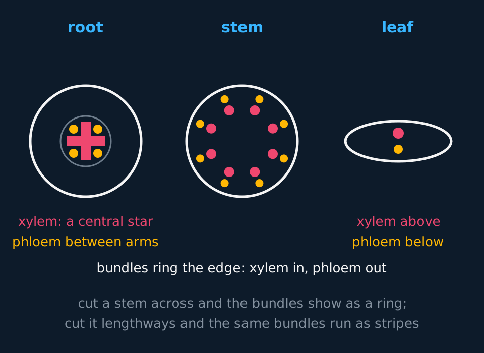
In the root, the vascular tissue is gathered into a central column called the stele. The xylem forms a star, usually drawn with four arms in section, and the phloem sits in separate patches between the arms. A root is loaded in tension: the shoot above levers it and wind tries to pull it out of the ground, and a rope resists pulling best when its strength runs down the middle.
In the stem, the vascular tissue is split into discrete vascular bundles arranged in a ring near the outside, each bundle with its xylem on the inner side, its phloem on the outer side, and a layer of dividing cambium between them. A stem is loaded in bending, and the same amount of material resists bending far better spread into a hollow cylinder than gathered into a solid rod, which is why scaffolding poles are tubes rather than bars. The ring of lignified bundles is the plant's version of the tube.
In the leaf, each vein is one bundle, seen most easily in the midrib. The xylem sits on the upper side of the bundle and the phloem on the lower side, and the reason is continuity rather than anything the leaf needs: the bundle runs out of the stem sideways into the leaf, and what was the inner face of the bundle in the stem becomes the upper face in the leaf. Use that continuity to determine the orientation of a leaf section.
| Organ | Xylem | Phloem | The mechanical logic |
|---|---|---|---|
| Root | A central star | In patches between the arms | Resists pulling, so the strength runs down the middle |
| Stem | Inner side of each bundle | Outer side of each bundle, ring of bundles near the edge | Resists bending, so the strength sits near the outside |
| Leaf | Upper side of each vein | Lower side of each vein | The stem's bundle carried out sideways, orientation kept |
The stem-cutting exercise
A student cuts one sunflower stem twice: once straight across, and once down its length through the centre. Describe what each cut shows of the vascular tissue, and explain why the longitudinal cut might show no vascular tissue at all.
Show the working
The transverse cut gives the map: a ring of separate vascular bundles near the edge of the circle, each with xylem towards the centre and phloem towards the outside. This is the view a plan diagram records, because every tissue's position shows at once.
The longitudinal cut turns each bundle it passes through into a pair of parallel stripes running the length of the stem, the xylem vessels showing as long open tubes. The same tissue, sliced along instead of across.
The bundles are discrete and lie near the edge. A lengthways cut through the centre of the stem passes between bundles and can miss all of them, showing only pith and cortex. This does not show that the stem has no vascular tissue; it shows that a single section plane is insufficient, which is why two cuts are made.
The airway wall in plan
The same convention reads animal organs. The trachea and the bronchus are tubes whose walls are built from concentric layers of tissue, which makes them natural subjects for a plan diagram: each layer becomes one band, and the drawing is a set of rings.
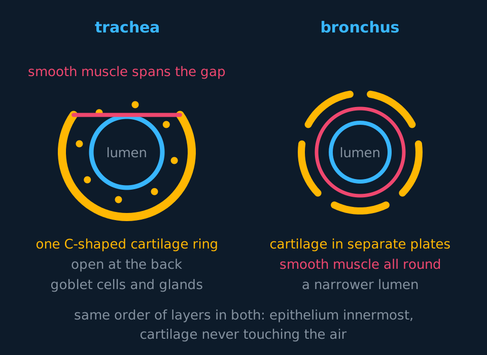
Work from the lumen outwards and label the layers in that order. The ciliated epithelium lines the airway, with goblet cells scattered through it, secreting and moving the mucus that traps particles. Beneath it lies looser tissue holding mucous glands, which add to what the goblet cells secrete, and blood vessels. Then smooth muscle and elastic fibres. Then the cartilage, the layer that keeps the tube open against the pressure changes of breathing, and outside that the connective tissue that ties the organ to its neighbours.
The cartilage is also the identification feature. In the trachea it forms a single C-shaped ring in each segment, incomplete at the back, with smooth muscle spanning the gap; the opening faces the oesophagus, which needs room to bulge as swallowed food passes. In a bronchus the C is gone, replaced by separate curved plates of cartilage with smooth muscle forming a complete ring, and the lumen is narrower. In a bronchiole there is no cartilage at all, only epithelium, smooth muscle and elastic fibres. Given an unlabelled airway section, look for the cartilage first: one C means trachea, plates mean bronchus, none means bronchiole.
Drawn as a plan diagram, the airway wall becomes five or six labelled concentric bands, which records the order of the layers without drawing any individual cell.
Four cells on a smear
A blood smear is made by putting one drop of blood at the end of a slide and drawing it out into a film one cell thick with the edge of a second slide, then staining it. The stains used for blood colour nuclei purple and cytoplasm pink or pale blue, which is why every identification below leans on the nucleus.
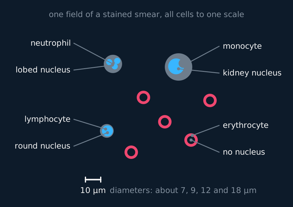
Erythrocytes dominate every field, outnumbering white cells by roughly seven hundred to one. Each is about 7 µm across, stains pink, and has no nucleus: the biconcave shape makes the middle of the disc thinner, so it stains paler and the cell appears as a ring. Any nucleated cell on the smear is a white cell, and three are named at this level.
A neutrophil is about 12 µm across with a nucleus in two to five connected lobes, and fine granules in its cytoplasm. It is the most abundant white cell and the first phagocyte to reach an infected tissue. A lymphocyte is smaller, about 9 µm, with a single round nucleus so large that only a thin rim of cytoplasm shows around it; these are the B and T cells of the specific immune response. A monocyte is the largest cell on the smear, up to about 18 µm, with a kidney-shaped or horseshoe nucleus pushed to one side; it leaves the blood to become a macrophage.
| Cell | Size | Nucleus | Job |
|---|---|---|---|
| Erythrocyte | about 7 µm | None: pale-centred disc | Carries oxygen bound to haemoglobin |
| Neutrophil | about 12 µm | Lobed, two to five lobes | Phagocytosis, first on the scene |
| Lymphocyte | about 9 µm | Round, almost fills the cell | The specific immune response |
| Monocyte | up to 18 µm | Kidney shaped, to one side | Becomes a macrophage in the tissues |
- Blood smear
- A single-cell-thick film of blood drawn across a slide and stained, used to examine and identify blood cells.
- Neutrophil
- A phagocytic white blood cell about 12 µm across with a lobed nucleus and granular cytoplasm.
- Lymphocyte
- A white blood cell about 9 µm across whose round nucleus almost fills the cell, responsible for the specific immune response.
- Monocyte
- The largest white blood cell of a smear, up to about 18 µm, with a kidney-shaped nucleus, which matures into a macrophage.
TRY IT: Naming a cell from two facts
On a stained smear a student finds a cell 13 µm across whose nucleus is drawn out into four connected lobes, and a second cell, 9 µm across, that is almost entirely nucleus. Identify both cells, and state one further feature of the field that needs no stain to see.
Check your answer
The first cell is a neutrophil: the diameter fits, and it is the only named cell with a lobed nucleus. The number of lobes need not be counted, since two to five all indicate the same cell type.
The second is a lymphocyte. A round nucleus filling the cell with only a rim of cytoplasm identifies it, and a diameter of 9 µm excludes a monocyte independently of nucleus shape.
The feature needing no stain is the erythrocytes: they are the overwhelming majority of cells in any field, small pale discs with no nucleus, paler in the centre where the biconcave disc is thinnest.
In the exam
- State the convention when asked what a plan diagram shows: 'tissue boundaries only, in proportion, with no individual cells'.
- Learn the three vascular arrangements as positions rather than as pictures: root central star, stem ring of bundles with xylem inner, leaf vein with xylem upper.
- If a section shows vascular bundles in a ring, identify it as a stem and give the evidence: discrete bundles, near the edge, xylem towards the centre. Name the organ and state the features it was identified from.
- For airway sections, the cartilage answers the identification: one C-shaped ring is the trachea, separate plates a bronchus, no cartilage a bronchiole.
- Give two features per cell when identifying blood cells: diameter and nucleus. 'About 18 µm with a kidney-shaped nucleus' identifies a monocyte; a size description alone does not.
- Quote the sizes with their unit: 7, 9, 12 and 18 µm. A relative description, such as a monocyte being two to three times the diameter of an erythrocyte, is an acceptable alternative.
Check yourself
A student is handed a transverse section of a plant organ. The vascular tissue forms a single central star of xylem with patches of phloem between the arms. Identify the organ, describe how the student should record the tissue layout, and explain why a section of the same plant's stem would give a different drawing.
Answer
The organ is a root. A central stele with the xylem as a star and the phloem between its arms is the root's map and no other organ's; a stem would show a ring of separate bundles and a leaf a flat blade with veins.
The layout should be recorded as a low-power plan diagram: sharp single lines marking the boundaries between tissues, the regions in proportion, measured against the eyepiece graticule rather than judged, no individual cells and no shading, ruled label lines touching each tissue, and the magnification stated.
The stem arranges the same two tissues differently because it carries a different load. A root resists being pulled, so its strength is gathered into a central rope; a stem resists bending, so its lignified tissue is spread into a ring of bundles near the outside, each with xylem on the inner face and phloem on the outer.
The stem drawing would therefore show a circle with eight or so discrete bundles near its edge and pith in the middle. A central star drawn for a stem records the arrangement of a different organ.
Questions
Question 15 marks
Describe the layers of the wall of the trachea in order from the lumen outward, naming the tissue found in each layer.
Mark scheme
- B1 ciliated epithelium, with goblet cells among it, lining the lumen
- B1 loose tissue beneath it, containing mucous glands and blood vessels
- B1 smooth muscle and elastic fibres
- B1 cartilage, as an incomplete C-shaped ring
- B1 an outer layer of connective tissue, attaching the trachea to its neighbours
Question 24 marks
Describe the distribution of xylem and phloem in a transverse section of the root and of the stem of a herbaceous dicotyledonous plant.
Mark scheme
- B1 in the root the vascular tissue forms a single central stele
- B1 the root's xylem forms a central star, with the phloem in separate patches between the arms
- B1 in the stem the vascular tissue is split into discrete bundles arranged in a ring near the outside
- B1 within each stem bundle the xylem lies on the inner side and the phloem on the outer side
Question 34 marks
A transverse section of a herbaceous dicot stem is 4.0 mm across, and its ring of vascular bundles lies 0.50 mm in from the epidermis. A student draws a low-power plan diagram of the section as a circle 120 mm across. Calculate the magnification to be written on the student's plan, calculate how far in from its outer line the ring of vascular bundles must be drawn, and state which rule of the plan diagram convention the second calculation serves.
Mark scheme
- M1 magnification = size of drawing ÷ actual size, both in millimetres: 120 ÷ 4.0
- A1 ×30, written on the plan with no unit, because a plan diagram must state its magnification or carry a scale bar
- A1 0.50 mm × 30 = 15 mm in from the outer line of the drawing
- B1 the tissues of a plan diagram must be drawn in their correct proportions, so the position of each boundary is measured, with an eyepiece graticule where needed, and scaled rather than estimated
Question 44 marks
State the four named blood cells of a stained smear in order of increasing diameter, giving an approximate size in micrometres for each.
Mark scheme
- B1 erythrocyte, about 7 µm
- B1 lymphocyte, about 9 µm
- B1 neutrophil, about 12 µm
- B1 monocyte, up to about 18 µm
Question 53 marks
A root carries its xylem as a central column, while a stem carries its lignified vascular bundles in a ring near its surface. Suggest why each arrangement suits the forces that organ must withstand.
Mark scheme
- B1 a root is loaded in tension, pulled by the shoot it anchors and by wind acting on the plant above
- B1 a central column resists pulling in the way a rope does, so strength down the middle suits the root
- B1 a stem is loaded in bending, and a hollow cylinder of strong material near the outside resists bending far better than the same material in the centre
Question 63 marks
Identify each of the following cells seen on a stained blood smear, giving a reason for each: cell A is 12 µm across with a nucleus of three connected lobes; cell B is 18 µm across with a kidney-shaped nucleus; cell C is 7 µm across and has no nucleus.
Mark scheme
- B1 A is a neutrophil, because no other cell you have to name has a lobed nucleus
- B1 B is a monocyte, the largest cell on a smear, with the kidney-shaped nucleus that separates it from a lymphocyte
- B1 C is an erythrocyte, because mature erythrocytes are the only blood cells with no nucleus
Question 73 marks
On a micrograph of a blood smear taken at a magnification of 2000 times, a monocyte measures 36 mm across. Calculate the actual diameter of the monocyte in micrometres, and use your answer and the nucleus to explain how the monocyte is told apart from the lymphocyte beside it.
Mark scheme
- M1 actual size = image size ÷ magnification, with 36 mm as 36 000 µm: 36 000 ÷ 2000 = 18 µm
- B1 at about 18 µm the monocyte is the largest cell on the smear, about twice the diameter of a lymphocyte at about 9 µm
- B1 the monocyte's nucleus is kidney or horseshoe shaped and pushed to one side, whereas a lymphocyte's round nucleus almost fills the cell, leaving only a thin rim of cytoplasm
Question 83 marks
Sections are taken through three different airways. Describe how the cartilage seen in each section allows the trachea, a bronchus and a bronchiole to be told apart.
Mark scheme
- B1 the trachea shows a single C-shaped ring of cartilage, incomplete at the back where smooth muscle spans the gap
- B1 a bronchus shows cartilage broken into separate curved plates with gaps between them
- B1 a bronchiole shows no cartilage at all, only epithelium, smooth muscle and elastic fibres
Question 93 marks
Describe the distribution of xylem and phloem in a transverse section through the midrib of a leaf, and describe how this arrangement relates to the arrangement in the stem the leaf grew from.
Mark scheme
- B1 the xylem lies on the upper side of the vein and the phloem on the lower side
- B1 this is the same vascular bundle that ran through the stem, carried out sideways into the leaf
- B1 what was the inner face of the bundle in the stem, the xylem, becomes the upper face in the leaf, so the orientation is continuous rather than newly determined
Question 103 marks
A student cuts a sunflower stem twice, once transversely and once longitudinally through its centre, and the longitudinal section shows no vascular tissue at all. Explain why this section might show no vascular bundles, and explain why this does not mean the stem lacks vascular tissue.
Mark scheme
- B1 the vascular bundles are discrete and lie in a ring near the edge of the stem, rather than filling the whole cross-section
- B1 a longitudinal cut straight through the centre of the stem can pass entirely between two bundles, showing only pith and cortex
- B1 the transverse section already showed the ring of bundles, so the correct conclusion is that one section plane is insufficient on its own, not that the tissue is absent
Question 113 marks
Compare a low-power plan diagram with a high-power biological drawing, referring to what each records and how many cells each shows.
Mark scheme
- B1 a plan diagram records the positions, boundaries and proportions of whole tissues without drawing any individual cell, whereas a high-power drawing records the structure of a small number of representative cells in detail
- B1 a plan diagram is drawn at low magnification so a whole organ or section fits in view, whereas a high-power drawing is made at a magnification high enough to show detail within one or a few cells
- B1 combining the two, drawing cellular detail inside a plan diagram, breaks the convention, because a plan diagram's purpose is the tissue map rather than cell structure
Question 123 marks
A cross-section is cut through a leaf blade away from the midrib, between two of the smaller veins. Suggest why this section is much less useful than a section through the midrib for showing the position of xylem relative to phloem.
Mark scheme
- B1 away from the midrib a vein is much smaller, with the xylem and phloem forming only a tiny bundle among the mesophyll, difficult to make out clearly
- B1 a section between two small veins might miss the vascular tissue almost entirely, showing mostly mesophyll and epidermis
- B1 the midrib is a fixed, predictable location carrying the largest vein, where the tissues are clearly separated and the upper-xylem, lower-phloem arrangement is easy to see
Question 132 marks
State two rules of the low-power plan diagram convention, and state what a plan diagram records that a high-power drawing does not.
Mark scheme
- B1 any two of: tissue boundaries only with no individual cells, no shading, sharp unbroken lines, regions in correct proportion, ruled label lines touching the structure, magnification or scale bar stated
- B1 it records the positions and proportions of whole tissues within the organ, rather than the detail of any cell
Question 142 marks
Explain why the cartilage in the tracheal wall forms an incomplete C-shape rather than a complete ring.
Mark scheme
- B1 the gap faces the oesophagus, which lies immediately behind the trachea
- B1 leaving the ring incomplete, with smooth muscle spanning the gap, allows the oesophagus room to bulge as swallowed food passes down it
Question 152 marks
State the typical ratio of erythrocytes to white blood cells seen in a field of a blood smear, and state one feature of an erythrocyte's appearance that follows from its biconcave shape.
Mark scheme
- B1 roughly 700 erythrocytes for every 1 white blood cell
- B1 the biconcave disc is thinner in the middle, so that part of the cell stains more palely and the cell appears as a ring with a paler centre
Worth remembering
- A plan diagram shows tissue boundaries in proportion and never shows individual cells.
- Root: central xylem star, phloem between the arms. Stem: ring of bundles, xylem inner, phloem outer. Leaf: veins with xylem upper.
- Cut across for the map; cut lengthways and tubes become parallel lines, and a cut between bundles can miss them entirely.
- Airway walls read from the lumen outwards, and the cartilage names the organ: C-ring trachea, plates bronchus, none bronchiole.
- On a smear: no nucleus, erythrocyte; lobed nucleus, neutrophil; round filling nucleus, lymphocyte; kidney nucleus and largest cell, monocyte.
CHECK YOUR PROGRESS
Rate how confident you are with each objective for this lesson. Ratings are saved in this browser, on this device, unless you sign in.
- State the rules of the low-power plan diagram convention, and say what a plan diagram deliberately leaves out.
- Draw the distribution of xylem and phloem in cross-sections of a root, a stem and a leaf of a herbaceous dicot.
- Predict what transverse and longitudinal cuts through a stem will each show, and explain why one cut is not enough.
- Describe the wall of the trachea and of a bronchus as layers in plan, and use the cartilage to tell the two apart.
- Identify an erythrocyte, a neutrophil, a lymphocyte and a monocyte on a stained blood smear from size and nucleus alone.
Open the full revision checklist to see every objective in the curriculum in one place.
Practise this lesson
WORKBOOK
The written questions from this topic, on paper with room to work, and a separate book of mark schemes. The question that is set on a diagram stays on this page, where the diagram can be drawn. Free to use; please do not redistribute or sell.