Biology › Gene regulation, genomics and biotechnology › Mutation, gene expression and cancer
Mutation, gene expression and cancer
Mutations can alter a coding sequence, a splice site or a regulatory region. Their effects range from none to major changes in protein amount or function. Cancer develops when accumulated genetic and epigenetic changes disrupt control of cell division, DNA repair and apoptosis. Benign and malignant tumours are distinguished, and established and experimental uses of stem cells are evaluated.
Before this The genetic code: triplet, degenerate, non-overlapping · Transcription factors and the promoter region · DNA methylation and histone acetylation · The cell cycle and its checkpoints · Stem cells and levels of potency
COMMON MISCONCEPTION
A base substitution that does not change the amino acid is a mutation that never really happened.
It happened: the base sequence changed, every daughter cell copies the change, and it can be passed on. 'Silent' means only that the new triplet codes for the same amino acid, and even then the change is not always harmless, since some silent changes disturb splicing.
What you should be able to do
- Name the types of gene mutation and give the consequence of each at the level of the polypeptide.
- Explain why many base substitutions change nothing about the protein, and why a deletion of one base usually changes everything.
- Distinguish a mutation in a gene from one in a control sequence, and give an example of each.
- Explain the difference between a proto-oncogene and a tumour suppressor gene, including how many alleles must be affected.
- Explain how methylation of a promoter can silence a tumour suppressor gene without altering a single base.
- Describe the therapeutic uses of stem cells and set out the ethical arguments on both sides of the embryonic case.
- Distinguish a chromosome mutation from a gene mutation, and explain why a balanced translocation carrier is usually healthy but can produce unbalanced gametes.
- Explain how a faulty CFTR channel makes mucus thick and sticky, and how that mucus impairs the gaseous exchange, digestive and reproductive systems.
The kinds of mutation, and what each does to a protein
A mutation is a change in the base sequence of DNA. The two commonest kinds arise as copying errors during replication. They are set out below with their consequences for the polypeptide. Prerequisite: the genetic code and transcription.
A substitution replaces one base with another and affects one triplet only. Three outcomes are possible. If the new triplet codes for the same amino acid, which happens often because the code is degenerate and triplets sharing an amino acid usually differ in the third base, the mutation is silent and the polypeptide is unchanged. If it codes for a different amino acid the mutation is missense, and the effect ranges from none to complete loss of function, depending on where in the protein the changed residue sits. If it produces a stop codon the mutation is nonsense, translation ends early, and the truncated polypeptide is usually non-functional.
An insertion or a deletion within a coding sequence, of a number of bases not divisible by three, shifts the reading frame, because the code is non-overlapping and read in threes from a fixed start. Every triplet downstream is read from a new position, which is a frame shift, so the amino acid sequence after the mutation differs from the original and a stop codon usually arises early by chance. The effect depends on the position of the change within the transcript and on how the transcript is processed: a frame shift near the start of a coding sequence usually gives a non-functional protein, while one near the end may not. A deletion of three bases removes one amino acid and shifts nothing, which is why the commonest cystic fibrosis allele, a deletion of three bases removing a single phenylalanine, produces a protein that is complete but misfolds: the cell's quality control holds it back in the endoplasmic reticulum and degrades most of it, so almost none reaches the membrane.
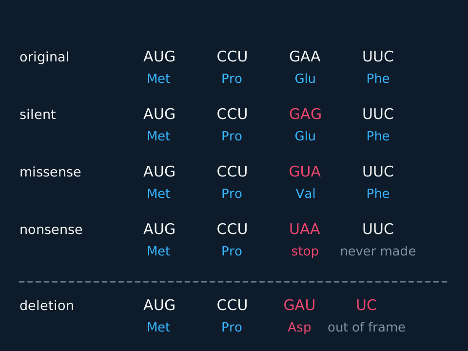
Describe the drawing
A comparison of four versions of the same short messenger RNA sequence, written as four codons in a row with the amino acid each one specifies printed underneath. The first row is the original, AUG CCU GAA UUC, giving methionine, proline, glutamic acid and phenylalanine. In the second row the third codon has become GAG, which still gives glutamic acid. In the third it has become GUA, giving valine instead. In the fourth it has become UAA, a stop codon. Below a dashed line, a fifth row shows a single base deleted, after which the codons no longer line up with those above and every one that follows is read differently.
| Mutation | What changes in the DNA | Effect on the protein |
|---|---|---|
| Substitution, silent | One base swapped; new triplet codes for the same amino acid | None to the amino acid sequence |
| Substitution, missense | One base swapped; new triplet codes for a different amino acid | One amino acid different; effect depends on where it is |
| Substitution, nonsense | One base swapped; new triplet is a stop codon | Polypeptide cut short, almost always non-functional |
| Deletion or insertion | One or two bases lost or gained | Frame shift: every triplet after it is misread |
| Duplication | A section is repeated | Frame shift unless the repeat is a multiple of three |
| Inversion | A section is reversed within the chromosome | The reversed section codes for a different sequence entirely |
| Translocation | A section moves to a different chromosome | Genes at both break points may be disrupted or newly switched on |
A human inherits roughly sixty to seventy new point mutations that neither parent had, most of them from the father, since sperm come from many more rounds of division than eggs do; almost all land in non-coding DNA and do nothing. The rate rises with exposure to mutagens: ultraviolet light, which links adjacent thymine bases; ionising radiation, which breaks strands; and chemicals such as the benzopyrene in tobacco smoke, which distorts the helix so that polymerase misreads it.
Chromosome mutations: changes to structure rather than sequence
Everything in the section above is a gene mutation: a change to the base sequence of one gene, affecting one triplet or a few. A chromosome mutation is a change on a far larger scale, to the structure of a chromosome or to the number of chromosomes in a cell, and it can lose, repeat or move hundreds of genes at once. The bases inside a moved section may be exactly as they were. What has changed is where the section sits, how many copies of it the cell has, or which way round it lies. Changes in number arise by non-disjunction in meiosis and are taught with meiosis; this section deals with structure, and Pearson Edexcel Biology B examines it through translocations.
A structural change begins with breaks in the DNA of a chromosome that the cell's repair enzymes rejoin wrongly. Four outcomes are recognised.
| Change | What happens to the chromosome | Usual consequence |
|---|---|---|
| Deletion | A section breaks off and is lost | Every gene in the section is lost from that chromosome; cri du chat syndrome results from loss of part of chromosome 5 |
| Duplication | A section is copied, so it appears twice | Extra copies of the genes in it; a duplicated gene is also raw material for evolution, since one copy can change while the other keeps working |
| Inversion | A section breaks out, turns through 180° and rejoins | The genes are all present, in reverse order; a gene cut at a break point is disrupted |
| Translocation | A section moves to a non-homologous chromosome | Explained below |
In a reciprocal translocation two non-homologous chromosomes each break, and the broken-off pieces swap places. Nothing is lost and nothing is gained, so a person whose cells carry it still has two copies of every gene and is usually healthy. Such a person is a balanced carrier. The exceptions are a break that falls inside a gene, which disrupts it, and a break that places a gene beside a different promoter, which changes how much of it is transcribed; either can alter the phenotype although no DNA has gone missing.
The difficulty appears at meiosis. A carrier has one normal and one translocated copy of each of the two chromosomes involved, and each gamete receives one version of each. If the two pairs are treated as separating independently, four kinds of gamete are made in equal numbers: one with both normal chromosomes, one with both translocated chromosomes, which is balanced like the parent, and two that each carry one normal and one translocated chromosome. Those two are unbalanced: each carries one section twice and lacks another. An embryo formed from an unbalanced gamete has three copies of some genes and only one of others, and most such pregnancies end in miscarriage. In reality the four chromosomes pair up together as a cross-shaped group in prophase I, so segregation is not independent and the proportions vary from one translocation to another. A balanced translocation in one partner is among the causes looked for when a couple have had several miscarriages.
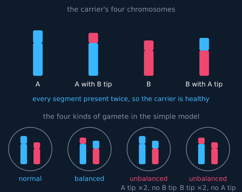
Describe the drawing
At the top, four chromosomes drawn as vertical bars, coloured cyan for material from chromosome A and coral for material from chromosome B. From left to right they are a normal A, an A whose tip has been replaced by the tip of B, a normal B, and a B whose tip has been replaced by the tip of A, so the carrier has every segment twice and is healthy. Below, four circles stand for the four kinds of gamete, each holding one version of A and one of B: a normal gamete, a balanced gamete with both swapped chromosomes, and two unbalanced gametes, one with the tip of A twice and no tip of B, the other the reverse.
Two translocations are met by name. In roughly one case of Down syndrome in twenty-five, the extra chromosome 21 material is attached to another chromosome, most often chromosome 14, rather than present as a separate third chromosome. One parent may be a balanced carrier, which raises the chance of a second affected child, so a karyotype of the child is used to tell the two kinds apart. And in chronic myeloid leukaemia the cancer cells carry a translocation between chromosomes 9 and 22 that joins part of a gene called BCR to part of the ABL gene. The fused gene codes for a permanently active enzyme that signals the cell to divide, so a proto-oncogene has become an oncogene by being moved, with no base substitution involved. That translocation arises in a single blood stem cell during a person's life and is not inherited; the drug imatinib, which blocks the fused enzyme, is the standard treatment.
- Chromosome mutation
- A change in the structure of a chromosome, or in the number of chromosomes, that can affect many genes at once.
- Translocation (chromosome)
- A chromosome mutation in which a section of one chromosome breaks off and joins a non-homologous chromosome.
- Balanced translocation carrier
- A person with a reciprocal translocation who has lost and gained no genes, is usually healthy, but can produce unbalanced gametes.
Mutations in coding sequences and in regulatory sequences
The genome comprises genes together with the sequences that determine when and how much each gene is transcribed: promoters, enhancers, the boundaries between introns and exons, and the genes coding for the transcription factors themselves. A change in any of those alters the amount of protein made, or when it is made, without altering its amino acid sequence.
Lactase persistence. Most adult humans stop making lactase after weaning, which is the ancestral condition. The variant that keeps it switched on in people of northern European descent is a single base change, and it is not in the lactase gene. It lies about fourteen thousand base pairs away, inside an intron of a neighbouring gene, in a sequence that acts as an enhancer for lactase. The lactase protein of a lactose-tolerant adult has the same amino acid sequence as anyone else's. The change is in a regulatory sequence.
β-thalassaemia. Many of the alleles causing it are not in the coding sequence of the β-globin gene at all. Some sit in the promoter and reduce transcription; others sit at an intron boundary, so the spliceosome cuts in the wrong place and the mature mRNA carries intron sequence. Either way the cell makes too little normal β-globin.
Tumours. A heavily methylated promoter can silence a gene whose sequence is intact. That case is treated in the next section.
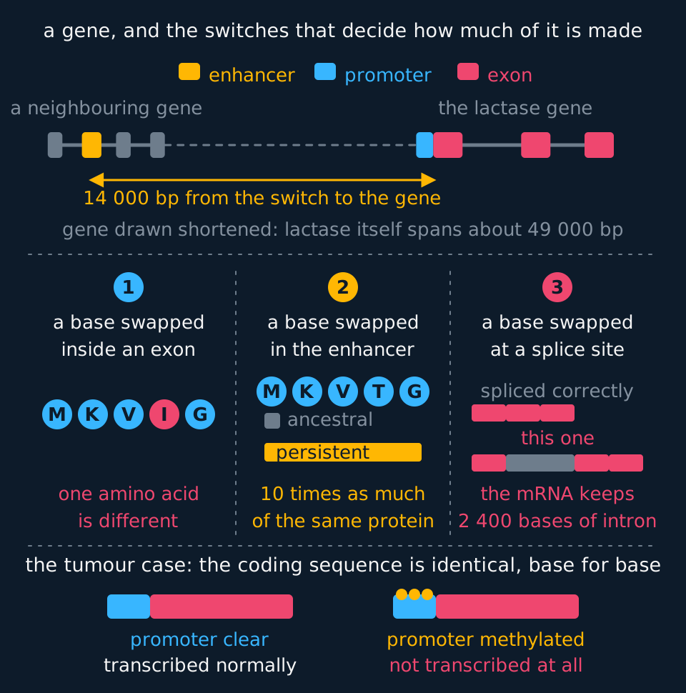
Describe the drawing
A map along the top drawn to a base-pair scale shows two genes on one stretch of DNA: on the left a neighbouring gene whose long intron contains an amber block, the enhancer for lactase, and on the right the lactase gene itself with a cyan promoter and three coral exons separated by introns. A measured span between them is labelled fourteen thousand base pairs; the lactase gene is drawn shortened, since it really spans about forty-nine thousand. Beneath, three panels take the same event, one base swapped, in three places. In the first the chain of amino acids has one bead coloured differently, because one residue has changed. In the second the chain is unchanged bead for bead and two bars beside it show ten times as much protein being made. In the third, two messenger RNA bars are compared and the lower one is longer by exactly the length of the first intron, which the spliceosome has failed to remove. A band at the foot draws a healthy promoter and a methylated one on genes with identical coding sequence, the first transcribed normally and the second not transcribed at all.
- Mutation
- A change in the base sequence of DNA. Whether the protein changes depends on which base changes and where in the transcript it lies.
- Silent mutation
- A base substitution that produces a triplet coding for the same amino acid, so the primary structure of the polypeptide is unchanged.
- Frame shift
- The displacement of the reading frame caused by insertion or deletion of a number of bases that is not a multiple of three.
- Control sequence
- A region of DNA that is not translated but determines whether, when and how strongly a gene is transcribed.
Cystic fibrosis: one faulty channel, three organ systems
Pearson Edexcel Biology A builds its second topic round cystic fibrosis and asks how the mutation impairs the gaseous exchange, digestive and reproductive systems. The explanation runs through one protein, the CFTR protein (cystic fibrosis transmembrane conductance regulator), and one substance, mucus. Prerequisite: osmosis and the transport of ions across membranes.
CFTR is a channel protein in the cell-surface membrane of epithelial cells, on the side facing the lumen of the tube they line. When it is opened, which requires ATP, chloride ions leave the cell through it into the lumen. Sodium ions follow the chloride, attracted by its negative charge, passing between the cells. The extra ions lower the water potential of the liquid outside the cells, so water leaves the cells by osmosis. In the airways this keeps a layer of liquid deep enough for the cilia to beat in, with runny mucus on top of it. CFTR also restrains a sodium channel in the same membrane that takes sodium ions back into the cell.
In cystic fibrosis both alleles are faulty. With the commonest allele the protein is made but misfolds and is destroyed before it reaches the membrane, as described above; other alleles give a channel that reaches the membrane but rarely opens, or no protein at all. In every case too few chloride ions are secreted. Sodium ions are absorbed faster than normal, because the restraint on the sodium channel has gone, and water follows them into the cells by osmosis. Liquid is drawn off the epithelial surface, and the mucus loses water and becomes thick and sticky. Every tube lined by this kind of epithelium is affected in the same way; the three systems differ in what the blocked tube was for.
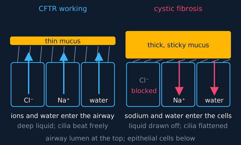
Describe the drawing
Two panels of airway lining side by side, each a row of three epithelial cells with a layer of liquid, cilia and mucus above them. On the left, with CFTR working, arrows show chloride ions, sodium ions and water all moving into the airway, the liquid layer is deep, the cilia stand upright, and the mucus on top is a thin band. On the right, in cystic fibrosis, chloride movement is marked as blocked, arrows show sodium ions and water moving into the cells, the liquid layer is almost gone, the cilia lie flattened, and the mucus is a thick, sticky band.
The gaseous exchange system. Mucus normally traps dust and bacteria, and the cilia sweep it up the airways to the throat. Dehydrated mucus is too thick for the cilia to move, and with the liquid layer gone they have no room to beat, so mucus collects in the bronchioles. Where it blocks a bronchiole completely, the alveoli beyond it are no longer ventilated, so the surface area available for gas exchange falls and less oxygen diffuses into the blood. Bacteria trapped in the stagnant mucus multiply, and repeated chest infections follow, Pseudomonas aeruginosa being a common cause. Each infection brings inflammation, and repeated inflammation leaves scar tissue that damages the lungs progressively. People with cystic fibrosis are short of breath, cough persistently, and do physiotherapy every day to clear the mucus.
The digestive system. The pancreas secretes amylase, lipase and proteases such as trypsin into the pancreatic duct, which carries them to the duodenum. In most people with cystic fibrosis the secretion in the duct is so thick that the duct becomes blocked, often before birth. The enzymes cannot reach the small intestine, so starch, fats and proteins are only partly digested and their products are not absorbed: the person struggles to gain weight however much they eat, and undigested fat passes out in the faeces. Trapped behind the blockage, the enzymes damage the pancreas itself, which becomes scarred and forms fluid-filled cysts, the fibrosis and cysts that gave the disease its name. Scarring can eventually reach the islets that secrete insulin, and diabetes is common in adults with the condition. Mucus lining the small intestine is also thicker, which slows absorption, and in some newborn babies the intestine is blocked outright. Most patients take capsules of pancreatic enzymes with every meal.
The reproductive system. In almost all men with cystic fibrosis the vas deferens, the tube that carries sperm from the testis towards the urethra, is blocked by thick secretion or is absent, having failed to develop before birth. Sperm are made normally but cannot leave, so the man is infertile, although sperm can be collected from the testis by a small operation and used in IVF. In women the mucus at the cervix is thicker than usual and sperm pass through it less easily, so fertility is reduced; many women with cystic fibrosis conceive without help.
| System | Where the thick mucus collects | Effect on function |
|---|---|---|
| Gaseous exchange | Bronchioles | Fewer alveoli ventilated, so less oxygen absorbed; trapped bacteria cause repeated infection and scarring |
| Digestive | Pancreatic duct and intestinal lining | Enzymes do not reach the duodenum, so food is poorly digested and absorbed; the pancreas is damaged |
| Reproductive | Vas deferens in men; cervix in women | Sperm cannot leave the testis; sperm pass the cervix less easily |
Drugs called CFTR modulators now act on the protein itself. Some help a misfolded protein reach the membrane and others hold the channel open for longer, and combinations of the two are licensed for people with at least one copy of the commonest allele. They improve lung function substantially, but they cannot rebuild tissue that has already been destroyed.
- CFTR protein
- A chloride ion channel in the cell-surface membrane of epithelial cells; when it is faulty, too little chloride and water are secreted and mucus becomes thick and sticky.
Oncogenes and tumour suppressor genes
A tumour is a mass of cells produced by uncontrolled division. A benign tumour remains localised and does not metastasise. A malignant tumour invades surrounding tissue and may spread through blood or lymph to form secondary tumours. Growth rate, degree of differentiation and encapsulation can provide supporting evidence, but they are not the defining distinction. Spread, metastasis, is what makes a cancer difficult to treat surgically, because there is no single site to remove.
Two classes of gene are involved, and they are altered in opposite directions.
Describe the drawing
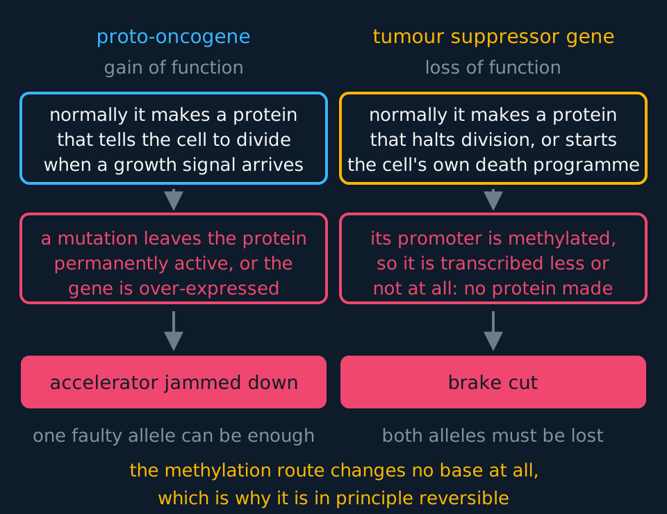
Two columns setting the two classes of cancer gene side by side. The left column follows a proto-oncogene, which normally makes a protein instructing the cell to divide when a growth signal arrives; a mutation leaves that protein permanently switched on, which the figure labels an accelerator jammed down, and one faulty allele suffices. The right column follows a tumour suppressor gene, which normally makes a protein that halts division or starts the cell's own death programme; heavy methylation of its promoter stops it being transcribed, which the figure labels a cut brake, and both alleles must be lost before it matters.
A proto-oncogene is a perfectly normal gene coding for a protein that stimulates cell division when a growth signal arrives: a growth factor, a receptor for one, or a protein that relays the signal inside the cell. A mutation can convert it into an oncogene: a version that is permanently active, or one that is present in too many copies, or one that has been moved next to a promoter that never switches off. The cell then receives a constant stimulus to divide whether or not any growth factor is present. One faulty allele is enough, because a permanently active protein does its damage regardless of the normal protein made by the other allele.
A tumour suppressor gene codes for a protein that does the opposite: it halts the cell cycle at a checkpoint, or triggers apoptosis in a cell whose DNA is damaged beyond repair. Loss of function removes that control, and in most cases both alleles must be lost, because one working copy still makes the protein. The two-copy requirement is the common case rather than a universal rule; for a few suppressor genes, loss of one copy already reduces the protein below the amount required. That accounts for inherited predisposition: someone born with one faulty BRCA1 allele does not have cancer, but every cell requires only one further event to lose the control, rather than the two events required in someone with two working alleles.
A third route involves no change to a base sequence. A tumour suppressor gene whose promoter becomes heavily methylated is transcribed less, or not at all, and the control is lost as it would be if the gene had been deleted, although the sequence is unaltered. This occurs in many tumours: the mismatch repair gene MLH1 is silenced this way in a substantial fraction of colorectal cancers, which explains why cancer is described as a disease of gene expression and not only a disease of mutation. Methylation can go wrong the other way too: hypomethylation of a proto-oncogene's promoter increases its transcription, so the growth-promoting protein is over-produced and the cell is stimulated to divide, as it would be by an oncogene.
- Proto-oncogene
- A normal gene whose product stimulates cell division in response to a growth signal.
- Oncogene
- A mutated or over-expressed proto-oncogene whose product stimulates division continuously.
- Tumour suppressor gene
- A gene whose product slows the cell cycle or triggers apoptosis; loss of both alleles removes that control.
- Metastasis
- The spread of cells from a malignant tumour through the blood or lymph to form secondary tumours.
Oestrogen has a role that should be stated precisely. Breast tissue is a target for oestrogen, and in a cell where a proto-oncogene lies downstream of an oestrogen response element, more oestrogen means more transcription of a gene that stimulates division. After the menopause the ovaries stop producing oestrogen but adipose tissue continues to, and higher concentrations of oestrogen in post-menopausal women are associated with a higher risk of breast cancer. That is an association supported by a plausible mechanism. It is not equivalent to the claim that oestrogen causes breast cancer in the way that loss of a tumour suppressor gene does. State which kind of claim an answer is making.
Reading methylation data from a tumour
Samples of healthy colon tissue and of tumour tissue from the same patient are analysed. The promoter of a tumour suppressor gene is found to be methylated at 8% of its cytosine sites in the healthy tissue and at 74% in the tumour. Sequencing shows the coding sequence of the gene is identical in both samples. Explain what has happened and why the cells divide uncontrollably.
Show the working
The gene has not been mutated: the sequences are identical, so any answer built on a changed base is ruled out by the data given. What has changed is the methylation of the promoter, which has risen roughly ninefold.
A heavily methylated promoter cannot be bound by the transcription factors and RNA polymerase that would normally start transcription, and the methyl groups also attract proteins that recruit histone deacetylases, so the chromatin around the gene condenses further. Transcription falls to nothing, no mRNA is made, and the tumour suppressor protein is not synthesised.
Without that protein, the checkpoint it enforces in the cell cycle is not applied. Cells with damaged DNA are neither held for repair nor sent into apoptosis, so they continue through the cycle, divide, and pass their damage on. Division becomes uncontrolled and a tumour forms.
One further inference the data supports: because no base has changed, the silencing is in principle reversible, which is why drugs that inhibit methylation or deacetylation are used in some cancers. A deleted gene offers no such possibility.
Stem cells, and the argument that surrounds them
Cancer is a failure of control over division. Stem cell medicine is an attempt to use controlled division deliberately, and the two sit in the same unit for that reason. You met the levels of potency earlier in the course; what matters here is where the cells come from and what they are being used for.
| Source | Potency | Where it stands |
|---|---|---|
| Embryo, four to five days old | Pluripotent: any cell type of the body | Widest use in principle; the source of the ethical objection |
| Umbilical cord blood | Multipotent, mainly blood-forming | Routinely banked; used in the same way as bone marrow |
| Adult tissue, such as bone marrow | Multipotent, limited to that tissue's range | Established in treatment; supply and range both limited |
| Induced pluripotent, from the patient's own cells | Pluripotent, after reprogramming with transcription factors | No embryo used, far lower rejection risk; still largely at trial stage |
One use is genuinely routine: bone marrow transplantation replaces a patient's blood-forming stem cells after their own have been destroyed by leukaemia or its treatment, and has been standard care for decades. A second is licensed: sheets grown from a patient's own limbal stem cells restore the corneal surface after chemical burns. Beyond those, trials are running for macular degeneration, Parkinson's disease, type 1 diabetes and spinal cord injury, some with encouraging early results, and none of them is established treatment. When evaluating a stem cell claim, distinguish a treatment in clinical trial from one in routine use.
The ethical arguments are set out below rather than resolved. The disagreement turns on a question biology does not answer: what moral status a five-day-old human embryo has.
Those who oppose the use of embryonic stem cells generally hold that a human embryo has the full moral status of a person from fertilisation onwards, since that is when a genetically distinct human organism begins and no later point marks a non-arbitrary threshold. On that view, destroying an embryo to obtain its cells is the deliberate destruction of a human life, and the good that might be done with the cells does not license it. The Roman Catholic Church argues this position formally, and it is held by many people on secular grounds as well.
Those who support it generally hold that moral status develops gradually, and that an embryo of about a hundred cells with no nervous system, no capacity for experience and no individuality yet fixed does not have the interests that make killing a person wrong. On that view the suffering of identifiable patients with untreatable conditions carries more weight than the destruction of a blastocyst, particularly when the embryos used are surplus from IVF and would otherwise be discarded.
A middle position accepts the use of surplus IVF embryos while rejecting the creation of embryos specifically for research, on the ground that there is a moral difference between using something that will be destroyed anyway and bringing it into existence in order to destroy it. British law reflects a version of this compromise: research on human embryos requires a licence from the Human Fertilisation and Embryology Authority, and no embryo may be kept beyond fourteen days.
Induced pluripotent stem cells alter the terms of the argument without resolving it. They are made by treating an adult cell, usually a skin or blood cell, with a small set of transcription factors that switch previously silent genes back on, returning the cell to a pluripotent state. No embryo is involved and, because the cells come from the patient, tissue grown from them carries a much lower risk of rejection, reduced rather than abolished, since immune reactions to reprogrammed cells have been reported. That removes the objection raised against embryonic sources. Three limitations remain: reprogrammed cells can carry mutations acquired during the adult cell's life, their tendency to form tumours has to be controlled, and the fact that they can in principle be turned into gametes raises questions of its own.
TRY IT: Evaluating a claim about stem cell treatment
A private clinic advertises an injection of the patient's own bone marrow stem cells as a treatment for multiple sclerosis, a disease in which the myelin sheaths around neurones are destroyed. The advertisement says that because the cells are the patient's own there is no ethical objection and no risk of rejection. Evaluate the claim biologically, and say what you would want to see before accepting it.
Check your answer
The rejection point is essentially sound. Cells taken from the patient carry the patient's own antigens, so the immune system does not identify them as non-self, and rejection of the kind seen with donor tissue is not expected. The ethical point about embryos is also sound as far as it goes: no embryo is destroyed, so the objection raised against embryonic stem cells does not apply.
The biological problem is potency. Bone marrow stem cells are multipotent, and the range they can produce is the blood cells: red cells, the various white cells and platelets. They are not pluripotent, so there is no reason to expect them to produce the oligodendrocytes that make myelin in the central nervous system. The advertisement asserts a benefit without a mechanism.
The claim about ethics is also too narrow. Absence of an embryo is not absence of ethical difficulty: selling an unproven treatment to people with an incurable disease raises questions about consent, about exploitation of hope and about the money involved.
Before accepting the claim you would want published results from randomised controlled trials with a placebo group, since multiple sclerosis relapses and remits on its own and testimonials cannot distinguish a treatment effect from a remission; a proposed mechanism by which the injected cells reach the central nervous system and produce myelin; and regulatory approval rather than an advertisement.
In the exam
- Name the type of mutation and then state its effect on the protein. 'A mutation occurs' names neither; 'a substitution produces a stop codon, so the polypeptide is shortened' gives both.
- Name the property of the code as well as the outcome: silent mutations arise because the code is degenerate, frame shifts because it is non-overlapping and read in threes.
- A deletion of three bases does not cause a frame shift. Check whether the number of bases is a multiple of three before using the term.
- One faulty allele can be enough for an oncogene; in most cases both alleles must be lost for a tumour suppressor gene.
- State that hypermethylation of a promoter silences a gene without changing its sequence. Methylation data alongside identical coding sequences point to that mechanism.
- For oestrogen and breast cancer, distinguish association from cause. Data showing a correlation support a hypothesis; they do not establish a cause.
- In an ethics question, state each position and the reason given for it. Answer both sides before reaching a judgement.
- Name the class of mutation. A translocation is a chromosome mutation and changes no base sequence; a substitution is a gene mutation.
- For cystic fibrosis, give the chain in order: too little chloride secreted, sodium and water absorbed, mucus dehydrated, tube blocked, then the consequence for that particular system.
Check yourself
A gene codes for a protein 480 amino acids long. In one patient a single base is deleted at base 30. In a second, a single base is substituted at base 1437, near the end of the coding sequence. In a third, three bases are deleted at position 900. Predict which of the three is likely to be most severely affected, and explain your reasoning fully.
Answer
The patient with the single base deletion at base 30 is likely to be worst affected by a wide margin.
Base 30 is inside the tenth triplet, so nine amino acids are translated normally and everything after that is read in a shifted frame. Because the code is non-overlapping and read in threes from a fixed start, removing one base pulls every subsequent base one place forward, so all of the remaining amino acids are potentially wrong. A shifted frame also throws up stop codons at random, so the polypeptide is usually cut short as well as altered in sequence. A polypeptide of nine correct amino acids followed by an unrelated sequence will not fold into a functional protein, so the cell has effectively no product from that allele.
The substitution at base 1437 falls in the 479th triplet, near the very end of a 480-residue protein, and changes one amino acid. That may do nothing at all: the code is degenerate, so the new triplet may specify the same amino acid, and even if it does not, a single change at the end of a chain rarely disturbs an active site or a binding site. The most likely outcome is a protein that works normally.
The three-base deletion at position 900 removes exactly one amino acid and shifts nothing, because three bases make exactly one triplet. Every triplet after it is read as before. The result is a protein 479 amino acids long with the rest of its sequence intact. It may still be faulty, since the commonest cystic fibrosis allele is precisely this kind of mutation and produces a protein the cell fails to deliver to the membrane, but it is a protein, and it is far closer to normal than a frame-shifted one.
The ranking is therefore: frame shift near the start, worst; three-base deletion in the middle, intermediate; substitution near the end, probably harmless. What decides severity is how much of the protein is affected and where, not how many bases changed.
Questions
Question 16 marks
Explain how a mutation in the CFTR gene impairs the functioning of the gaseous exchange system and of the digestive system in a person with cystic fibrosis.
Mark scheme
- B1 the CFTR protein is a chloride ion channel in the cell-surface membrane of epithelial cells; when it is missing or faulty, too few chloride ions are secreted from the cells
- B1 sodium ions are absorbed into the cells and water follows by osmosis, rather than leaving the cells, so the mucus is dehydrated and becomes thick and sticky
- B1 cilia cannot move the thick mucus, so it collects in and blocks bronchioles, and the alveoli beyond are not ventilated
- B1 the surface area available for gas exchange is reduced, so less oxygen diffuses into the blood; bacteria trapped in the mucus cause repeated infections that scar the lungs
- B1 thick mucus blocks the pancreatic duct, so pancreatic enzymes such as lipase, amylase and proteases do not reach the duodenum
- B1 food is incompletely digested and the products are poorly absorbed, made worse by thick mucus lining the small intestine, so the person struggles to gain weight
Question 24 marks
Explain why the deletion of a single base near the start of a gene usually destroys the protein, while the deletion of three bases at the same point usually does not.
Mark scheme
- B1 the code is non-overlapping and read in triplets from a fixed starting point
- B1 losing one base moves every base after it one place forward, so the reading frame is shifted and every triplet downstream is read from a new position
- B1 all the amino acids after the deletion are therefore potentially wrong, and a stop codon usually appears early by chance, so the polypeptide is shortened as well as scrambled and will not fold into anything
- B1 three is a multiple of three, so the frame is not shifted: one amino acid is removed and every triplet after it is read exactly as before
Question 34 marks
Compare a proto-oncogene with a tumour suppressor gene, referring to what the product of each normally does and to how many alleles must be affected before division is lost from control.
Mark scheme
- B1 a proto-oncogene codes for a protein that stimulates cell division when a growth signal arrives, whereas a tumour suppressor gene codes for a protein that halts the cell cycle at a checkpoint or triggers apoptosis
- B1 a proto-oncogene does harm when a mutation makes it permanently active or present in too many copies, whereas a tumour suppressor gene does harm when its product is lost
- B1 one faulty allele is enough for an oncogene, because a permanently active protein acts whatever the other allele makes, whereas both alleles of a tumour suppressor gene must go, because one working copy still makes the protein
- B1 in both cases the outcome is the same: cells pass checkpoints they should not and divide repeatedly, so a mass of cells accumulates
Question 44 marks
A research group wishes to obtain stem cells from embryos left over from IVF treatment. Evaluate the case for allowing this work.
Mark scheme
- B1 for: embryonic stem cells are pluripotent and can give rise to any cell type of the body, so they offer a range of treatment that multipotent adult stem cells cannot
- B1 for: the embryos are surplus from IVF and would otherwise be discarded, and many people hold that there is a moral difference between using such an embryo and creating one in order to destroy it
- B1 against: those who hold that a human embryo has the full moral status of a person from fertilisation onwards regard its destruction as the deliberate ending of a human life, which the good done with the cells does not license
- B1 judgement: British law takes a middle position, licensing such research through the Human Fertilisation and Embryology Authority up to fourteen days; induced pluripotent cells narrow the disagreement, since they are pluripotent and need no embryo, though they can carry mutations acquired in the adult cell and a tendency to form tumours
Question 54 marks
A person inherits about 70 new mutations that neither parent carried. A human body cell contains 6.2 × 10⁹ base pairs of DNA in total, and about 1.5% of the genome codes for protein. Calculate the rate of new mutation per base pair per generation, and calculate how many of the 70 new mutations would be expected to fall within protein-coding DNA.
Mark scheme
- M1 the rate is the number of new mutations divided by the number of base pairs in which one could occur, so 70 ÷ (6.2 × 10⁹)
- A1 1.1 × 10⁻⁸ per base pair per generation
- M1 the number expected in coding DNA is 1.5% of 70, so 0.015 × 70
- A1 about 1 mutation, with the other 69 falling in non-coding DNA, where most change no polypeptide at all
Question 64 marks
The table shows part of the coding strand of a gene, and the same region of four mutant alleles of it, each carrying a single mutation. Identify the type of mutation in each mutant allele, and identify in each case whether the reading frame of the triplets that follow it is shifted.
| Allele | Base sequence of part of the coding strand |
|---|---|
| original | GCATACGGTATCCAT |
| mutant 1 | GCATACGATATCCAT |
| mutant 2 | GCATACGGTAATCCAT |
| mutant 3 | GCATGGCATATCCAT |
| mutant 4 | GCATACTACGGTATCCAT |
Mark scheme
- B1 mutant 1 is a substitution: the eighth base has been replaced and the length is unchanged, so the frame is not shifted and only that one triplet is altered
- B1 mutant 2 is an insertion of a single base, an extra A after the ninth base, so the sequence is one base longer and every triplet after the insertion is read from a new position, which is a frame shift
- B1 mutant 3 is an inversion: the six bases from the fourth to the ninth read as the original six reversed, so the length is unchanged and the frame is not shifted, but the reversed region codes for a different sequence of amino acids
- B1 mutant 4 is a duplication of the triplet TAC, which may also be described as an insertion of three bases; three is a multiple of three, so the frame is not shifted and one extra amino acid is added
Question 74 marks
The table gives five-year survival for patients with a tumour of the same tissue, grouped by how far the tumour had spread when it was found; the figures are constructed for this question rather than measured. Suggest why survival falls so steeply from the first group to the third, referring to what distinguishes a benign tumour from a malignant one.
| Extent of the tumour when it was found | Number of patients | Number alive after five years |
|---|---|---|
| contained within a capsule in the tissue where it arose | 400 | 372 |
| invading the surrounding tissue, no secondary tumours found | 400 | 244 |
| secondary tumours present in other organs | 400 | 48 |
Mark scheme
- B1 survival falls from 372 of 400, which is 93%, to 244 of 400, which is 61%, and then to 48 of 400, which is 12%
- B1 the first group's tumour behaves as a benign one: it has remained localised and has not metastasised, and the supporting features are that it is contained, often slow growing and made of cells much like those of the tissue it came from, so removing it removes the disease
- B1 the second group's tumour is malignant: its cells have invaded the surrounding tissue, they have often lost the appearance and the function of that tissue, and surgery may leave cells behind from which the tumour regrows
- B1 the third group's tumour has metastasised, shedding cells into the blood and lymph which have established secondary tumours in other organs, so there is no single site to remove and any treatment has to reach the whole body
Question 84 marks
The base change responsible for lactase persistence lies inside an intron of a neighbouring gene, not inside the lactase gene itself. Explain how it can still change how much lactase is made, without changing the lactase protein's amino acid sequence.
Mark scheme
- B1 the base change lies within an intron of a neighbouring gene, in a sequence that acts as an enhancer for the lactase gene rather than inside the lactase gene's own coding sequence
- B1 because the change is not in the lactase gene's own coding sequence, it does not alter a single codon, so the amino acid sequence of lactase is identical whether or not a person carries it
- B1 what the enhancer change affects is how much lactase mRNA is transcribed, keeping the lactase gene switched on into adulthood rather than being silenced after weaning
- B1 this shows that a mutation in a control sequence changes how much protein is made rather than changing what the protein is
Question 94 marks
In some cases of β-thalassaemia the mutation lies at the boundary between an intron and an exon of the β-globin gene rather than in its coding sequence. Suggest how this could reduce the amount of normal β-globin made, and suggest why sequencing only the exons of the gene might miss this mutation.
Mark scheme
- B1 a mutation at an intron-exon boundary can disrupt the sequence the spliceosome uses to recognise where to cut, so splicing occurs at the wrong position
- B1 the mature mRNA then retains intron sequence or loses exon sequence, most likely shifting the reading frame or introducing a premature stop codon, so translation gives a shortened or non-functional polypeptide, or none at all
- B1 because the mutation lies at the very edge of an intron rather than inside an exon, sequencing that reads only exon sequence could fail to cover the position where the change lies
- B1 the coding sequence read on its own would then appear completely normal, so the cause of the reduced β-globin would be missed unless the intron-exon boundary itself were sequenced
Question 104 marks
Explain why a person born with one faulty BRCA1 allele does not have cancer at birth, but is at a greatly increased lifetime risk of developing it.
Mark scheme
- B1 BRCA1 is a tumour suppressor gene, and in most cells one working allele still makes enough functional protein to enforce the checkpoint or trigger the apoptosis it is responsible for
- B1 every cell in that person's body already carries one faulty allele, so only a single further mutation, in the one remaining working allele of any one cell, removes the protein from that cell completely
- B1 someone born with two working alleles would need that same single event to happen twice, independently, in the same cell, which is far less likely during a lifetime
- B1 the inherited faulty allele does not itself cause cancer, but it removes one of the two events needed and greatly raises the chance that some cell in the body will eventually lose both
Question 114 marks
Compare an embryonic stem cell with an induced pluripotent stem cell, referring to potency, the need for an embryo, and the risk of tissue rejection.
Mark scheme
- B1 an embryonic stem cell is naturally pluripotent, taken from an embryo about four to five days old, whereas an induced pluripotent stem cell is made by treating an adult, differentiated cell with a small set of transcription factors that return it to a pluripotent state
- B1 obtaining embryonic stem cells destroys the embryo they came from, whereas producing induced pluripotent stem cells uses no embryo at all
- B1 tissue grown from a patient's own induced pluripotent stem cells carries a much lower risk of rejection, since it shares the patient's own antigens, whereas embryonic stem cells come from a different individual and can trigger an immune response
- B1 both are pluripotent in principle, but induced pluripotent cells can carry mutations acquired during the donor adult cell's life and have a tendency to form tumours; embryonic stem cells also form tumours if undifferentiated cells are transplanted, so both need the same safety controls
Question 124 marks
A tumour has lost function of both copies of a tumour suppressor gene: one copy silenced by promoter hypermethylation and the other carrying a point mutation in its coding sequence. Evaluate whether treating this tumour with a drug that inhibits DNA methylation alone would restore normal control of cell division.
Mark scheme
- B1 for: removing methyl groups from the hypermethylated copy could restore transcription of that allele and let it make functional protein again, exactly as it does in a tumour where methylation silences both copies
- B1 against: the second copy carries a point mutation in its coding sequence, so even once it is transcribed and translated normally the protein produced could still have the wrong primary structure and remain non-functional, since demethylation cannot correct a changed base
- B1 against: with only one of the two alleles genuinely restored, the cell may still make too little functional protein to enforce its checkpoint fully if, as for a few suppressor genes, one copy is not enough
- B1 judgement: the drug would very likely restore control from the demethylated allele alone, since one working copy is usually enough, but the control would be less secure than in a tumour silenced by methylation in both copies, because one further mutation in that allele would remove it again
Question 134 marks
A healthy woman has had several miscarriages. Her karyotype shows a reciprocal translocation between chromosome 4 and chromosome 11. Explain why she is healthy, and why her pregnancies often end in miscarriage.
Mark scheme
- B1 in a reciprocal translocation, sections of two non-homologous chromosomes swap places, so no genetic material is lost or gained
- B1 she still has two copies of every gene, so she is a balanced carrier and her phenotype is normal, provided no break point falls inside a gene
- B1 at meiosis each gamete receives one version of chromosome 4 and one of chromosome 11, so some gametes receive one normal and one translocated chromosome
- B1 those gametes are unbalanced, carrying one section twice and lacking another, so an embryo formed from one has extra copies of some genes and only one copy of others, and usually does not survive
Question 143 marks
A drug that inhibits the enzymes adding methyl groups to DNA is being trialled in patients whose tumours carry an intact but heavily methylated tumour suppressor gene. Suggest why the drug might restore control of division in those cells, and suggest one reason it may also harm healthy cells.
Mark scheme
- B1 with fewer methyl groups on the promoter, transcription factors and RNA polymerase can bind to it again, so the gene is transcribed and mRNA is made
- B1 the tumour suppressor protein is then synthesised and the checkpoint it enforces is applied again, so cells with damaged DNA are held for repair or sent into apoptosis instead of dividing; no base had changed, so nothing needed repairing
- B1 the drug reduces methylation across the whole genome and in every cell it reaches, so genes that were properly silenced in healthy cells are switched on as well
Question 153 marks
Outline the difference between an established use of stem cells and a use that remains at the clinical-trial stage, giving one named example of each.
Mark scheme
- B1 bone marrow transplantation, replacing a patient's blood-forming stem cells destroyed by leukaemia or its treatment, is an established use that has been standard care for decades
- B1 sheets of limbal stem cells restoring the corneal surface after a chemical burn are also licensed and established
- B1 treatments for conditions such as macular degeneration, Parkinson's disease, type 1 diabetes or spinal cord injury remain in clinical trials, with some encouraging early results but none yet in routine use
Question 163 marks
Tests show that a man with cystic fibrosis produces normal sperm in his testes but has no sperm in his semen. Suggest an explanation, and suggest how he could still become the biological father of a child.
Mark scheme
- B1 the vas deferens, which carries sperm from the testis towards the urethra, is blocked by thick mucus or is absent, having failed to develop
- B1 so sperm are made but cannot leave the testis and join the semen
- B1 sperm could be collected directly from the testis by a minor operation and used to fertilise an egg by IVF, for example by injecting a sperm into the egg
Question 173 marks
Most people with cystic fibrosis take capsules containing pancreatic enzymes with every meal. Explain why.
Mark scheme
- B1 thick, sticky mucus blocks the pancreatic duct
- B1 so lipase, amylase and proteases from the pancreas cannot reach the small intestine, and fats, starch and proteins are not fully digested
- B1 the capsules supply the enzymes with the food, so it is digested and the products can be absorbed, allowing the person to gain weight normally
Question 183 marks
In chronic myeloid leukaemia, a translocation between chromosomes 9 and 22 joins part of the BCR gene to part of the ABL gene, which codes for an enzyme involved in signalling cells to divide. No base in either gene is substituted. Suggest how the translocation causes uncontrolled cell division, and suggest why the leukaemia is not passed on to the patient's children.
Mark scheme
- B1 the fused gene codes for a hybrid enzyme that is permanently active, or is transcribed under the control of the BCR promoter rather than ABL's own
- B1 so the cell receives a constant signal to divide; the ABL proto-oncogene has become an oncogene by being moved rather than by a change to its base sequence
- B1 the translocation arose in a blood stem cell during the patient's life, a somatic cell, so it is not present in the cells that give rise to gametes
Question 192 marks
Name the kind of substitution that produces a stop codon, and name the property of the genetic code that allows a different substitution to change no amino acid at all.
Mark scheme
- B1 a nonsense substitution, which ends translation early
- B1 the code is degenerate, so more than one triplet codes for the same amino acid
Question 202 marks
State the defining difference between a benign tumour and a malignant tumour.
Mark scheme
- B1 a malignant tumour's cells invade the surrounding tissue
- B1 and may spread through the blood or the lymph to form secondary tumours elsewhere in the body, which a benign tumour does not do
Question 212 marks
Compare a gene mutation with a chromosome mutation.
Mark scheme
- B1 a gene mutation changes the base sequence within one gene, while a chromosome mutation changes the structure or number of whole chromosomes
- B1 a gene mutation affects one triplet or a few, while a chromosome mutation can lose, duplicate or move many genes at once, possibly without changing any base sequence
Worth remembering
- Substitutions affect one triplet; insertions and deletions that are not multiples of three shift the whole reading frame.
- Silent, missense and nonsense are the three outcomes of a substitution, and each has a distinct effect on the polypeptide.
- A mutation in a control sequence changes how much protein is made without changing what the protein is.
- An oncogene is a permanently active proto-oncogene; a tumour suppressor gene is one whose product restrains division, and in most cases both alleles must be lost.
- Methylating the promoter of a tumour suppressor gene silences it without altering a single base.
- Bone marrow and limbal stem cell treatments are established; most other stem cell therapies are still in trials.
- A chromosome mutation changes the structure or number of chromosomes; a balanced translocation carrier is usually healthy but can produce unbalanced gametes.
- In cystic fibrosis too little chloride and water are secreted, so mucus thickens and blocks the bronchioles, the pancreatic duct and the vas deferens.
CHECK YOUR PROGRESS
Rate how confident you are with each objective for this lesson. Ratings are saved in this browser, on this device, unless you sign in.
- Name the types of gene mutation and give the consequence of each at the level of the polypeptide.
- Explain why many base substitutions change nothing about the protein, and why a deletion of one base usually changes everything.
- Distinguish a mutation in a gene from one in a control sequence, and give an example of each.
- Explain the difference between a proto-oncogene and a tumour suppressor gene, including how many alleles must be affected.
- Explain how methylation of a promoter can silence a tumour suppressor gene without altering a single base.
- Describe the therapeutic uses of stem cells and set out the ethical arguments on both sides of the embryonic case.
- Distinguish a chromosome mutation from a gene mutation, and explain why a balanced translocation carrier is usually healthy but can produce unbalanced gametes.
- Explain how a faulty CFTR channel makes mucus thick and sticky, and how that mucus impairs the gaseous exchange, digestive and reproductive systems.
Open the full revision checklist to see every objective in the curriculum in one place.
Practise this lesson
WORKBOOK
The written questions from this topic, on paper with room to work, and a separate book of mark schemes. The 2 questions that are set on a diagram stay on this page, where the diagram can be drawn. Free to use; please do not redistribute or sell.
Used later in Monohybrid inheritance and genetic diagrams