Biology › Inheritance and population genetics › Monohybrid inheritance and genetic diagrams
Monohybrid inheritance and genetic diagrams
A monohybrid cross follows one gene through a generation, set out line by line: allele symbols, parents, gametes, offspring genotypes and the ratio of phenotypes. The standard layout, the ratios it produces, codominance, multiple alleles, sex linkage and the reading of pedigree diagrams are covered here.
Before this Meiosis and the origin of variation · The genetic code and transcription · Mutation and cancer
COMMON MISCONCEPTION
A 3 : 1 ratio means that a family of four children will have exactly three showing the dominant characteristic.
A genetic ratio is an expected probability, not a guaranteed family outcome. Each fertilisation is independent. Large numbers of offspring tend to approach the expected ratio; small families can differ substantially by chance.
What you should be able to do
- Use the terms gene, allele, locus, genotype and phenotype with their standard meanings.
- Set out a complete monohybrid genetic diagram, from parental phenotypes to the offspring ratio.
- Predict and interpret ratios for complete dominance, codominance and multiple alleles.
- Work out blood group possibilities from parental genotypes and the reverse.
- Handle a sex-linked cross, including why sons and daughters are affected at different rates.
- Name the gene and the protein behind albinism, sickle cell anaemia, haemophilia A, Huntington's disease and stem length in peas.
- Explain why an allele is dominant or recessive from what its protein does.
- Read and draw a pedigree diagram, and deduce from it whether an allele is dominant or recessive and whether its gene is autosomal or on the X chromosome.
The words, used precisely
A gene is a length of DNA coding for a polypeptide or a functional RNA. An allele is one of the alternative versions of that gene. The locus is the fixed position on the chromosome where the gene sits, the same position on both members of a homologous pair, which is why you carry two alleles of every autosomal gene.
- Genotype
- The alleles an organism carries, for the gene or genes under discussion.
- Phenotype
- The observable characteristics of an organism, produced by its genotype interacting with its environment.
- Homozygous
- Carrying two identical alleles of a gene.
- Heterozygous
- Carrying two different alleles of a gene.
- Dominant
- An allele whose effect appears in the phenotype even when only one copy is present.
- Recessive
- An allele whose effect appears only when no dominant allele is present: in a homozygote, or in a male for a gene on the X chromosome.
- Carrier
- An individual heterozygous for a recessive allele, so not showing the characteristic but able to pass the allele on.
- F1 and F2
- The first filial generation, the offspring of the original parental cross, and the second, the offspring of F1 individuals crossed with each other.
- Test cross
- A cross between an organism of dominant phenotype and a homozygous recessive, to find whether the first is homozygous or heterozygous.
Two conventions apply to allele symbols. Choose a letter whose capital and lower-case forms look different, since T and t are distinguishable in handwriting where S and s and C and c are not, and use the same letter for both alleles of a gene rather than one letter for the dominant and a different letter for the recessive. Define the symbols before using them: a line reading 'let T be the allele for tall and t the allele for dwarf' is part of a complete genetic diagram.
The environment appears in the definition of phenotype because it contributes to it. Two genetically identical plants grown in different light reach different heights; monozygotic twins differ in mass. Where a question asks why two organisms of the same genotype differ in appearance, state the environmental influence on the characteristic concerned.
The layout of a genetic diagram
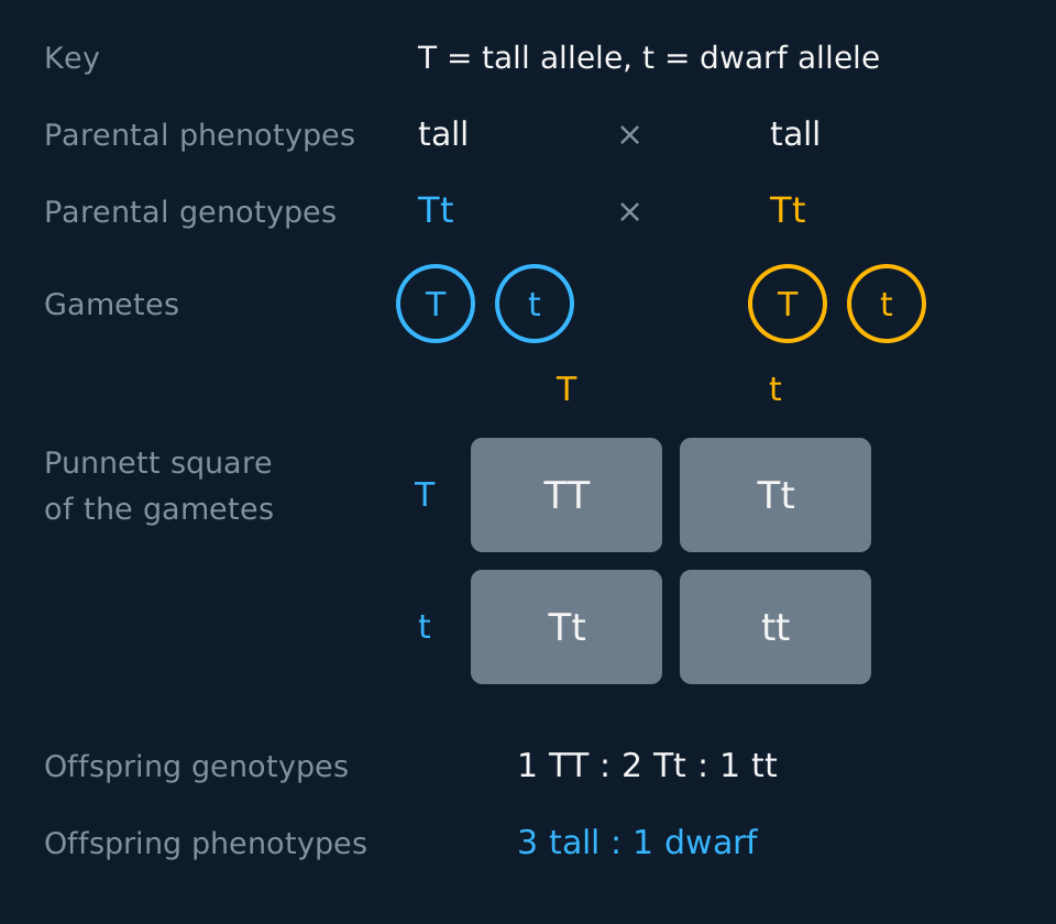
A complete genetic diagram defines the allele symbols and shows parental phenotypes and genotypes, gametes, offspring genotypes, and the expected phenotype ratio. A Punnett square displays the possible fertilisations. The ratio is a probability distribution and is approached most closely when many offspring are produced.
Describe the drawing
A monohybrid cross written out as a labelled column of stages. A key at the top defines capital T as the allele for tall and lower-case t as the allele for dwarf. The parental phenotypes are two tall plants; their genotypes are capital T lower-case t and capital T lower-case t; each parent's two gametes are drawn inside circles, carrying capital T or lower-case t. A two-by-two Punnett square below combines them, with one parent's gametes along the top and the other's down the side, and its four cells read capital T capital T, capital T lower-case t, capital T lower-case t and lower-case t lower-case t. The offspring genotype ratio one to two to one and phenotype ratio three tall to one dwarf are written underneath.
The order is: parental phenotypes, parental genotypes, gametes, offspring genotypes, offspring phenotypes and ratio. Setting the offspring out in a Punnett square rather than as crossed lines makes each fertilisation explicit. Read the ratio from the square rather than recalling it, since a remembered ratio belongs to a particular cross.
Three crosses cover almost everything at this level. Two heterozygotes give 1 TT : 2 Tt : 1 tt, and so 3 tall : 1 dwarf. A heterozygote crossed with a homozygous recessive gives 1 Tt : 1 tt, and so 1 : 1. A homozygous dominant crossed with anything gives all dominant offspring, because every offspring receives at least one dominant allele. The first of those separates the two ratios: 1 : 2 : 1 by genotype against 3 : 1 by phenotype.
The second cross has a name and a purpose. A test cross pairs an organism showing the dominant phenotype with a homozygous recessive, to find out whether the first one is homozygous or heterozygous. If any offspring shows the recessive phenotype, the unknown parent must have carried a recessive allele and is heterozygous. If a large number of offspring all show the dominant phenotype, the unknown parent is very probably homozygous, and 'very probably' is the accurate word, because a heterozygote could produce eight dominant offspring in a row by chance about once in every 256 attempts.
Working back from the offspring to the parents
Two pea plants, both tall, are crossed. Of 160 offspring, 118 are tall and 42 are dwarf. Deduce the genotypes of the parents and explain your reasoning.
Show the working
Dwarf offspring appear from two tall parents, so dwarf must be recessive and each parent must carry a dwarf allele. Both parents are tall, so neither can be homozygous dwarf. Both are therefore heterozygous, Tt.
Check against the numbers. Tt × Tt predicts 3 tall : 1 dwarf, which for 160 offspring is 120 tall and 40 dwarf. The observed 118 and 42 are close to that, and the small difference is what chance produces.
State the reasoning explicitly: a phenotype appears that neither parent shows, which establishes that the phenotype is recessive and that both parents are heterozygous.
Codominance, incomplete dominance and multiple alleles
Complete dominance is common but is not the only relation between two alleles. Two others occur. Where two alleles are codominant, both are expressed in the heterozygote and the phenotype shows both fully, side by side. Where dominance is incomplete, the heterozygote shows a blended, intermediate phenotype instead. In either case neither allele is subordinate, so they cannot be written as a capital and a lower-case letter; the convention is a capital letter for the gene with a raised letter for each allele, such as CR and CW.
Snapdragon flower colour is the standard example of incomplete dominance. CRCR plants are red, CWCW are white, and CRCW are pink, a blend rather than both colours at once. Roan cattle are the clean example of codominance: red hairs and white hairs sit side by side in the same coat, each fully expressed.
Cross two heterozygotes in either case and the offspring come out 1 : 2 : 1, because the heterozygote is distinguishable and the phenotype ratio is the genotype ratio. That means the ratio alone cannot tell the two relations apart; look at the heterozygote itself. A blend points to incomplete dominance, both phenotypes at once to codominance. Some textbooks file both cases under codominance, and if your board does, use its wording in the exam.
Human sickle-cell anaemia works the same way at the molecular level. The gene is HBB, which codes for the beta-globin polypeptide of haemoglobin, and someone with genotype HbAHbS makes both normal haemoglobin and sickle haemoglobin, so both alleles are expressed and the alleles are codominant. Whether the person is described as having the disease is a separate question from which alleles are expressed: heterozygotes express both polypeptides but do not usually show the clinical condition.
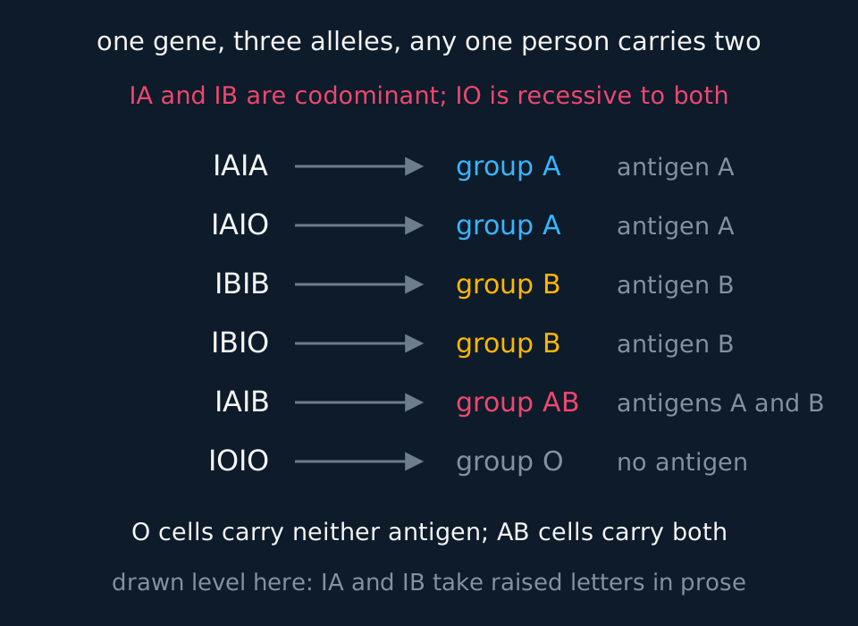
A gene can also have more than two alleles in the population, which is called having multiple alleles. Any one individual still carries only two, because there are only two copies of the locus, one on each chromosome of the homologous pair. The human ABO blood group system is the example every board uses, and it combines multiple alleles with codominance in one gene.
Describe the drawing
Six genotypes listed down the left, each with an arrow across to the blood group it produces and the antigen on the red cell. I A with I A and I A with I O both give group A carrying antigen A; I B with I B and I B with I O both give group B carrying antigen B; I A with I B gives group AB carrying both antigens; I O with I O gives group O with no antigen. A line above states that I A and I B are codominant and I O is recessive to both.
The three alleles are IA, IB and IO. IA codes for antigen A on the red cell surface and IB for antigen B; IO codes for neither. IA and IB are codominant with each other, and both are dominant to IO. Some textbooks and some boards write the third allele as a lower-case i rather than IO; the biology is identical, so use whichever notation your specification uses and define it at the top of your answer.
TRY IT: A question of parentage
A woman of blood group O has a child of blood group B. She names a man of blood group AB as the father. A second man, of blood group A, is also considered. Determine whether each man could be the father, showing your reasoning.
Check your answer
The mother is group O, so her genotype is IOIO and every egg she produces carries IO. The child is group B, so the child's genotype must be IBIO, and the IB must have come from the father.
The group AB man is IAIB. Half his sperm carry IB, so he could be the father.
The group A man is either IAIA or IAIO. Neither genotype contains IB, so he could only give the child IA or IO, producing a group A or group O child. He cannot be the father.
Blood groups can exclude a man but cannot identify one: many other men are also group AB. State that limitation as part of the conclusion.
Genes on the X chromosome
The sex chromosomes break the symmetry the rest of genetics relies on. In humans a female is XX and a male XY, and the Y is much smaller than the X and carries far fewer genes. A gene present on the X with no equivalent on the Y is sex-linked. A female carries two alleles of it and can be homozygous or heterozygous; a male carries one, and whichever allele he has is expressed, because there is no second copy to mask it.
That asymmetry accounts for the inheritance pattern. Recessive sex-linked conditions such as haemophilia A, red-green colour blindness and Duchenne muscular dystrophy are far more common in males, because a male needs only one recessive allele to show the condition while a female needs two. A female heterozygote is a carrier and shows nothing.
Haemophilia A is the one to know by its gene. The gene is F8, it sits on the X chromosome, and it codes for factor VIII, one of the proteins in the cascade that converts fibrinogen to fibrin and clots blood. The recessive allele produces a version of factor VIII that does not work, or too little of it, so the cascade stalls, clotting is slow and bleeding into joints and muscles follows minor injuries. A person carrying one working copy of F8 makes enough functional factor VIII to clot normally, which is exactly why the allele is recessive.
Describe the drawing
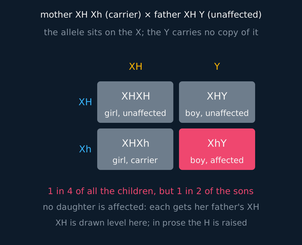
A two-by-two Punnett square for a sex-linked cross. The mother's two gametes, X carrying the dominant allele and X carrying the recessive allele, run down the side; the father's, X carrying the dominant allele and Y, run along the top. The four cells read: an unaffected girl, an unaffected boy, a carrier girl, and an affected boy, the last one filled in coral. Text beneath notes that this is one child in four but one son in two.
Write the allele as a raised letter on the X, as XH and Xh, and include the Y, so a male genotype reads XhY rather than a bare h. The X and Y are what show the gene to be sex-linked; without them the diagram does not distinguish sex linkage from autosomal inheritance.
Two rules follow from tracing the alleles through a family. A father passes his X to every daughter and never to a son, so an affected father cannot pass a sex-linked condition to his sons at all, and every one of his daughters is at least a carrier. A son gets his single X from his mother, so an affected son inherited the allele from his mother, whatever the father's phenotype. Those two sentences answer most pedigree questions on the topic before you draw anything.
Working a carrier cross
Haemophilia is caused by a recessive allele on the X chromosome. A woman whose father had haemophilia marries a man who does not have it. Calculate the probability that their first child is a boy with haemophilia, and the probability that a daughter is a carrier.
Show the working
The woman's father was XhY, and he gave his only X to every daughter, so she must have received Xh. She does not have haemophilia, so her genotype is XHXh. Her husband is unaffected, so he is XHY.
The four equally likely offspring are XHXH, XHXh, XHY and XhY. The probability of a boy with haemophilia is 1 in 4, or 0.25.
Of the two possible daughters, one is a carrier, so the probability that a daughter is a carrier is 1 in 2. The two answers differ because they are taken over different populations of children: all children in the first case, daughters only in the second.
The first step carries the deduction. The woman's genotype was not given; it follows from her father's phenotype and the rule that a father gives his X to every daughter.
Pedigree diagrams
Nobody can set up a test cross between two people, so human genetics works from the families that already exist. A pedigree diagram, or family tree, records one characteristic through several generations of a family, and it is the genetic diagram used where breeding experiments are impossible.
Questions on one ask for one of three things: whether the allele is dominant or recessive, whether the gene is on an autosome or on the X chromosome, and the genotype of one person or the probability for a child not yet born.
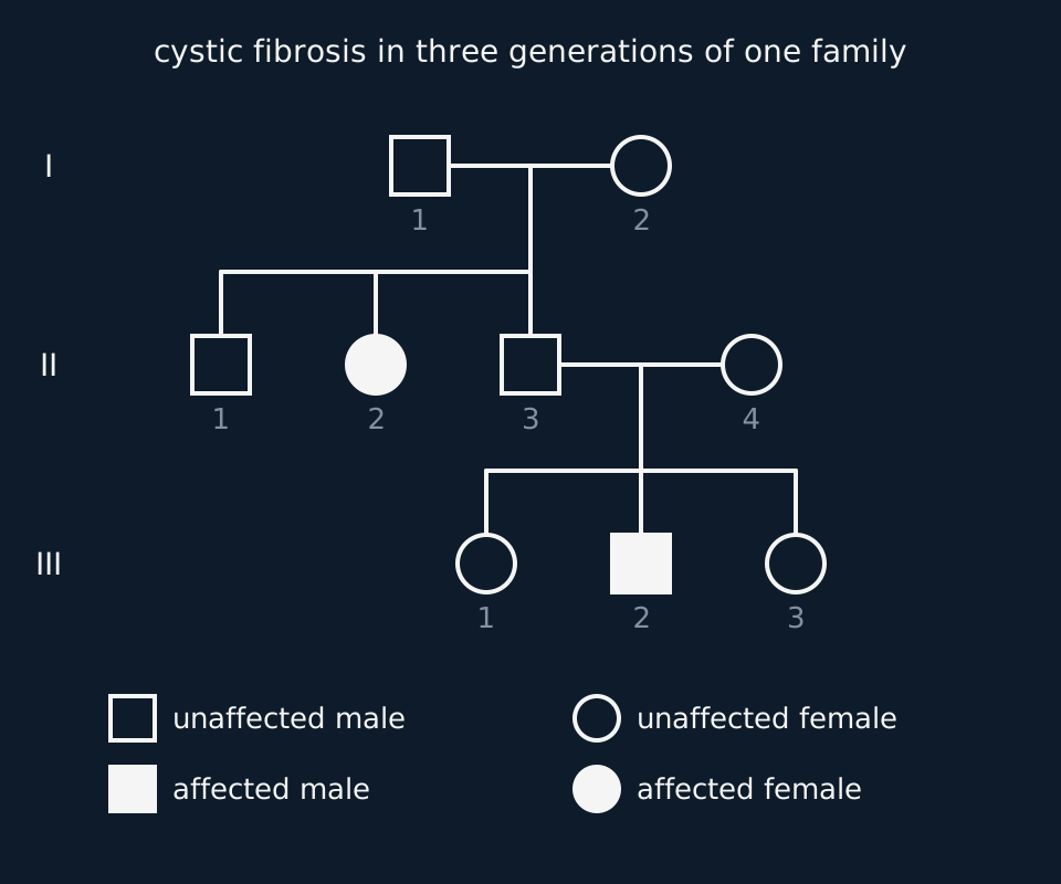
The symbols are standard. A square is a male and a circle a female. A shaded symbol is a person who shows the characteristic, described as affected, and an open symbol is a person who does not.
A horizontal line joining a square and a circle marks a couple who have had children together. A vertical line drops from the middle of it to a second horizontal line, and each child hangs from that line, eldest on the left.
Generations are numbered down the page in Roman numerals, and the people in each generation are numbered across it from left to right, so II-3 is the third person in the second generation. Someone who married into the family is drawn on the same row as their partner, with no line up to the generation above. Some diagrams mark a known carrier with a half-shaded symbol or a dot; an exam paper always prints a key, so read it before reading the tree.
Describe the drawing
A family tree in three rows, numbered I, II and III down the left. In generation I an unaffected man, 1, and an unaffected woman, 2, are joined by a horizontal line, and a vertical line drops from it to their three children in generation II: an unaffected son, 1, an affected daughter, 2, drawn as a filled circle, and an unaffected son, 3. Son 3 is joined to an unaffected woman, 4, who married into the family, and their three children in generation III are an unaffected daughter, 1, an affected son, 2, drawn as a filled square, and an unaffected daughter, 3. A key at the bottom shows squares as males, circles as females and filled symbols as people with cystic fibrosis.
To draw a pedigree from a written account, give each generation its own row, put each couple side by side with the line between them, hang the children in birth order, shade only the people the account says are affected, and add a key. Number the generations and the individuals so that every person in an answer can be named.
The deduction runs in a fixed order. The first question is dominant or recessive. Look for two unaffected parents with an affected child. The child received an allele for the characteristic from each parent, yet neither parent shows it, so the allele was hidden in both: it is recessive, and both parents are heterozygous.
The opposite pattern, two affected parents with an unaffected child, shows the allele is dominant, because each parent must have carried a recessive allele to pass on. If neither pattern appears in the tree, the question cannot be settled from it; say which is more likely and why.
The second question is autosomal or sex-linked, and it rests on the two rules from the section above: a father passes his X to every daughter and to no son, and a son's single X comes from his mother.
For a recessive allele, an affected daughter with an unaffected father rules out the X chromosome, since her father would have had to give her an X carrying the allele and would then have shown the condition himself. An affected mother with an unaffected son rules it out too, because both of her X chromosomes carry the allele and the son's only X came from her.
For a dominant allele, an affected father with an unaffected daughter rules out the X. A son who can only have received the allele from his father, such as the affected son of an affected father and an unaffected mother when the allele is dominant, shows the gene is autosomal, because a son receives his father's Y and not his X. With a recessive allele, an affected father and an affected son prove nothing about the X on their own, since the mother may be a carrier. A tree in which mostly males are affected suggests X-linked recessive inheritance but does not prove it.
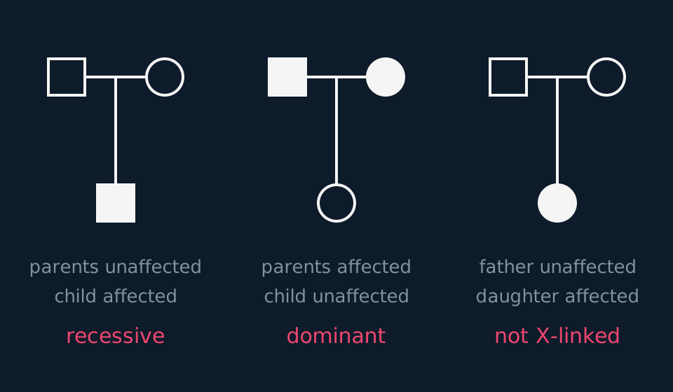
Describe the drawing
Three small family trees side by side, each a father and a mother joined by a line with one child below. Left: both parents unaffected and a son affected, labelled recessive. Middle: both parents affected, drawn filled, and a daughter unaffected, labelled dominant. Right: an unaffected father and mother with an affected daughter, labelled not X-linked, because a father passes his X to every daughter and an unaffected father has no recessive allele on it to give.
The third step is the genotypes, starting with the people whose genotype is fixed by what they show. For a recessive condition every affected person is homozygous recessive. An unaffected parent of an affected child must be heterozygous, and so must an unaffected child of an affected parent. The people left over could be either, and are written that way, as FF or Ff.
For the unaffected child of two carriers there is a number to put on it. Of the four equally likely outcomes of Ff × Ff, one is ff, and that one is ruled out because the child is unaffected. That leaves FF, Ff and fF, so the probability that the child is a carrier is 2/3, not 1/2.
Reading the cystic fibrosis pedigree
Use the pedigree in the figure above. Cystic fibrosis is caused by an allele of the CFTR gene. Deduce whether the allele is dominant or recessive and whether the gene is autosomal or on the X chromosome, give the genotypes of I-1, I-2, II-3 and II-4, and calculate the probability that II-1 is a carrier and the probability that the next child of II-3 and II-4 has cystic fibrosis.
Show the working
I-1 and I-2 are unaffected and their daughter II-2 is affected, so the allele is recessive. II-3 and II-4 with their affected son III-2 show the same thing.
II-2 is an affected daughter of an unaffected father. If the gene were on the X, I-1 would have given her an X carrying the allele and would have had cystic fibrosis himself. He does not, so the gene is autosomal.
Let F be the normal allele and f the cystic fibrosis allele. I-1 and I-2 are unaffected parents of an affected child, so both are Ff. II-3 and II-4 are unaffected parents of an affected child, so both are Ff as well. II-4 married into the family, so the f allele in III-2 came from two different sources.
II-1 is an unaffected child of Ff × Ff. Once ff is ruled out, two of the three remaining outcomes are Ff, so the probability that he is a carrier is 2/3.
II-3 and II-4 are both known to be Ff, so each of their children has a probability of 1/4 of being ff, whatever their earlier children were.
TRY IT: A dominant condition passed from father to son
Huntington's disease is caused by a dominant allele. A man with Huntington's disease and a woman without it have four children: an affected son, an unaffected son, an affected daughter and an unaffected daughter. Explain what the family shows about the man's genotype and about where the gene is carried, and give the probability that a further child would inherit the allele.
Check your answer
The man has unaffected children, so he must have passed a non-disease allele to them: he is heterozygous. The woman is unaffected, and with a dominant allele that means she is homozygous for the normal allele.
His affected son received his father's Y chromosome, not his X, yet inherited the disease allele from him. So the allele cannot be on the X chromosome, and the gene is autosomal. His unaffected daughter shows the same thing: had the dominant allele been on his X, every daughter would have received it.
Each child receives one of the man's two alleles at random, so the probability that a further child inherits the disease allele is 1/2. Earlier children do not change it.
- Pedigree diagram
- A family tree showing how one characteristic is inherited through several generations, with males as squares, females as circles and affected people shaded.
- Affected
- Showing the characteristic a pedigree follows; drawn as a shaded symbol.
- Autosomal
- Carried on an autosome, any chromosome other than the sex chromosomes, so inherited in the same way by sons and daughters.
Named genes and the proteins they code for
The crosses above use letters chosen for convenience. Cambridge International also requires the gene, the protein it codes for, and the effect of the altered allele on that protein.
A phenotype can result from one or many genes and may also be influenced by the environment. Where a named example is used, explain the relevant gene product and how the allele changes its amount or function; do not present every phenotype as a single protein that either works or fails.
Oculocutaneous albinism type 1 is one such example. The gene is TYR, and it codes for tyrosinase, the enzyme that catalyses the first step in converting the amino acid tyrosine into the pigment melanin. The recessive allele carries a mutation that gives a non-functional enzyme, so a person homozygous for it makes little or no melanin and has white or very pale hair, pale skin and pale irises. A heterozygote has one working copy of TYR, makes tyrosinase from it and is normally pigmented, which is why the allele is recessive: one functional copy of an enzyme gene is usually enough. Albinism as a whole is genetically heterogeneous, and other genes give other forms of it, so name the type rather than treating TYR as the single cause.
Sickle cell anaemia gives a different relation between the alleles. The gene is HBB, coding for the beta-globin polypeptide of haemoglobin. A single base substitution changes one codon, so valine appears at the sixth amino acid position where glutamic acid should be, and the resulting haemoglobin S sticks to itself when it gives up its oxygen, forming long fibres that pull the red cell out of shape. A heterozygote makes both polypeptides, because both alleles are transcribed and translated, so both are expressed and the alleles are codominant rather than one being masked.
Haemophilia A is set out above: the gene is F8 on the X chromosome, the protein is factor VIII, and a non-functional version slows the clotting cascade. Its position on the X, rather than the chemistry of the protein, is what gives it a different pattern of inheritance from the other three.
Huntington's disease differs from the others in the kind of mutation involved. The gene is HTT and the protein is huntingtin. The mutation is not a substitution but an expansion: a short sequence of three bases, CAG, is repeated far more times than usual, so the protein carries an abnormally long run of the amino acid it codes for. That altered protein damages neurones in the brain, and it does so whether or not a normal huntingtin is also present, which is why the allele is dominant. A single copy is enough, every child of an affected parent has a one in two chance of inheriting it, and the symptoms usually appear between the ages of thirty and fifty, so people often have children before they know. Dominant means only that the allele's effect appears in a heterozygote; it says nothing about how common the allele is.
| Gene | Protein | What the altered allele does | Inheritance |
|---|---|---|---|
| TYR | Tyrosinase | No working enzyme, so no melanin is made | Autosomal recessive |
| HBB | Beta-globin | Valine replaces glutamic acid, so haemoglobin S polymerises when deoxygenated | Autosomal; alleles codominant, disease only in HbSHbS |
| F8 | Factor VIII | The clotting cascade stalls, so clotting is slow | Sex-linked recessive |
| HTT | Huntingtin | An expanded CAG repeat gives a protein that damages neurones | Autosomal dominant |
| Le | A gibberellin-forming enzyme | The le allele gives no working enzyme, so little active gibberellin is made | Autosomal recessive |
The last row names the gene behind the standard pea example. The tall and dwarf phenotypes of the crosses above are controlled by the gene Le. The functional allele, Le, codes for an enzyme that carries out the last step in making an active gibberellin, the growth substance that lengthens the internodes of a stem. The allele le codes for a version of that enzyme which does not work, so a plant of genotype le le makes very little active gibberellin, its internodes stay short and it is a dwarf. A heterozygote, Le le, has one working copy, makes enough of the enzyme and grows tall, so tall is dominant for the same reason albinism is recessive.
A testable prediction follows. Supplying active gibberellin to a dwarf pea plant causes it to grow tall, because the plant lacks the product rather than the ability to respond to it. Supplying it to a tall plant has little effect, since the plant already makes sufficient gibberellin. Supplying tyrosine does not restore pigmentation in oculocutaneous albinism type 1, because tyrosine is the substrate of the missing enzyme: only a product beyond the blocked step could bypass it, as gibberellin does for the dwarf pea.
TRY IT: Explaining dominance from the protein
Explain, in terms of the proteins involved, why the oculocutaneous albinism type 1 allele of TYR is recessive while the Huntington's disease allele of HTT is dominant.
Check your answer
Start with albinism. TYR codes for tyrosinase, an enzyme, and enzymes are catalysts, so they are not used up and a small quantity converts a great deal of substrate. A heterozygote has one functional allele, transcribes and translates it, and makes enough tyrosinase to convert tyrosine to melanin at a normal rate. The altered allele contributes no functional enzyme, but it does not interfere with the product of the working copy, so the phenotype is normal and the allele is recessive.
Huntington's is a different kind of fault. The expanded CAG repeat does not merely fail to produce a working huntingtin; it produces an altered protein that actively damages neurones. The normal protein made from the other allele cannot undo that damage, so the presence of one faulty allele is enough to give the phenotype and the allele is dominant.
Dominance is therefore a property of what the protein does, not of the allele's importance or its frequency. A loss-of-function allele is usually recessive, because one working copy supplies enough product; an allele whose product is itself damaging is usually dominant, because the normal product does not compensate for it.
In the exam
- Define the allele symbols before using them, in one line stating which letter stands for which allele.
- Show the gametes as a separate line, circled. A genetic diagram is credited line by line, and the gamete line is one of them.
- Codominance and incomplete dominance both give three phenotypes and a 1 : 2 : 1 ratio from two heterozygotes. If a cross produces three phenotypes, stop calling either allele dominant, and tell the two apart from the heterozygote: side-by-side phenotypes mean codominance, a blend means incomplete dominance.
- Write sex-linked genotypes with the X and the Y included, as XhY rather than a bare h.
- Where a question names a gene, use the gene symbol and the protein: TYR and tyrosinase, HBB and beta-globin, F8 and factor VIII, HTT and huntingtin, Le and the enzyme that makes active gibberellin.
- Explain dominance through the protein. A loss-of-function allele is usually recessive because one working copy makes enough; an allele whose product does damage is dominant because the normal protein cannot undo it.
- Read whether the question asks for a proportion of all offspring or a proportion of sons. The two proportions differ by a factor of two, so state which denominator the answer uses.
- In a pedigree question, name the individuals that are the evidence: 'I-1 and I-2 are unaffected but their daughter II-2 is affected' rather than 'two unaffected parents have an affected child'.
- The unaffected child of two carriers is a carrier with probability 2/3, not 1/2, because the affected outcome has already been ruled out.
Check yourself
In cattle, coat colour is controlled by a single gene with two codominant alleles: CRCR cattle are red, CWCW are white, and CRCW are roan, a mixture of red and white hairs. A farmer crosses a roan bull with a roan cow and gets 24 calves. Set out the cross and state how many calves of each colour you would expect. The farmer then crosses the same bull with a white cow: state the expected ratio for that cross.
Answer
Roan × roan is CRCW × CRCW. Each parent produces gametes carrying CR or CW in equal numbers, so the offspring are 1 CRCR : 2 CRCW : 1 CWCW.
That is 1 red : 2 roan : 1 white, so of 24 calves you would expect 6 red, 12 roan and 6 white.
Roan × white is CRCW × CWCW. The white cow can only give CW, so the offspring are half CRCW and half CWCW: 1 roan : 1 white, with no red calves at all.
The roan coat itself shows the alleles to be codominant rather than incompletely dominant: it consists of red hairs and white hairs side by side, both alleles expressed, rather than a pink blend.
Questions
Question 15 marks
In tomatoes, red fruit is dominant to yellow fruit. Complete a full genetic diagram for a cross between a heterozygous red-fruited plant and a yellow-fruited plant, showing the parental genotypes, the gametes, the offspring genotypes and the expected phenotype ratio.
Mark scheme
- B1 the symbols are defined, for example R for the allele for red fruit and r for the allele for yellow fruit, using one letter for both alleles
- B1 parental genotypes given as Rr for the red-fruited plant and rr for the yellow-fruited plant
- B1 gametes shown as circled single alleles rather than pairs: R and r from the red-fruited parent, r and r from the yellow-fruited parent
- B1 offspring genotypes obtained from the gametes as Rr and rr, in equal numbers
- A1 offspring phenotypes and ratio given as 1 red-fruited : 1 yellow-fruited
Question 24 marks
A breeder has a tall pea plant and needs to know whether it is homozygous or heterozygous. Explain how a test cross would answer the question, and explain why a result suggesting the plant is homozygous can never be certain.
Mark scheme
- B1 cross the tall plant with a homozygous recessive dwarf plant, which can only contribute a recessive allele to every offspring
- B1 if any offspring is dwarf, the tall parent must have supplied a recessive allele, so it is heterozygous
- B1 if a large number of offspring are all tall, the tall parent is very probably homozygous, because a heterozygote would be expected to give a 1 : 1 ratio
- A1 each fertilisation is independent, so a heterozygote can give a run of dominant offspring by chance: eight in a row happens about once in every 256 attempts
Question 34 marks
Cystic fibrosis is caused by a recessive allele. Two parents are both carriers and neither has the condition. Calculate the probability that their first child has cystic fibrosis, and calculate the probability that their first two children both have it.
Mark scheme
- M1 both parents are heterozygous, so the cross is Ff × Ff and the offspring genotypes are 1 FF : 2 Ff : 1 ff
- A1 one of the four equally likely outcomes is homozygous recessive, so the probability is 1 in 4, or 0.25
- M1 the two fertilisations are independent events, so multiply the separate probabilities: 0.25 × 0.25
- A1 1 in 16, or 0.0625
Question 44 marks
Haemophilia is caused by a recessive allele carried on the X chromosome. Explain why the condition is far more common in males than in females, and explain why a man with haemophilia can never pass it to his sons.
Mark scheme
- B1 a male is XY, and the Y chromosome is much smaller and carries no equivalent of the gene
- B1 a male therefore carries only one allele of the gene and expresses it whichever it is, because there is no second copy to mask it, so one recessive allele is enough
- B1 a female is XX and needs two copies of the recessive allele to show the condition; with one she is an unaffected carrier, and inheriting two is much less likely
- A1 a father passes his X to every daughter and his Y to every son, so his sons receive no allele of the gene from him at all and every daughter is at least a carrier
Question 54 marks
A woman of blood group AB has children with a man of blood group O. Explain why none of their children can be blood group AB, and explain why none of them can be blood group O.
Mark scheme
- B1 the woman's genotype is IAIB, so every egg carries either IA or IB and never both
- B1 the man's genotype is IOIO, so every sperm carries IO
- B1 every child is therefore IAIO or IBIO, giving blood group A or blood group B, because IA and IB are each dominant to IO
- A1 group AB would need IA and IB together, which the father cannot supply, and group O would need two copies of IO, which the mother cannot supply
Question 64 marks
Explain why the albinism allele of TYR is recessive while the Huntington's disease allele of HTT is dominant, referring to the proteins the two genes code for.
Mark scheme
- B1 TYR codes for the enzyme tyrosinase, and an enzyme is a catalyst that is not used up, so a small quantity converts a great deal of tyrosine into melanin
- B1 a heterozygote transcribes and translates its one functional allele and makes enough tyrosinase for normal pigmentation, and the faulty allele contributes nothing but does no harm, so it is recessive
- B1 the expanded HTT allele produces an altered huntingtin that actively damages neurones rather than simply failing to work
- A1 the normal huntingtin made from the other allele cannot undo that damage, so one copy of the allele is enough to give the phenotype and it is dominant
Question 74 marks
A woman of blood group A whose mother was group O has children with a man of blood group B whose father was group O. Complete the two empty cells of the Punnett square, and give the blood groups of the children with the ratio in which they are expected.
Mark scheme
- M1 the two empty cells are AO and OO, taking one allele from the gamete above the column and one from the gamete beside the row
- B1 AB gives blood group AB, because A and B are codominant and both are expressed
- B1 BO gives group B and AO gives group A, because O is recessive to both
- A1 OO gives group O, so the four groups AB, B, A and O are expected in a 1 : 1 : 1 : 1 ratio
Question 84 marks
Red-green colour blindness is caused by a recessive allele on the X chromosome. A colour-blind man has children with a woman who is homozygous for normal colour vision. Calculate the probability that a daughter of this couple is colour-blind, and calculate the probability that a son is colour-blind.
Mark scheme
- M1 the man is XbY and the woman is XBXB, so daughters receive Xb from their father and XB from their mother, and sons receive XB from their mother and Y from their father
- A1 every daughter is XBXb, a carrier, so the probability that a daughter is colour-blind is 0
- M1 every son is XBY, because a son never receives his father's X chromosome
- A1 the probability that a son is colour-blind is also 0, since no son can inherit the father's Xb
Question 94 marks
The pedigree shows the inheritance of a condition in two families. Explain, using evidence from the pedigree, whether the allele causing the condition is dominant or recessive, and whether the gene is on an autosome or on the X chromosome.
Mark scheme
- B1 the allele is recessive
- B1 I-1 and I-2 are unaffected but their daughter II-1 is affected (or I-3 and I-4 with II-4), so both parents carried the allele without showing it
- B1 the gene is on an autosome
- B1 II-1 is an affected daughter of an unaffected father, I-1; a father gives his X to every daughter, so if the gene were on the X he would have given her the allele and would be affected himself
Question 104 marks
The condition in the pedigree is caused by a recessive allele of an autosomal gene. II-2 and II-3 plan to have a child. Calculate the probability that the child will have the condition.
Mark scheme
- M1 I-1 and I-2 are both heterozygous, because their daughter II-1 is affected, so the unaffected II-2 is a carrier with probability 2/3 (ff is ruled out, leaving FF, Ff and fF)
- M1 I-3 and I-4 are both heterozygous, because their son II-4 is affected, so II-3 is also a carrier with probability 2/3
- M1 the child is affected only if both parents are carriers and it receives the recessive allele from each: 2/3 × 2/3 × 1/4
- A1 1/9, or 0.11
Question 114 marks
The pedigree shows the inheritance of a condition through three generations. Explain, using evidence from the pedigree, why the condition is caused by a dominant allele of a gene on an autosome, and explain what the genotypes of II-3 and II-4 must be.
Mark scheme
- B1 II-3 and II-4 are both affected but their daughter III-1 is unaffected, so each parent carried an allele that does not cause the condition and was masked: the allele causing it is dominant
- B1 I-1 is an affected father with an unaffected daughter, II-2, or I-1 and the unaffected I-2 have an affected son, II-1
- B1 a father gives his X to every daughter and to no son, so a dominant allele on his X would have made II-2 affected, or could not have reached II-1 from him; the gene is autosomal
- B1 II-3 and II-4 are both heterozygous, because each must have given III-1 the allele that does not cause the condition
Question 124 marks
The condition in the pedigree is caused by a recessive allele on the X chromosome. Identify the two females in the pedigree who must be carriers, giving your reasons, and calculate the probability that III-2 is a carrier.
Mark scheme
- B1 I-2 is a carrier: her son II-1 is affected, a son's single X comes from his mother, and she is unaffected so she is heterozygous
- B1 II-3 is a carrier: her son III-1 is affected and she is unaffected, for the same reason
- M1 with XA as the normal allele, II-3 is XAXa and II-4 is XAY; every daughter receives XA from her father and either X from her mother
- A1 the probability that III-2 is a carrier is 1/2
Question 133 marks
Name the protein coded for by each of the genes TYR, F8 and HTT, and state the phenotype associated with a faulty allele of each.
Mark scheme
- B1 TYR codes for tyrosinase, and an allele giving no working enzyme means no melanin is made, which is albinism
- B1 F8 codes for factor VIII, and a faulty allele stalls the clotting cascade, which is haemophilia A
- B1 HTT codes for huntingtin, and an allele with an expanded CAG repeat gives a protein that damages neurones, which is Huntington's disease
Question 143 marks
In peas, the allele Le codes for a working enzyme that makes an active gibberellin and the allele le codes for a version that does not work. Suggest why plants of genotype le le are dwarf, and suggest what would happen if active gibberellin were applied to such a plant.
Mark scheme
- B1 a le le plant has no working copy of the enzyme, so very little active gibberellin is made
- B1 gibberellin lengthens the internodes of a stem by promoting cell division and cell elongation between the nodes, so without it the internodes stay short and the plant is dwarf
- A1 applying active gibberellin supplies the product the plant could not make, so the internodes lengthen and the plant grows tall; a Le plant treated the same way changes little, because it already has enough
Question 153 marks
In codominance, both alleles are fully expressed in the heterozygote, so the products of both appear side by side. In incomplete dominance, the heterozygote has a phenotype intermediate between those of the two homozygotes. Roan cattle, from a cross between red and white parents, have a coat of red hairs mixed with white hairs. Pink snapdragons come from a cross between red-flowered and white-flowered plants. Explain which relationship each example shows, and explain why a cross between two heterozygotes gives a 1 : 2 : 1 phenotype ratio in both cases.
Mark scheme
- B1 roan cattle show codominance, since each hair is fully red or fully white, while pink snapdragons show incomplete dominance, since the colour is intermediate; both are needed for the mark
- B1 each heterozygous parent passes either allele to half its gametes and gametes fuse at random, so the offspring genotypes are 1 homozygous : 2 heterozygous : 1 homozygous for the other allele
- B1 in both relationships the heterozygote looks different from either homozygote, so each genotype has its own phenotype and the genotype ratio appears unchanged as the phenotype ratio
Question 163 marks
A person with genotype HbAHbS makes both normal haemoglobin and sickle haemoglobin. Explain, in terms of transcription and translation, why both alleles are expressed in this heterozygote, and explain why the alleles are described as codominant rather than one being dominant and the other recessive.
Mark scheme
- B1 both alleles of the HBB gene are present in the nucleus of every red-cell precursor and both are transcribed into mRNA and translated into their respective beta-globin polypeptides, since neither allele is switched off by the presence of the other
- B1 the result is that the heterozygote's red cells contain both normal haemoglobin and sickle haemoglobin, made from the two different alleles at once
- B1 this is codominance rather than complete dominance because both phenotypes, both proteins, are simultaneously present and detectable in the heterozygote, rather than one allele's effect being completely masked by the other
Question 173 marks
A student writes a genetic diagram for a monohybrid cross using the symbols S for the dominant allele and s for the recessive allele, without stating what either allele stands for. Suggest two reasons why this is poor practice, even though S and s are legitimate genetic symbols.
Mark scheme
- B1 without a stated definition, a reader cannot know what characteristic S and s refer to, so the diagram cannot be understood or marked on its own
- B1 S and s are hard to tell apart in handwriting, unlike letters such as T and t whose capital and lower-case forms look clearly different, so the diagram risks being misread even once the meaning is known
- B1 defining the symbols in a stated line, such as 'let S represent...', is treated as part of a complete genetic diagram, so omitting it loses a mark
Question 183 marks
State what each of the following shows in a pedigree diagram: an open circle, a shaded square, and the label III-2.
Mark scheme
- B1 an open circle is a female who does not show the characteristic, an unaffected female
- B1 a shaded square is a male who shows the characteristic, an affected male
- B1 III-2 is the second person from the left in the third generation
Question 193 marks
A student says that the pedigree proves the allele is on the X chromosome, because only males are affected. Suggest why the pedigree on its own does not prove this.
Mark scheme
- B1 the pedigree is also consistent with an autosomal recessive allele: I-1 and I-2 could both be heterozygous, and so could II-3 and II-4
- B1 the autosomal explanation needs II-4, who married into the family, to be a carrier as well; that is less likely for a rare allele but not impossible, so the pedigree makes X-linkage more probable without proving it
- B1 only two people are affected, so both being male could be chance; with an autosomal allele each affected child is male with probability 1/2, so both are male with probability 1/4
Question 202 marks
State what is meant by the term allele, and state what is meant by describing someone as a carrier of a recessive condition.
Mark scheme
- B1 an allele is one of the alternative versions of a gene, found at the same locus on a chromosome
- B1 a carrier is an individual heterozygous for a recessive allele, so they do not show the condition but can pass the allele on
Question 212 marks
State the difference between a gene and an allele, and state what is meant by the locus of a gene.
Mark scheme
- B1 a gene is a length of DNA coding for a polypeptide or a functional RNA, whereas an allele is one of the alternative versions of that gene
- B1 the locus is the fixed position on a chromosome at which a particular gene is found, the same position on both members of a homologous pair
Worth remembering
- Parental phenotypes, parental genotypes, gametes, offspring, ratio: in that order, every time.
- 3 : 1 from two heterozygotes, 1 : 1 from a test cross, all dominant from a homozygous dominant parent.
- Codominance and incomplete dominance both give 1 : 2 : 1 and three phenotypes; the genotype ratio and the phenotype ratio are the same.
- Three alleles in a population, two in any one individual: IA and IB codominant, both dominant to IO.
- A male has one allele of a sex-linked gene and expresses it; a father gives his X to every daughter and to no son.
- Name the gene, name the protein, then say what the faulty allele does to it: TYR, HBB, F8, HTT, and Le for tall and dwarf peas.
- In a pedigree, unaffected parents with an affected child mean recessive; affected parents with an unaffected child mean dominant.
- An affected daughter of an unaffected father rules out X-linked recessive inheritance; a son who can only have had the allele from his father rules out the X altogether.
CHECK YOUR PROGRESS
Rate how confident you are with each objective for this lesson. Ratings are saved in this browser, on this device, unless you sign in.
- Use the terms gene, allele, locus, genotype and phenotype with their standard meanings.
- Set out a complete monohybrid genetic diagram, from parental phenotypes to the offspring ratio.
- Predict and interpret ratios for complete dominance, codominance and multiple alleles.
- Work out blood group possibilities from parental genotypes and the reverse.
- Handle a sex-linked cross, including why sons and daughters are affected at different rates.
- Name the gene and the protein behind albinism, sickle cell anaemia, haemophilia A, Huntington's disease and stem length in peas.
- Explain why an allele is dominant or recessive from what its protein does.
- Read and draw a pedigree diagram, and deduce from it whether an allele is dominant or recessive and whether its gene is autosomal or on the X chromosome.
Open the full revision checklist to see every objective in the curriculum in one place.
Practise this lesson
WORKBOOK
The written questions from this topic, on paper with room to work, and a separate book of mark schemes. The 6 questions that are set on a diagram stay on this page, where the diagram can be drawn. Free to use; please do not redistribute or sell.
Used later in Population genetics and Hardy-Weinberg equilibrium · Genome analysis, genetic screening and gene therapy · Continuous and discontinuous variation · Artificial selection and selective breeding