Biology › Hormonal communication, plant responses and homeostasis › The liver: detoxification, deamination and the making of urea
The liver: detoxification, deamination and the making of urea
Excretion is the removal of the waste products of metabolism, principally carbon dioxide from respiration and nitrogen from amino acids that cannot be stored. The liver receives blood from two vessels, detoxifies absorbed substances, deaminates surplus amino acids and converts the resulting ammonia to urea.
Before this Excretion of urea at the kidney, and how urine is made · Deamination met in passing as a source of respiratory substrate · The hepatic portal vein and absorption from the ileum
COMMON MISCONCEPTION
Urea is made in the kidney, which is why it leaves the body in the urine.
The liver makes urea and the kidneys excrete it. Hepatocytes remove amino groups from surplus amino acids. In the urea cycle, one nitrogen enters as ammonia, the second comes from aspartate, and the carbon comes from bicarbonate. Urea then travels in the blood to the kidneys.
What you should be able to do
- Define excretion, name the two waste products it mainly deals with, and say which organ removes each.
- Name the liver's three blood vessels and state what the blood in each of them is carrying.
- Describe a liver lobule, and relate sinusoids, hepatocytes and Kupffer cells to what the organ does.
- Explain how the liver detoxifies a substance, using the breakdown of alcohol through ethanal as the worked case.
- Describe deamination and the ornithine cycle in order, saying where in the cell each step happens.
- Explain why an animal spends ATP turning ammonia into urea rather than excreting the ammonia itself.
Excretion, and the two things it is mostly about
Excretion is the removal from the body of the waste products of metabolism: substances made by the body's own reactions that would do harm if they accumulated. The definition excludes most of the material that leaves in the faeces, which is undigested food, bacteria and shed gut lining; passing it out is egestion. Faeces are therefore not an excretory product in that sense.
Two waste products dominate, and both come out of reactions you have already studied. Carbon dioxide is made continuously by respiring cells, is carried to the lungs mostly as hydrogencarbonate ions, and is excreted at the alveoli. Left in the blood it forms carbonic acid and the pH falls, which changes the charge on the R groups of every protein in the body, so a rise in carbon dioxide is dangerous long before it is a shortage of anything.
Nitrogenous waste is the second. A meal containing more protein than the body needs cannot simply be stored: there is no store for amino acids in the way there is glycogen for glucose and fat for lipid. The surplus is broken up instead, the nitrogen-containing part is stripped off in the liver, and it leaves the body as urea in the urine. Urea is made in the liver and excreted by the kidney.
A third excretory pathway also runs through the liver. Old red blood cells are broken down and the haem group is converted into bile pigments, which leave in the bile, pass into the gut and colour the faeces. Faeces therefore contain some body-derived material, including bile pigments and shed cells, as well as undigested food.
- Excretion
- The removal from the body of the waste products of metabolism, such as carbon dioxide and urea.
- Egestion
- The removal of undigested food and other material that has never taken part in the body's metabolism.
- Deamination
- The removal of the amino group from an amino acid, producing ammonia and a keto acid.
- Detoxification
- The conversion by the liver of a toxic substance into one that is less toxic and can be excreted.
An organ with two blood supplies
Everything the liver does depends on what arrives in its blood, and it is one of very few organs fed by two vessels rather than one. Between them they deliver roughly a quarter of everything the heart pumps while you are resting.
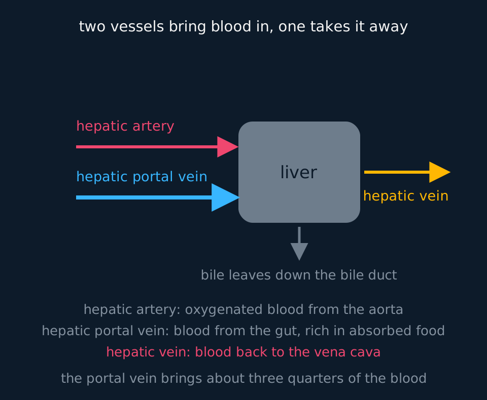
The hepatic artery branches from the aorta and brings oxygenated blood, which the liver needs as much as any tissue: its cells respire hard and it is one of the warmest organs in the body. The hepatic portal vein is the unusual one. It carries blood that has already passed through the capillaries of the stomach, small intestine and large intestine, so it is deoxygenated but loaded with everything that has just been absorbed: glucose, amino acids, mineral ions, and any drug, alcohol or toxin that came in with the meal. About three quarters of the liver's blood arrives this way.
One consequence follows directly. Every molecule absorbed from the gut reaches the liver before it reaches anywhere else, so the liver sets the concentration the rest of the body sees. That is why blood glucose is regulated there, and why a drug swallowed rather than injected may be largely broken down before it has been anywhere near the tissue it was meant for.
The hepatic vein takes blood away, back to the inferior vena cava and so to the heart. And one further vessel leaves the liver carrying something that is not blood: the bile duct, which takes bile to the gall bladder and from there to the duodenum.
The lobule, and the cells inside it
Cut the liver and it looks uniform, but under a microscope it is built from a repeated unit a millimetre or so across called a lobule. Drawn in section a lobule is roughly hexagonal, with a vein running down its axis and blood vessels at its corners.
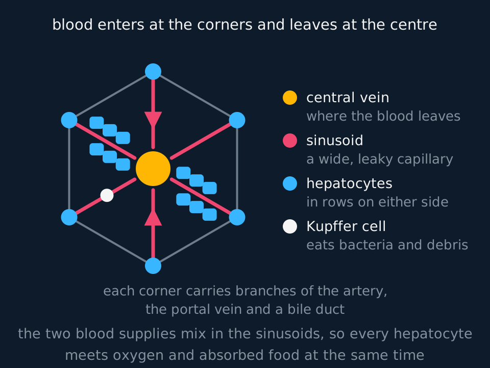
At each corner of the hexagon sit branches of the hepatic artery and of the hepatic portal vein, together with a bile ductule. Blood from both vessels runs from the corners towards the middle along wide, leaky capillaries called sinusoids, mixing as it goes, so oxygen and absorbed food reach the same cells at the same time. What is left drains into the central vein running down the middle of the lobule, and the central veins join to form the hepatic vein.
The cells packed either side of every sinusoid are the hepatocytes, and they do nearly all of the work. They are cubical with a large nucleus, and their specialisation lies in their contents rather than their shape: a great deal of smooth endoplasmic reticulum, where detoxification happens, abundant rough endoplasmic reticulum and Golgi for the plasma proteins they secrete, dense mitochondria for the ATP all of it consumes, and granules of stored glycogen. Their surface facing the sinusoid carries microvilli, so exchange with the blood is rapid.
Scattered along the sinusoids are Kupffer cells, which are macrophages fixed in place. They engulf bacteria arriving from the gut and break down worn-out red blood cells, and it is their handling of haem that produces the bile pigments. Bile itself is secreted by the hepatocytes into fine channels called bile canaliculi that run between them, out to the bile ductules at the corners of the lobule. The two flows run in opposite directions: blood inwards to the central vein, bile outwards to the corners.
- Lobule
- The repeating structural unit of the liver: a roughly hexagonal block of hepatocytes with a central vein at its axis.
- Sinusoid
- A wide, leaky capillary running from the edge of a lobule to its central vein, carrying mixed blood from the hepatic artery and hepatic portal vein.
- Hepatocyte
- A liver cell, carrying out detoxification, deamination, storage and the secretion of bile.
- Kupffer cell
- A macrophage fixed in a sinusoid, which engulfs bacteria and breaks down old red blood cells.
Detoxification, and what alcohol does to a liver
A toxin is a molecule the body cannot use and cannot afford to leave circulating. The liver's answer is nearly always the same: change it chemically, usually by oxidising it, until it is less harmful and soluble enough for the kidney to excrete. Enzymes in the smooth endoplasmic reticulum of hepatocytes, the cytochrome P450 group among them, handle most drugs and many hormones this way; the liver breaks down insulin, oestrogen and testosterone once they have done their work, which is part of why hormonal signals fade. Catalase in the same cells decomposes the hydrogen peroxide that metabolism keeps producing, and hepatocytes carry more of it than almost any other cell.
Alcohol is the standard worked example, and is a two-step oxidation. Ethanol is oxidised by ethanol dehydrogenase to ethanal, and ethanal is oxidised in turn by ethanal dehydrogenase to ethanoate, which is converted into acetyl coenzyme A and fed into respiration. Both steps hand hydrogen to NAD, so both produce reduced NAD.
The accumulation of reduced NAD is what causes the damage. A liver dealing with a great deal of alcohol accumulates reduced NAD, and a cell with plenty of reduced NAD has no need to oxidise fatty acids: it synthesises them instead. Fat therefore builds up in the hepatocytes, which is fatty liver, and it is reversible if the drinking stops. Sustained over years, the cells are damaged and die, fibrous scar tissue replaces them, and the organ becomes hard and shrunken with far fewer working cells. That is cirrhosis, and it is not reversible, because although the liver regenerates lost tissue remarkably well, it cannot replace tissue that has been turned into scar.
Why a drug label warns about liver disease
A drug is broken down entirely by enzymes in the smooth endoplasmic reticulum of hepatocytes. Its label warns that people with cirrhosis should take a smaller dose. Explain why.
Show the working
Start with what the liver is doing to the drug. Detoxification converts it into a form that can be excreted, so the concentration of the active drug in the blood falls at a rate set by how much of that enzyme is available.
In cirrhosis, working hepatocytes have been replaced by fibrous scar tissue, so there are fewer cells and less enzyme. The same dose is therefore broken down more slowly.
The drug consequently stays in the blood longer and reaches a higher concentration, especially if doses are repeated, so a normal dose can become an overdose. The general principle is that a dose is a statement about a rate of removal as much as about a quantity given.
Deamination and the ornithine cycle
Amino acids surplus to requirements are broken down in the hepatocytes, and the first step is deamination: the amino group is removed and leaves as ammonia, while the rest of the molecule becomes a keto acid. The keto acid is useful and goes nowhere unpleasant: it is respired for ATP, or converted into glucose or into fat for storage. It is the ammonia that is the problem.
Ammonia is small, extremely soluble and very toxic. It raises the pH of solutions it enters and interferes with the reactions of respiration in the brain, and a concentration high enough to be dangerous is a very low one. An animal living in fresh water can simply let it diffuse out across the gills, because there is unlimited water outside to dilute it. A land animal cannot excrete ammonia this way.
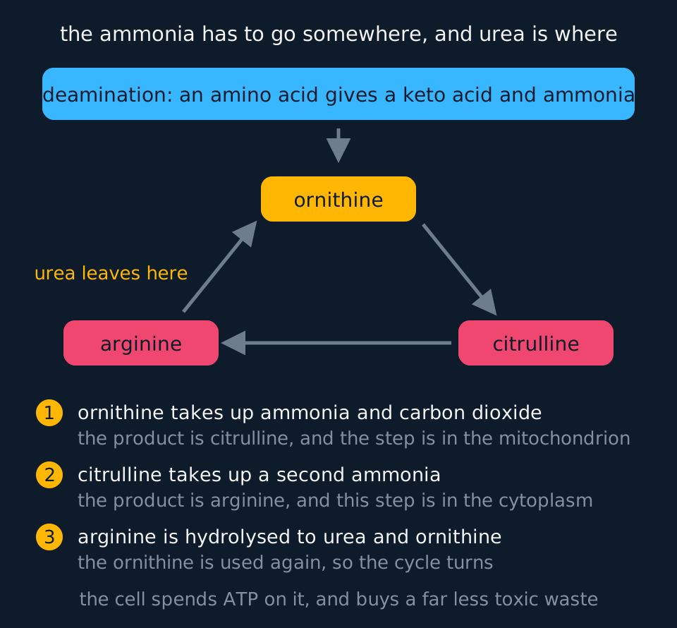
The ornithine cycle, also called the urea cycle, runs in the hepatocytes. State where each step takes place as well as what joins and leaves.
In the first step, inside a mitochondrion, ammonia combines with bicarbonate, derived from carbon dioxide, and the product joins ornithine to give citrulline and water. In the second, in the cytoplasm, citrulline acquires the second nitrogen from aspartate and becomes arginine. In the third, also in the cytoplasm, the arginine is hydrolysed to urea and ornithine. The ornithine returns to the start of the sequence and is used again, which is what makes it a cycle, and the urea diffuses into the blood.
Overall, one nitrogen enters as ammonia, the second comes from aspartate, and the carbon comes from bicarbonate, giving one molecule of urea per turn at a cost in ATP. Several specifications simplify this to two molecules of ammonia and one of carbon dioxide; use the form your specification states, and keep the source of each atom consistent within an answer.
| Step | Where | What joins or leaves |
|---|---|---|
| Ornithine to citrulline | Mitochondrion | Ammonia and bicarbonate supply the first nitrogen and the carbon; water is released |
| Citrulline to arginine | Cytoplasm | Aspartate supplies the second nitrogen |
| Arginine to ornithine | Cytoplasm | Water joins; urea leaves and the ornithine is reused |
The remaining question is why the ATP is spent at all, when ammonia could be excreted directly.
Because ammonia would have to be excreted in enormous volumes of water. To keep the concentration in the blood below the level that damages the nervous system, a mammal would need to produce urine at a rate no land animal could drink to replace. Urea is far less toxic and still soluble, so it can be carried in the blood at concentrations tens of times higher and concentrated further in the urine. The ATP is spent in order to conserve water. Freshwater fish excrete ammonia directly across the gills at no metabolic cost; birds, reptiles and insects convert nitrogen instead to uric acid, which is almost insoluble and can be excreted as a paste, which is what makes a shelled egg possible in the first place, since a developing embryo sealed in a shell cannot afford either water loss or a soluble poison.
TRY IT: Reading a liver failure case
A patient with advanced liver disease has a blood urea concentration well below normal and a blood ammonia concentration well above it. They are confused and drowsy. Explain all three observations, and say why measuring urea alone could give a misleading impression of kidney function.
Check your answer
Urea is made in the liver, not in the kidney. With most of the hepatocytes destroyed there are too few working ornithine cycles, so less urea is produced and its concentration in the blood falls. A low blood urea in liver disease reflects reduced production, not unusually efficient excretion by the kidney.
The ammonia rises for the same reason. Deamination of surplus amino acids continues in whatever liver tissue is left, and bacteria in the gut release ammonia that arrives along the hepatic portal vein, but the cycle that would convert it is not there in sufficient quantity, so ammonia accumulates in the blood.
The confusion and drowsiness follow from the ammonia. It is toxic to nervous tissue, altering pH and interfering with the reactions that supply neurones with ATP, so brain function is disturbed before any other tissue shows much sign of it.
Blood urea is used to judge renal excretion on the assumption that the rate of production is steady. Here production has fallen, so a normal or low value gives no information about kidney function, and the ammonia measurement is the informative one.
In the exam
- Excretion is the removal of the waste products of metabolism. Faeces are egested rather than excreted, apart from the bile pigments and shed cells they contain.
- The liver makes urea and the kidney excretes it. Urea is not made in the kidney.
- Name the vessels precisely. Hepatic artery from the aorta with oxygen, hepatic portal vein from the gut with absorbed food, hepatic vein leaving for the vena cava.
- For the ornithine cycle, give the three molecules in order and state what joins at each step: ornithine, citrulline, arginine and back to ornithine, with the first step in the mitochondrion.
- The reason for making urea is that ammonia is too toxic and too soluble to be carried or excreted without large volumes of water. State what the ATP achieves as well as that it is used.
- In alcohol questions, the intermediate is ethanal, and the damage comes from the reduced NAD that accumulates, so fatty acids are synthesised rather than respired.
Check yourself
A person eats a meal containing far more protein than their body needs. Describe what happens to the surplus amino acids, naming the organ and the processes involved, and explain why the nitrogen leaves the body as urea rather than as ammonia. Then state which blood vessel carried the amino acids to that organ.
Answer
The surplus cannot be stored, because there is no store for amino acids as there is glycogen for glucose. It is broken down in the hepatocytes of the liver, beginning with deamination: the amino group is removed and leaves as ammonia, and the remainder of the molecule is a keto acid, which is respired or converted into glucose or fat.
The ammonia enters the ornithine cycle in the same cells. It combines with bicarbonate and joins ornithine to form citrulline in a mitochondrion; the citrulline acquires the second nitrogen from aspartate in the cytoplasm to become arginine; and the arginine is hydrolysed to urea and ornithine, so the ornithine is available again. One nitrogen enters as ammonia, the second comes from aspartate and the carbon comes from bicarbonate, giving one molecule of urea per turn at a cost in ATP.
Ammonia is why the expense is worth it. It is very soluble and very toxic, particularly to nervous tissue, so the concentration the blood could safely carry is minute and excreting it would need volumes of water a land animal cannot replace. Urea is much less toxic and still soluble, so it can be carried at a far higher concentration and concentrated again in the urine at the kidney.
The amino acids reached the liver along the hepatic portal vein, which carries blood from the capillaries of the gut straight to the liver, so everything absorbed from a meal passes through the liver before it reaches the rest of the body.
Questions
Question 15 marks
Describe what happens to an amino acid that the body does not need, from deamination through to the urea leaving the liver, naming where each step of the ornithine cycle takes place.
Mark scheme
- B1 deamination in a hepatocyte removes the amino group, which leaves as ammonia, and the remainder of the molecule is a keto acid that is respired or converted to glucose or fat
- B1 in the mitochondrion, the ammonia combines with carbon dioxide and joins ornithine to form citrulline
- B1 in the cytoplasm, the citrulline takes up a second ammonia and becomes arginine
- B1 also in the cytoplasm, the arginine is hydrolysed to urea and ornithine, so the ornithine is available to start the cycle again
- A1 overall two molecules of ammonia and one of carbon dioxide give one molecule of urea, which diffuses into the blood and is excreted by the kidney
Question 24 marks
Describe the structure of a liver lobule, and describe the direction in which blood and bile each travel through it.
Mark scheme
- B1 a lobule is a roughly hexagonal block of hepatocytes with a central vein running down its axis
- B1 at each corner sit branches of the hepatic artery and of the hepatic portal vein, together with a bile ductule
- B1 blood from both vessels flows inwards from the corners along wide leaky capillaries called sinusoids, mixing as it goes, and drains into the central vein
- A1 bile is secreted by the hepatocytes into bile canaliculi running between them and travels outwards to the ductules at the corners, so blood and bile move in opposite directions
Question 34 marks
Explain how the liver deals with the alcohol in a drink, and explain why heavy drinking over many years leads to fat accumulating in the liver.
Mark scheme
- B1 ethanol is oxidised by ethanol dehydrogenase to ethanal
- B1 ethanal is oxidised by ethanal dehydrogenase to ethanoate, which is converted to acetyl coenzyme A and fed into respiration
- B1 both steps pass hydrogen to NAD, so reduced NAD accumulates in the hepatocytes
- A1 a cell with plenty of reduced NAD synthesises fatty acids rather than oxidising them, so fat builds up in the hepatocytes, and sustained damage leads on to cirrhosis as scar tissue replaces the cells
Question 44 marks
Describe the role of Kupffer cells in the liver, and describe what happens to the haem group of an old red blood cell that they break down.
Mark scheme
- B1 Kupffer cells are macrophages fixed in place along the sinusoids of the liver
- B1 they engulf bacteria arriving from the gut in the blood, and they break down worn-out red blood cells
- B1 the haem group from those red blood cells is converted into bile pigments
- A1 the bile pigments leave the liver in the bile, pass into the gut and colour the faeces, which is why faeces contain some body-derived material as well as undigested food
Question 54 marks
Explain why a drug taken as a tablet by mouth may reach the rest of the body at a much lower concentration than the same dose given by injection.
Mark scheme
- B1 a swallowed drug is absorbed from the gut into the blood of the hepatic portal vein
- B1 the hepatic portal vein carries that blood, and everything dissolved in it, directly to the liver before it reaches any other tissue
- B1 hepatocytes detoxify a wide range of substances using enzymes such as those of the cytochrome P450 group, so a significant fraction of the drug can be broken down on this first pass through the liver
- A1 an injected drug enters the general circulation directly and is diluted throughout the whole blood volume before any of it reaches the liver, so a much higher fraction of the same dose reaches the rest of the body intact
Question 64 marks
A patient with cirrhosis is prescribed a drug that is normally broken down entirely by enzymes in the smooth endoplasmic reticulum of hepatocytes. Suggest why their doctor prescribes a lower dose than usual, and suggest what would happen if the normal dose were given repeatedly.
Mark scheme
- B1 in cirrhosis, many working hepatocytes have been replaced by fibrous scar tissue, so there is less of the enzyme available to detoxify the drug
- B1 the same dose is therefore broken down more slowly than in a person with a healthy liver
- B1 a lower dose is prescribed so that the concentration of active drug reaching the blood stays within a safe range despite the slower removal
- A1 if the normal dose were given repeatedly, the drug would not be cleared between doses, so its concentration in the blood would keep rising with each dose and could reach a toxic level even though each individual dose was the standard one
Question 74 marks
Birds and reptiles convert nitrogenous waste into uric acid rather than into urea. Explain why uric acid is a more suitable excretory product than urea for an animal that develops inside a shelled egg.
Mark scheme
- B1 uric acid is almost insoluble in water and can be excreted as a semi-solid paste, using very little water compared with a liquid solution of urea
- B1 a shelled egg is a sealed container: nothing can be added to it once it is laid, so the developing embryo inside cannot obtain more water to dilute a soluble waste product
- B1 if the embryo excreted urea instead, being soluble, it would accumulate as a solution inside the shell and its concentration would keep rising as development continued
- A1 because uric acid is insoluble, it can be stored inside the shell as a solid without raising the water potential of the fluid around the embryo, so development can continue without the embryo either poisoning itself or running out of water
Question 84 marks
Compare the excretion of carbon dioxide with the excretion of urea, referring to where each waste is produced, the organ where each is processed for removal, and the organ where each actually leaves the body.
Mark scheme
- B1 carbon dioxide is produced by respiration in essentially every living cell, whereas urea is produced specifically in the liver, from the deamination of surplus amino acids
- B1 carbon dioxide needs no processing before excretion, being carried in the blood mostly as hydrogencarbonate ions, whereas the more toxic ammonia from deamination is processed in the liver into far safer urea first
- B1 carbon dioxide leaves the body at the lungs, across the alveolar epithelium, whereas urea leaves the body at the kidney, filtered out at the renal capsule and concentrated in the urine
- A1 both are true excretory products under the same definition, since both are waste products of the body's own metabolism, even though one requires a dedicated chemical conversion by an organ before it can be safely excreted and the other does not
Question 93 marks
Name the two blood vessels that carry blood into the liver and the vessel that carries blood away from it, stating what the blood in each of the incoming vessels contains.
Mark scheme
- B1 the hepatic artery, carrying oxygenated blood from the aorta
- B1 the hepatic portal vein, carrying deoxygenated blood from the gut that is rich in absorbed food and in anything else taken in with a meal
- B1 the hepatic vein, which carries blood away to the inferior vena cava
Question 103 marks
A freshwater fish excretes its nitrogenous waste as ammonia, while a mammal spends ATP converting the same ammonia into urea. Explain why the mammal does so.
Mark scheme
- B1 ammonia is extremely soluble and very toxic, particularly to nervous tissue, so only a very low concentration can be tolerated in the blood
- B1 excreting it would therefore require a large volume of water, which a fish surrounded by water can lose freely but a land mammal cannot replace
- A1 urea is far less toxic and still soluble, so it can be carried in the blood at a much higher concentration and concentrated further in the urine, and the ATP spent on the ornithine cycle is what makes that saving possible
Question 113 marks
A patient with advanced liver damage has a blood urea concentration below the normal range and a blood ammonia concentration above it. Suggest an explanation for both measurements, and suggest why the urea reading could give a misleading impression of how well the kidneys are working.
Mark scheme
- B1 urea is made in the liver, so with fewer working hepatocytes there are fewer ornithine cycles running and less urea is produced
- B1 deamination continues and ammonia also arrives from gut bacteria along the hepatic portal vein, but too little of the cycle remains to convert it, so ammonia accumulates
- A1 blood urea is usually read as a measure of kidney excretion on the assumption that production is steady; here production has fallen, so a low value says nothing reassuring about the kidneys
Question 123 marks
Hepatocytes contain unusually large amounts of the enzyme catalase. Explain what this enzyme does, and explain why a liver cell needs so much of it.
Mark scheme
- B1 catalase catalyses the decomposition of hydrogen peroxide into water and oxygen
- B1 hydrogen peroxide is toxic and is produced continuously as a by-product of the many metabolic reactions, including detoxification reactions, taking place in a hepatocyte
- A1 because hepatocytes carry out so much metabolism, they generate hydrogen peroxide faster than most cells, so they need a correspondingly large amount of catalase to break it down before it can damage the cell
Question 133 marks
An athlete eats a very high-protein diet for several weeks to build muscle, far more protein than their body needs. Suggest what happens to the extra amino acids, and suggest why their blood urea concentration is likely to be higher than average as a result.
Mark scheme
- B1 the surplus amino acids cannot be stored, since there is no store for amino acids as glycogen stores glucose, so they are deaminated in the liver: the amino group leaves as ammonia and the keto acid remaining is respired or converted to glucose or fat
- B1 the ammonia produced enters the ornithine cycle and is converted into urea, so more surplus protein being deaminated means more urea being produced per day than in someone eating a normal amount of protein
- A1 because the extra urea is made continuously while the high-protein diet continues, and the kidney takes time to filter and excrete each batch, the athlete's blood urea settles at a higher steady level than someone eating less protein, even with normally functioning kidneys
Question 142 marks
State what is meant by excretion, and state why the passing out of faeces is not an example of it.
Mark scheme
- B1 excretion is the removal from the body of the waste products of metabolism, such as carbon dioxide and urea
- B1 faeces consist of undigested food, bacteria and dead gut lining, which have never taken part in the body's own reactions, so passing them out is egestion
Question 152 marks
State where carbon dioxide is excreted from the body, and state what happens to blood pH if carbon dioxide is allowed to accumulate.
Mark scheme
- B1 carbon dioxide is excreted at the alveoli of the lungs
- B1 excess carbon dioxide forms carbonic acid in the blood, so the blood pH falls
Worth remembering
- Excretion removes the waste products of metabolism, mainly carbon dioxide at the lungs and urea at the kidney; faeces are egested.
- Two vessels supply the liver: the hepatic artery with oxygen and the hepatic portal vein with everything absorbed from the gut.
- In a lobule, blood runs inwards along the sinusoids to the central vein, and bile runs outwards along the canaliculi.
- Detoxification changes a toxin into something less harmful and more easily excreted; alcohol goes to ethanal, then to ethanoate.
- Deamination gives ammonia and a keto acid; in the ornithine cycle one nitrogen of urea comes from ammonia, the second from aspartate and the carbon from bicarbonate.
- Making urea costs ATP and conserves water, which is why land mammals excrete urea and freshwater fish excrete ammonia.
CHECK YOUR PROGRESS
Rate how confident you are with each objective for this lesson. Ratings are saved in this browser, on this device, unless you sign in.
- Define excretion, name the two waste products it mainly deals with, and say which organ removes each.
- Name the liver's three blood vessels and state what the blood in each of them is carrying.
- Describe a liver lobule, and relate sinusoids, hepatocytes and Kupffer cells to what the organ does.
- Explain how the liver detoxifies a substance, using the breakdown of alcohol through ethanal as the worked case.
- Describe deamination and the ornithine cycle in order, saying where in the cell each step happens.
- Explain why an animal spends ATP turning ammonia into urea rather than excreting the ammonia itself.
Open the full revision checklist to see every objective in the curriculum in one place.
Practise this lesson
WORKBOOK
The same questions as the player, on paper with room to work, and a separate book of mark schemes. Free to use; please do not redistribute or sell.