Biology › Pathogens, disease and immunity › The specific immune response
The specific immune response
The body carries millions of different lymphocytes, each with receptors of a single shape. An antigen selects the lymphocytes complementary to it, which then divide by mitosis to form a clone. The memory cells produced account for the faster and larger response to a second exposure to the same pathogen.
Before this Phagocytosis and antigen presentation · Protein tertiary structure and complementary shapes · Mitosis
COMMON MISCONCEPTION
Memory cells are the ones that make antibody quickly the second time you meet a pathogen.
Memory cells secrete no antibody while they circulate, and some persist for decades. On re-exposure they divide rapidly into plasma cells, and it is the plasma cells that secrete antibody. The speed comes from starting with thousands of the right cells.
What you should be able to do
- Explain what an antigen is and how a lymphocyte distinguishes self from non-self.
- Describe clonal selection and clonal expansion, and say which cell does which.
- Set out the roles of T-helper cells, T-killer cells, B cells, plasma cells and memory cells without confusing any two of them.
- Put the events from infection to circulating antibody into the right order.
- Account for the secondary response being faster, larger and longer-lasting than the primary one.
- Name cytokines as the signal by which an activated T-helper cell stimulates B cells in the humoral response and T-killer cells in the cell-mediated response.
Antigens, self and non-self
An antigen is a molecule the immune system can recognise, in practice a protein or glycoprotein sitting on a cell surface, where its shape is exposed. Proteins are used because there are effectively unlimited different shapes available, and shape is what recognition depends on.
Your own cells carry antigens too. During foetal development and shortly after birth, lymphocytes are produced with an enormous variety of receptor shapes, and any that happen to fit the antigens on your own cells either die or are suppressed. What survives is a population that responds to shapes your body does not contain. That is why the immune system tolerates you and attacks a transplanted kidney, and why autoimmune diseases such as type 1 diabetes and rheumatoid arthritis are described as a failure of that early screening rather than as an infection.
- Antigen
- A molecule, usually a surface protein or glycoprotein, that is recognised as self or non-self and can trigger an immune response.
- Self
- Antigens present on an organism's own cells, which its lymphocytes do not respond to.
- Non-self
- Antigens not present on the organism's own cells: pathogens, transplanted tissue, cancerous cells with altered surface proteins, and toxins.
- Lymphocyte
- A white blood cell carrying receptors of a single shape, produced in bone marrow; T cells mature in the thymus and B cells in the bone marrow.
The list of things counted as non-self is broader than 'germs'. A transplanted organ carries the donor's antigens, which is why recipients take immunosuppressants. Cancer cells often display abnormal surface proteins and are destroyed by the same mechanism, quietly and constantly. Toxins are foreign molecules in their own right and provoke antibodies against them without any cell being involved.
Selection, not instruction
Lymphocytes are not shaped by the infection. They exist already, in enormous variety, each with one receptor shape, and the antigen selects those complementary to it.
- Clonal selection
- The binding of an antigen to the lymphocytes whose receptors are complementary to it, activating those cells and no others.
- Clonal expansion
- The repeated division by mitosis of the selected lymphocyte, producing a large clone of genetically identical cells.
Selection is the binding; expansion is the mitosis that follows. State both: the lymphocyte with the complementary receptor is selected, then divides by mitosis to form a clone.
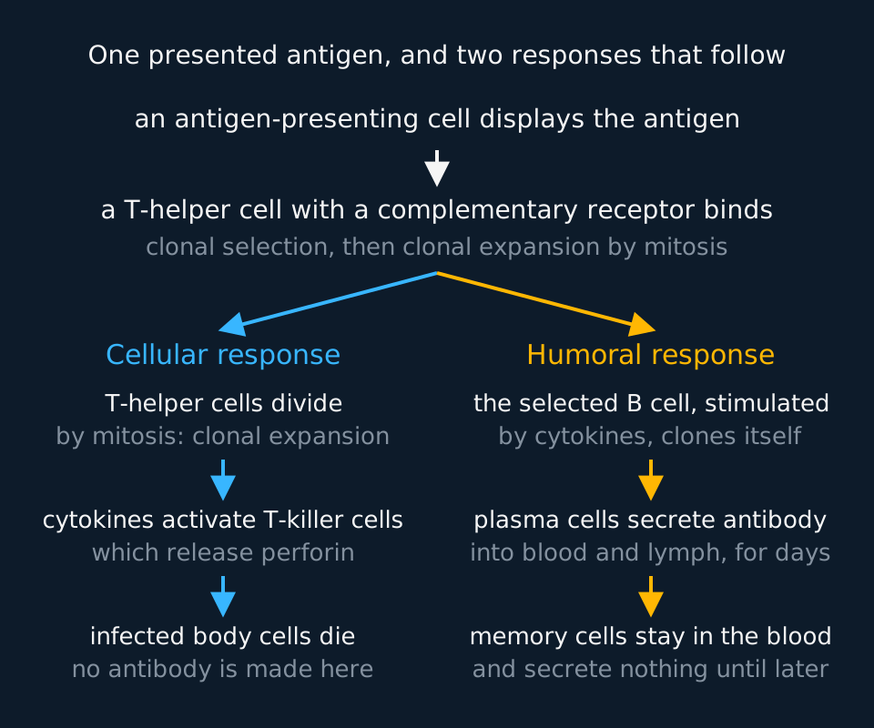
Describe the drawing
A branching flow chart. At the top an antigen-presenting cell displays an antigen, and a T-helper cell with a complementary receptor binds to it and is cloned. The flow then splits. On the left the cellular response: T-helper cells divide, cytokines from them activate T-killer cells, which release perforin and destroy infected body cells, with no antibody made. On the right the humoral response: the B cell whose antibody fits is selected and, stimulated by cytokines from the T-helper cell, clones itself; plasma cells secrete antibody, and memory cells remain in the blood secreting nothing.
The sequence begins with a phagocyte that has hydrolysed a pathogen and presented its antigens, not with the pathogen itself and not with a B cell.
The cellular response
T lymphocytes are made in the bone marrow and mature in the thymus. Each carries receptors of one shape, and what distinguishes the cellular response is that a T cell responds to antigen presented on a cell surface, not to free antigen floating in plasma.
A phagocyte or dendritic cell processes a pathogen and presents antigen fragments on its cell-surface membrane. A T-helper cell with a complementary receptor binds to the antigen-MHC complex and becomes activated. It divides by mitosis to form a clone of effector and memory cells. Activated T-helper cells release cytokines, including interleukins, that activate appropriate B cells and cytotoxic T cells; they do not differentiate into those cell types. They also stimulate phagocytes to engulf more actively.
Cytokines are the signal in every one of those activations. They are small proteins that one cell secretes and another cell detects with a receptor on its cell-surface membrane, and the ones T-helper cells release to stimulate other lymphocytes are mostly interleukins. The T-helper cell does not hand anything over by contact alone: it binds the cell it is activating, and the cytokines it releases bind to receptors on that cell and switch on its division and differentiation. In the cellular response, a T-killer cell whose receptor is complementary to the antigen on an infected or antigen-presenting cell is selected, but it needs cytokines from an activated T-helper cell nearby before it divides by mitosis into a clone of active T-killer cells and T memory cells.
- Cytokine
- A small signalling protein secreted by one cell that binds to receptors on another and changes its behaviour; T-helper cells release cytokines to activate B cells, T-killer cells and phagocytes.
- Interleukin
- A cytokine released by one white blood cell to act on others, such as the interleukins an activated T-helper cell releases to stimulate B cells and T-killer cells to divide.
The verb is activates. A T-helper cell and a T-killer cell are separate lineages, committed during maturation in the thymus: a helper carries CD4 and responds to antigen presented by another cell, a killer carries CD8 and binds infected cells. Neither differentiates into the other.
T-killer cells, or cytotoxic T cells, deal with host cells that are already infected. A cell containing a replicating virus displays viral antigens on its own surface, and a T-killer cell with the complementary receptor binds to it and releases perforin, a protein that makes pores in the cell-surface membrane. Through those pores it delivers enzymes called granzymes, which set off the cell's own death programme, apoptosis: the cell is dismantled tidily from the inside rather than simply leaking to death, and it dies before the virus inside it can finish replicating. It is a controlled loss: one of your own cells is sacrificed because it has already been taken over.
No cell of the cellular response secretes antibody. T cells activate other cells and destroy infected cells; antibody comes from plasma cells derived from B cells.
The importance of the T-helper cell is easiest to see in what happens when it is lost. HIV infects cells carrying the CD4 protein, using it as its way in, and replicates inside them. T-helper cells carry the most CD4 and matter the most, though macrophages and some other CD4-bearing cells are infected too. A few weeks after infection there is often a short illness like flu, followed by years with few symptoms. Over those years, untreated, the number of T-helper cells falls far enough that B cells are no longer activated properly and T-killer cells are no longer stimulated. The specific response stops working, and the infections that follow, such as pneumonia and tuberculosis, together with certain cancers, are what constitute AIDS. Death results from those opportunistic infections and cancers rather than from the virus directly.
The humoral response
B lymphocytes mature in the bone marrow, and each one carries thousands of copies of a single antibody embedded in its cell-surface membrane, acting as its receptor. Between them, the B cells in your body carry something of the order of ten million different antibody shapes, none of which was designed for any particular pathogen.
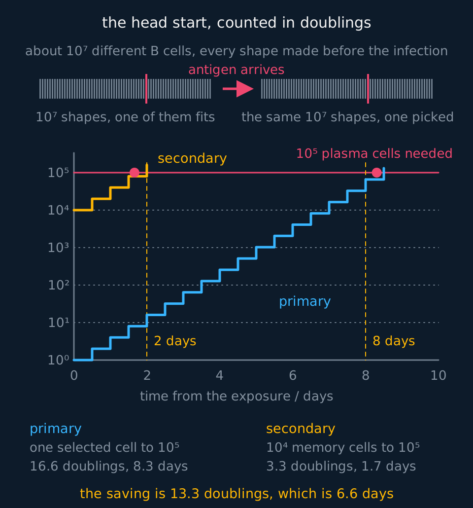
Describe the drawing
Two panels. The upper one draws the antibody repertoire twice as a row of small ticks, about ten million shapes, with one tick in coral; the second row is identical to the first, because the antigen picks a cell out of the repertoire rather than changing it. The lower one is a graph of cell number on a logarithmic vertical axis against days, carrying two doubling ladders that start together. The primary ladder climbs from a single selected cell and needs about sixteen and a half doublings, at twelve hours each, to reach the hundred thousand plasma cells an answer takes, which is a little over eight days. The secondary ladder starts from ten thousand memory cells and needs about three, under two days. Dashed lines mark the eight-day and two-day lags drawn on the antibody graph in this lesson.
When an antigen with a complementary shape binds to a B cell's surface antibody, the B cell takes it in, processes it, and presents it on its own surface. An activated T-helper cell with the matching receptor binds there and releases cytokines, interleukins among them, which stimulate the B cell to divide and its clone to differentiate. This is clonal selection again, applied to a different cell, and it is why the two responses cannot be separated: without T-helper cells, most B cells are never properly activated.
The clone that results differentiates into two kinds of cell: plasma cells, also called B effector cells, and memory cells.
- Plasma cell
- A differentiated B cell that secretes large quantities of a single antibody into blood and lymph. Most survive only a few days; a minority settle in the bone marrow and secrete for years.
- Memory cell
- A long-lived B or T lymphocyte produced by clonal expansion. It secretes no antibody, but divides rapidly if the same antigen is met again: a memory B cell into plasma cells, a memory T cell into T-helper or T-killer cells of its own type.
A plasma cell secretes on the order of two thousand antibody molecules a second and usually survives only a few days, which is why the antibody concentration in the blood falls once an infection is cleared. A minority of plasma cells settle in the bone marrow and continue secreting for years, which is part of why some antibody persists long after an infection. Memory cells secrete no antibody, and some persist for decades: immunity to measles after infection is usually lifelong, and it depends both on those memory cells and on bone-marrow plasma cells that keep measles antibody in the blood.
| Cell | Where it acts | What it does | How long it lasts |
|---|---|---|---|
| T-helper | On antigen-presenting cells | Releases cytokines that activate B cells, T-killer cells and phagocytes | Days; memory forms |
| T-killer | On infected body cells | Releases perforin, so the infected cell dies | Days |
| B cell | In lymph nodes and blood | Selected by antigen, then clones itself | Days |
| Plasma cell | Blood and lymph | Secretes antibody continuously | Usually a few days; some bone-marrow residents last years |
| Memory cell | Blood and lymph | Secretes nothing; divides fast on re-exposure | Years to decades |
Primary and secondary responses
The two responses are compared on the same axes, with a logarithmic vertical scale; on a linear scale the primary response is barely visible.
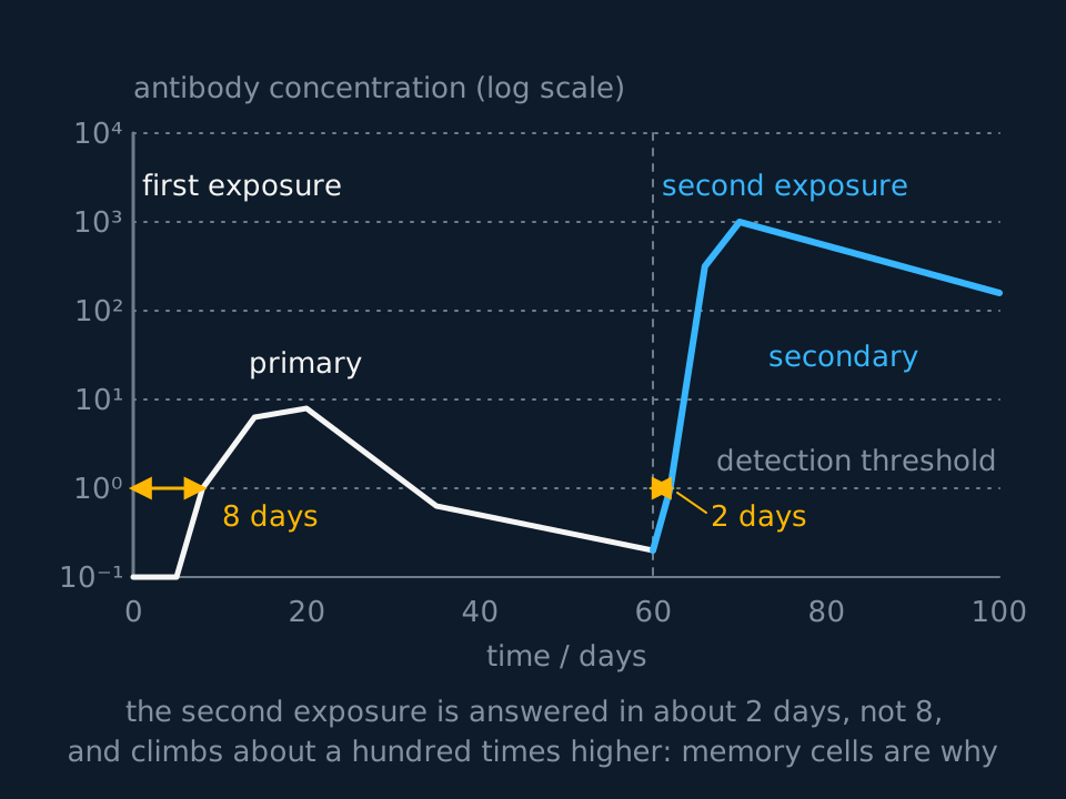
Describe the drawing
A graph of antibody concentration on a logarithmic vertical axis against time in days. After the first exposure at day zero the concentration stays undetectable for about eight days, rises to a low peak near ten units, then falls away below the detection threshold. A second exposure to the same antigen at day sixty is answered within about two days, and the concentration climbs roughly a hundred times higher and is still high forty days later.
The primary response is slow because almost everything has to happen from scratch. A phagocyte must present the antigen; the handful of lymphocytes with a complementary receptor must be found among millions; those cells must divide repeatedly before there are enough of them to matter; and only then do plasma cells appear. Eight days or so is typical before there is measurable antibody, and the person is ill for most of it.
The secondary response starts from a completely different position. Memory cells specific to that antigen are already circulating, in thousands rather than ones, and they are already committed. Each must still meet its antigen and be reactivated before it proliferates and differentiates into plasma cells, but with thousands of primed cells the antigen is bound quickly. Antibody appears within two or three days, reaches a concentration perhaps a hundred times higher, and stays high for longer because more memory cells are made in the process. The pathogen is usually destroyed before it has reproduced enough to cause symptoms, which is what people mean when they say they are immune. T memory cells, left by the T-helper and T-killer clones of the first response, speed up the cellular response in the same way.
Describing and explaining the two curves
Using the graph, describe two differences between the primary and secondary responses, and explain the difference in the time taken for antibody to become detectable.
Show the working
Describe with figures. Antibody becomes detectable about eight days after the first exposure and about two days after the second; the peak concentration is roughly a hundred times higher after the second exposure. 'Using the graph' requires figures read from the axes.
Explain with cells. After the first exposure, the antigen had to be presented, the few B lymphocytes with a complementary antibody had to be selected, and clonal expansion had to run for several generations before plasma cells existed in useful numbers.
After the second exposure, memory cells specific to that antigen were already present in large numbers. Clonal selection had already happened, so those cells divided rapidly and differentiated into plasma cells almost at once, and far more plasma cells were produced.
Read the axis carefully. Each gridline is a tenfold increase, so a peak two gridlines higher is a hundred times more antibody, not twice as much. Drawn on a linear scale, the primary response would lie along the bottom of the axis and the two could not be compared.
The sequence from infection to circulating antibody
The order of events, from a pathogen entering the body to antibody in the plasma, is as follows.
The pathogen enters and is engulfed by a phagocyte, which hydrolyses it and presents its antigens on its own cell-surface membrane. A T-helper cell with a complementary receptor binds to the presented antigen and is activated, which is clonal selection, and divides by mitosis. Meanwhile the same antigen binds to the surface antibody of the B cell that fits it, and the B cell processes and presents it. An activated T-helper cell binds to that B cell and releases cytokines that stimulate it. The B cell divides by mitosis, clonal expansion, and the clone differentiates into plasma cells, which secrete antibody, and memory cells, which do not.
Two points fix the sequence. It begins with the phagocyte and antigen presentation, not with the B cell. And memory cells secrete no antibody: they divide into plasma cells, and the plasma cells secrete.
TRY IT: Same pathogen, different patient
A patient receiving immunosuppressant drugs after a kidney transplant develops a bacterial infection. Their phagocytes are unaffected by the drugs, but their T-helper cells are greatly reduced in number. Predict how their antibody response to the infection will differ from a healthy person's, and explain why.
Check your answer
Phagocytosis will be normal. It is non-specific, needs no lymphocytes, and antigen will still be presented on the phagocytes' surfaces.
The antibody response will be much smaller and much slower. B cells can bind antigen with their surface antibody, but most of them are not fully activated until an activated T-helper cell with a complementary receptor binds to the antigen they are presenting. With few T-helper cells, that second signal rarely arrives.
So clonal expansion of B cells is limited, few plasma cells are produced, and antibody concentration stays low. Fewer memory cells are made as well, so a later exposure to the same bacterium will not produce a proper secondary response either.
The same reasoning applies to untreated HIV infection. The cause differs, a prescribed drug in one case and a virus infecting T-helper cells in the other, but the consequence for the specific response is similar.
In the exam
- Clonal selection and clonal expansion are separate events. Selection is the antigen binding the lymphocyte with a complementary receptor; expansion is that cell dividing by mitosis. State both.
- Use complementary rather than 'the same shape'. A receptor and an antigen fit together; they are not identical.
- Plasma cells secrete antibody; memory cells do not, until they have divided into plasma cells.
- HIV is the virus and AIDS is the condition that follows when enough T-helper cells have been destroyed. Use each term for its own referent.
- On a logarithmic axis, count gridlines: two decades is a hundredfold increase, and a fold-change is what such a scale is read for.
- Begin a sequence with the phagocyte and antigen presentation, not with the B cell, and give the order before the detail.
- Name the signal: the activated T-helper cell releases cytokines (interleukins) that stimulate the B cell or T-killer cell to divide. 'Stimulates' on its own does not say how.
Check yourself
A vaccine against a bacterium is given to a child, who develops a mild fever for a day and no other symptoms. Eighteen months later the child is exposed to the live bacterium and does not become ill at all. Explain what happened on each occasion, in terms of the cells involved.
Answer
The vaccine contained antigens from the bacterium. Phagocytes engulfed them and presented the antigens on their cell-surface membranes, and a T-helper cell with a complementary receptor was selected and divided by mitosis.
B cells whose surface antibody was complementary to the same antigen took it in and presented it, were stimulated by activated T-helper cells, and underwent clonal expansion. The clone differentiated into plasma cells, which secreted antibody over the following days, and memory cells, which remained. This was a primary response: slow, small and, because there was no live pathogen reproducing, symptom-free apart from the fever produced by the response itself.
Eighteen months later the memory cells were still circulating. On exposure, the same antigen bound to memory B cells that were already present in large numbers and already selected, so they divided rapidly into plasma cells. Antibody appeared within two or three days rather than eight, at a much higher concentration.
The bacteria were agglutinated and marked for phagocytosis before they had reproduced enough to cause symptoms. The child was never ill because the response was faster than the infection, not because the bacteria never entered.
Questions
Question 15 marks
Describe the sequence of events from a pathogen entering the body to antibody appearing in the blood plasma.
Mark scheme
- B1 the pathogen is engulfed by a phagocyte, which hydrolyses it and presents its antigens on its own cell-surface membrane
- B1 a T-helper cell with a complementary receptor binds to the presented antigen and is activated, which is clonal selection, and divides by mitosis
- B1 the same antigen binds to the surface antibody of the B cell that fits it, and that B cell takes the antigen in, processes it and presents it
- B1 an activated T-helper cell binds to the presenting B cell and stimulates it to divide by mitosis, which is clonal expansion
- B1 the clone differentiates into plasma cells, which secrete antibody, and memory cells, which secrete nothing
Question 25 marks
Compare the cellular response with the humoral response, referring to the lymphocytes involved, to what each of those lymphocytes recognises, and to what finally destroys or disables the pathogen.
Mark scheme
- B1 both begin with clonal selection, the antigen binding the lymphocytes whose receptors are complementary to it, and both continue with clonal expansion by mitosis and leave memory cells behind
- B1 the cellular response is carried out by T lymphocytes, which are made in the bone marrow but mature in the thymus, whereas the humoral response is carried out by B lymphocytes, which mature in the bone marrow itself
- B1 a T cell responds only to antigen presented on a cell surface, whereas a B cell binds free antigen in blood and lymph using the antibody in its own cell-surface membrane, although it still needs an activated T-helper cell before it divides
- B1 the cellular response acts on the body's own cells, a T-killer cell binding an infected cell and releasing perforin so that it dies, whereas the humoral response acts outside cells, plasma cells secreting soluble antibody that agglutinates pathogens and marks them for phagocytosis
- A1 so the cellular response is the one that can reach a virus already inside a host cell and the humoral response the one that deals with bacteria and toxins in the plasma, but they run at the same time and share the T-helper cell, so neither is a separate system
Question 34 marks
Explain why detectable antibody appears about eight days after a first exposure to an antigen but only about two days after a second exposure to the same antigen.
Mark scheme
- B1 on the first exposure the antigen must first be presented by a phagocyte, and the few lymphocytes with a complementary receptor must be selected out of millions
- B1 those selected cells must then divide repeatedly by mitosis before enough exist to matter, and only then do plasma cells appear and begin secreting
- B1 on the second exposure, memory cells specific to that antigen are already circulating in thousands rather than ones, so selection has already happened
- B1 the memory cells divide rapidly straight into plasma cells, so antibody is secreted in quantity within about two days
Question 44 marks
Explain why an untreated HIV infection eventually leaves a person unable to deal with infections that an intact immune system would handle easily.
Mark scheme
- B1 HIV infects T-helper cells specifically, using the CD4 protein on their surface as its way in, and replicates inside them
- B1 infected T-helper cells are killed, by the virus replicating inside them or by T-killer cells destroying them, so over years the number of T-helper cells falls
- B1 B cells are no longer properly activated, so few plasma cells are produced and little antibody is secreted
- B1 T-killer cells are no longer activated, so infected body cells are not destroyed; the infections that follow are what constitute AIDS
Question 54 marks
A virus is replicating inside the cells lining a person's airways. Suggest how T-killer cells limit the infection, and suggest why antibody alone would not be enough.
Mark scheme
- B1 a cell containing a replicating virus displays viral antigens on its own cell-surface membrane
- B1 a T-killer cell with a complementary receptor binds to that cell and releases perforin, which makes pores in the cell-surface membrane
- B1 granzymes enter through the pores and trigger apoptosis, so the infected cell dies before the virus inside it can finish replicating
- B1 antibody binds antigens outside cells, so it cannot reach virus particles already inside a host cell; only destroying the infected cell removes them
Question 64 marks
Lymphocytes carrying receptors that fit the body's own antigens are produced before birth but do not survive. Explain how this leaves a person tolerant of their own cells, and explain what has gone wrong in an autoimmune disease such as type 1 diabetes.
Mark scheme
- B1 lymphocytes are produced with an enormous variety of receptor shapes, generated before any antigen has been met, so some of them inevitably fit antigens carried on the body's own cells
- B1 during foetal development and shortly after birth, any lymphocyte whose receptor binds a self antigen dies or is suppressed, so the population left responds only to shapes the body does not contain
- B1 self antigens are therefore not treated as foreign and no response is mounted against them, while non-self antigens on a pathogen, a transplanted organ or a cell with altered surface proteins still provoke one
- B1 in an autoimmune disease, that screening has failed, so a lymphocyte specific to a self antigen has survived and been activated and the response is directed at the body's own tissue; in type 1 diabetes the cells that secrete insulin are destroyed
Question 74 marks
The table describes four cells taken from a person who is fighting an infection. Identify each of the four cells, and identify which of them are acting as antigen-presenting cells.
| Cell | What has been observed about it |
|---|---|
| W | has engulfed a pathogen, hydrolysed it with enzymes from a lysosome, and now carries fragments of the pathogen's antigens on its own cell-surface membrane |
| X | is a body cell in which a virus is replicating, and it carries viral antigens on its own cell-surface membrane |
| Y | carries thousands of copies of one antibody in its cell-surface membrane, and has taken in, processed and displayed the antigen those antibodies fit |
| Z | secretes about two thousand antibody molecules a second and displays no antigen on its surface |
Mark scheme
- A1 W is a phagocyte, and it is an antigen-presenting cell; its presentation is what begins the specific response
- A1 X is an infected body cell, which is presenting antigen too, and a T-killer cell with a complementary receptor binds to it
- A1 Y is a B cell, and it is also an antigen-presenting cell, presenting to an activated T-helper cell whose binding is what stimulates it to divide
- A1 Z is a plasma cell, which secretes antibody and presents nothing, so it is not an antigen-presenting cell
Question 84 marks
A new drug blocks the surface protein an activated T-helper cell uses to signal to a B cell that is presenting antigen, without affecting T-helper cell numbers or phagocytosis. Suggest how this would affect a person's ability to produce antibody against a new infection.
Mark scheme
- B1 phagocytosis and antigen presentation by phagocytes would be unaffected, since neither depends on this protein
- B1 T-helper cells could still be activated by antigen presented by a phagocyte, since the drug does not affect antigen recognition
- B1 but T-helper cells bound to a presenting B cell could no longer signal to it, so B cells would not receive the signal needed to divide
- A1 clonal expansion of B cells would be severely reduced, so few plasma cells and little antibody would be produced even though phagocytosis and T-helper cell activation are both normal
Question 94 marks
Describe the development of the cell-mediated immune response to a virus, including the role of cytokines.
Mark scheme
- B1 an antigen-presenting cell displays viral antigen on its cell-surface membrane
- B1 a T-helper cell with a complementary receptor binds, is selected and divides by mitosis
- B1 the activated T-helper cells release cytokines that stimulate T-killer cells with a complementary receptor to divide by mitosis
- B1 the T-killer cells bind to infected cells displaying the viral antigen and release perforin, so the infected cells die
Question 103 marks
T-helper cells and T-killer cells are separate lineages that are committed while they mature in the thymus. Explain why a T-helper cell cannot differentiate into a T-killer cell later in an infection.
Mark scheme
- B1 the two do different jobs: a T-helper cell releases cytokines that stimulate B cells, T-killer cells and phagocytes, whereas a T-killer cell destroys infected body cells itself
- B1 which job a T cell does is fixed during its maturation in the thymus, before it ever meets an antigen
- B1 when an antigen activates a T-helper cell, it divides by mitosis into a clone of genetically identical T-helper cells, so the response makes more T-helper cells and never T-killer cells
Question 113 marks
A plasma cell can secrete about 2000 antibody molecules a second. Calculate the number of antibody molecules one plasma cell secretes in 24 hours, giving your answer in standard form, and suggest one feature of the ultrastructure of a plasma cell that allows this rate of secretion.
Mark scheme
- M1 2000 × 60 × 60 × 24
- A1 1.7 × 10⁸ antibody molecules (accept 1.73 × 10⁸)
- B1 any one of: extensive rough endoplasmic reticulum, with many ribosomes, to make the antibody protein; a large Golgi apparatus to package it into vesicles; many mitochondria to supply ATP
Question 123 marks
A person vaccinated against hepatitis B as a baby is found, twenty years later, to have almost no antibody against the virus circulating in their blood, yet they remain protected against it. Suggest why.
Mark scheme
- B1 most plasma cells survive only a few days, so the concentration of antibody falls once the response to the vaccine is over
- B1 memory cells, unlike plasma cells, secrete no antibody but can persist for decades
- A1 on meeting hepatitis B antigen again, the memory cells would divide rapidly into plasma cells and mount a fast secondary response, so protection comes from the memory cells rather than from circulating antibody
Question 133 marks
Compare how a T cell recognises antigen with how a B cell recognises antigen.
Mark scheme
- B1 a T cell responds only to antigen presented on the surface of another cell, whereas a B cell can bind free antigen directly using the antibody in its own cell-surface membrane
- B1 despite binding free antigen, a B cell still needs an activated T-helper cell to bind to the antigen it then presents before it is fully activated and divides
- A1 so the two forms of recognition are not independent: a B cell's own recognition of antigen is not sufficient on its own for clonal expansion to proceed
Question 143 marks
Give three examples of things, other than a pathogen, that carry non-self antigens and so can provoke an immune response.
Mark scheme
- B1 a transplanted organ, which carries the donor's antigens on its cells
- B1 a cancer cell, which often displays abnormal surface proteins recognised as non-self
- B1 a toxin, which is a foreign molecule that can itself provoke antibodies without any cell being involved
Question 153 marks
Outline three things that cytokines released by an activated T-helper cell stimulate or activate.
Mark scheme
- B1 cytokines activate B cells that have presented the same antigen, triggering clonal expansion
- B1 cytokines activate T-killer (cytotoxic T) cells that have bound the same presented antigen
- B1 cytokines also stimulate phagocytes, making them engulf pathogens more actively
Question 163 marks
Explain the role of cytokines released by T-helper cells in the humoral immune response.
Mark scheme
- B1 a T-helper cell activated by antigen on an antigen-presenting cell binds to a B cell presenting the same antigen
- B1 it releases cytokines, such as interleukins, which bind to receptors on that B cell
- B1 the cytokines stimulate the B cell to divide by mitosis and the clone to differentiate into plasma cells, which secrete antibody, and memory cells
Question 172 marks
State what is meant by clonal selection and state what is meant by clonal expansion.
Mark scheme
- B1 clonal selection is the binding of an antigen to the lymphocytes whose receptors are complementary to it, activating those cells and no others
- B1 clonal expansion is the repeated division of each selected lymphocyte by mitosis, producing a large clone of genetically identical cells
Worth remembering
- Lymphocytes are selected by an antigen, not instructed by it: the shapes exist before the infection does.
- Clonal selection is the binding; clonal expansion is the mitosis that follows.
- T cells respond to antigen presented on a cell surface; B cells can bind free antigen, but still need a T-helper cell to be fully activated.
- Plasma cells secrete antibody for days. Memory B cells secrete nothing for years, then divide into plasma cells; memory T cells give a faster cellular response.
- The secondary response is faster, larger and longer-lasting because it starts with thousands of the right cells instead of a few.
- T-helper cells activate by releasing cytokines: B cells in the humoral response, T-killer cells in the cell-mediated one.
CHECK YOUR PROGRESS
Rate how confident you are with each objective for this lesson. Ratings are saved in this browser, on this device, unless you sign in.
- Explain what an antigen is and how a lymphocyte distinguishes self from non-self.
- Describe clonal selection and clonal expansion, and say which cell does which.
- Set out the roles of T-helper cells, T-killer cells, B cells, plasma cells and memory cells without confusing any two of them.
- Put the events from infection to circulating antibody into the right order.
- Account for the secondary response being faster, larger and longer-lasting than the primary one.
- Name cytokines as the signal by which an activated T-helper cell stimulates B cells in the humoral response and T-killer cells in the cell-mediated response.
Open the full revision checklist to see every objective in the curriculum in one place.
Practise this lesson
Practise the specific immune response with 17 original questions and point-by-point mark schemes
WORKBOOK
The written questions from this topic, on paper with room to work, and a separate book of mark schemes. The question that is set on a diagram stays on this page, where the diagram can be drawn. Free to use; please do not redistribute or sell.