Biology › Pathogens, disease and immunity › Pathogens and non-specific defences
Pathogens and non-specific defences
Four kinds of organism cause almost every infectious disease, and they damage the body in a small number of ways. Against all of them the body runs the same opening defence: barriers that keep pathogens out, chemistry that kills them at the entrances, and phagocytes that engulf anything foreign.
Before this Cell membranes and membrane proteins · Cell ultrastructure: lysosomes and enzyme hydrolysis · Water potential and osmosis
COMMON MISCONCEPTION
White blood cells fight infection by recognising the germ and producing the right antibody for it.
Phagocytes recognise broad pathogen-associated molecular patterns rather than one unique antigen. They engulf pathogens and can present antigen fragments. Antigen-specific recognition occurs when a lymphocyte with a complementary receptor is selected.
What you should be able to do
- Name the four groups of pathogen and give a disease caused by each.
- Explain how a pathogen damages its host, using toxins and cell lysis as the two standard exam routes.
- Distinguish a physical barrier from a chemical one, with named examples of both.
- Describe phagocytosis as an ordered sequence, ending with antigen presentation.
- Explain why an antibiotic that cures tuberculosis does nothing whatever for influenza.
- Distinguish an exotoxin from an endotoxin, with Staphylococcus and Salmonella as the examples, and set both beside invasion of tissue by Mycobacterium tuberculosis.
- Describe the transmission, mode of infection and pathogenic effect of the stem rust fungus, Puccinia graminis, on wheat.
- Explain how interferon released by a virus-infected cell protects the cells around it.
What counts as a pathogen
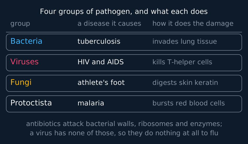
A pathogen is a biological agent that causes disease in a host. Bacteria, fungi and protoctists are cellular pathogens; viruses are acellular pathogens. Pathogens can damage tissue directly, release toxins or provoke damaging host responses. The body first opposes them with non-specific physical, chemical, biological and cellular defences. The four groups are defined by their effect on a host and are not closely related to one another.
Describe the drawing
A four-row table of pathogen groups. Bacteria cause tuberculosis by invading lung tissue. HIV, a virus that leads to AIDS, kills T-helper cells. Fungi cause athlete's foot by digesting skin keratin. Protoctista cause malaria by bursting red blood cells. A note underneath says antibiotics attack bacterial walls, ribosomes and enzymes, none of which a virus has.
Bacteria are prokaryotes: no nucleus, a peptidoglycan cell wall, 70S ribosomes, and a metabolism of their own. Mycobacterium tuberculosis causes tuberculosis, Vibrio cholerae causes cholera, and various staphylococci turn an ordinary wound into an infected one. Because bacteria have structures and enzymes that your cells do not, they can be attacked selectively, which is what an antibiotic does.
Viruses have no cytoplasm, no ribosomes and no metabolism. A virus is nucleic acid in a protein coat, and it can do nothing at all until it is inside a host cell whose machinery it can use. Influenza, measles and HIV are viruses. Be careful with the last one: HIV is the virus, and AIDS is the condition that develops years later when the infection has destroyed enough T-helper cells that the specific immune response can no longer be co-ordinated.
Fungi that infect humans are usually superficial. Athlete's foot and ringworm are both forms of tinea, caused by dermatophyte fungi such as Trichophyton that grow in the keratin of skin and nails, digesting tissue outside their own cells and absorbing the products. In plants the fungal diseases are far more destructive: black sigatoka in banana crops and stem rust in wheat, described below, are the standard examples.
Protoctista are eukaryotic and mostly single-celled. Plasmodium, carried between people by female Anopheles mosquitoes, causes malaria; Phytophthora infestans caused the potato blight of the 1840s. Both are eukaryotes, which is precisely why drugs against them are harder to design than antibiotics: their cells work much the way yours do.
- Pathogen
- A biological agent that causes disease in a host, including cellular organisms such as bacteria, fungi and protoctists and acellular agents such as viruses.
- Antigen
- A molecule, usually a surface protein or glycoprotein, that is recognised as self or non-self and can trigger an immune response.
- Non-specific defence
- A defence that acts against any pathogen, without recognising which one it is, and acts at the same speed every time.
Most microorganisms are not pathogens. Resident microbiota provide a biological, competitive defence by occupying space and using resources that might otherwise be available to pathogens, which is why the bacteria living on your skin and in your large intestine count as part of your defences rather than a threat to them. A course of broad-spectrum antibiotics kills a good many of those too, and the digestive upset that follows is the consequence.
How a pathogen causes disease
Two mechanisms of damage account for most of the examples at A level.
The first is toxins. Many bacteria release proteins that interfere with a host process. Cholera toxin binds to the cells lining the small intestine and hijacks their own signalling: the level of a messenger molecule, cyclic AMP, rises and holds open a chloride channel called CFTR, so chloride ions pour into the gut lumen. That lowers the water potential of the lumen below that of the cells, water follows by osmosis, and the resulting diarrhoea can remove several litres a day. Death in cholera results from dehydration rather than from the bacterium directly, which is why oral rehydration salts are so effective a treatment. Tetanus toxin does something different again, blocking the signals that would normally stop a motor neurone firing, so muscles contract and cannot relax.
The second is cell lysis. A virus enters a host cell, its nucleic acid is transcribed and translated using the host's ribosomes, enzymes and nucleotides, and hundreds of new virus particles are assembled. Many viruses are released by bursting the cell open; others, HIV among them, bud out through the membrane, and the infected cell declines more gradually, with much of the loss caused by the immune response eliminating infected cells. Repeat the damage across an epithelium and you have a sore throat; repeat it in the T-helper cells of an untreated HIV infection and, over years, you have AIDS. Plasmodium lyses red blood cells outright, and the bursting of infected cells in synchrony is why the fever of malaria comes in waves rather than continuously.
Not every disease fits the two boxes. Mycobacterium tuberculosis survives inside the very macrophages that engulf it, and much of the lung damage in tuberculosis is done by the host's own sustained inflammatory response, which walls infected cells into masses called granulomas, rather than by a released toxin. The two exam routes are the common cases, not a complete classification.
Fungi work another way again, secreting hydrolytic enzymes onto the tissue they grow in and absorbing what dissolves, which is why athlete's foot itches and flakes. Several symptoms of an infection are produced by the host rather than by the pathogen: fever, swelling, aching and tiredness are effects of the immune response itself.
Reading a toxin question properly
Cholera toxin causes chloride ions to be secreted from intestinal epithelial cells into the lumen of the gut. Explain how this leads to severe diarrhoea, and suggest why oral rehydration solution contains both salts and glucose.
Show the working
Start with the water potential. Chloride ions accumulating in the lumen lower the water potential there, so it becomes more negative than the water potential of the epithelial cells and the tissue fluid behind them.
Water therefore moves out of the cells and into the lumen down a water potential gradient, by osmosis, through the partially permeable cell-surface membranes. Large volumes of watery faeces follow, and the patient loses both water and ions.
The glucose has a specific role. Sodium ions are absorbed from the gut by co-transport with glucose, so putting glucose in the solution drives sodium uptake, which raises the water potential in the lumen relative to the cells and lets water be absorbed again. Salts alone are absorbed far more slowly.
The answer requires no knowledge of the bacterium itself. Explain that water moves down a water-potential gradient by osmosis, and that sodium is absorbed by co-transport with glucose.
Exotoxins, endotoxins and invasion
Bacterial toxins come in two kinds, and the difference is where the toxin comes from. An exotoxin is a protein that a living bacterium secretes into its surroundings. Cholera toxin and tetanus toxin are both exotoxins. An endotoxin is part of the bacterium itself: the lipopolysaccharide (LPS) of the outer membrane that covers the thin peptidoglycan wall of Gram-negative bacteria. Small amounts are shed as the bacteria grow, and a large amount is released when they die and their walls break down.
- Exotoxin
- A toxic protein secreted by a living bacterium, acting on a specific target in the host; Staphylococcus species are the standard example.
- Endotoxin
- The lipopolysaccharide of the outer membrane of a Gram-negative bacterium, released mainly when the bacterium dies; it causes fever and inflammation. Salmonella species are the standard example.
- Toxoid
- An exotoxin treated so that it is no longer toxic but keeps its antigens, used as a vaccine against diseases such as tetanus and diphtheria.
Staphylococcus species are the standard exotoxin producers. Staphylococcus aureus lives harmlessly on the skin and in the nose of many people, but in a wound or in food it secretes several exotoxins with different targets. Some make pores in host cell-surface membranes, so the cells lyse. Others are enterotoxins: a strain growing in food left at room temperature secretes toxin into the food, and eating it causes vomiting within a few hours, because the toxin is already made and nothing has to grow in the gut first. The toxin behind toxic shock syndrome overstimulates T cells, which release cytokines in such quantity that blood pressure falls dangerously. The staphylococcal food toxins are unusually stable to heat for proteins, so reheating food can kill the bacteria and leave the toxin active.
Salmonella species are the standard example of endotoxin. They are Gram-negative rods; most cases of Salmonella food poisoning come from undercooked poultry and eggs, and one serotype, Salmonella Typhi, causes typhoid. The bacteria swallowed in food have to multiply and invade the cells lining the intestine before anything happens, so symptoms usually begin between 12 hours and 3 days after eating. As the bacteria are killed by the host's defences, lipopolysaccharide is released. Its lipid part binds to receptors on macrophages and other cells, which respond by releasing cytokines. The result is inflammation of the gut lining, with diarrhoea and abdominal cramps, and fever. If large amounts of endotoxin reach the blood, widespread dilation of blood vessels can lead to septic shock. The damage is done by the host's response to a part of the bacterium.
| Exotoxin | Endotoxin | |
|---|---|---|
| What it is | Protein | Lipopolysaccharide of the outer membrane |
| How it is released | Secreted by living bacteria | Mainly when the bacterium dies and its wall breaks down |
| Bacteria that make it | Gram-positive and Gram-negative | Gram-negative only |
| Effect | Specific target; potent in tiny amounts | Fever, inflammation, shock; less potent |
| Vaccine | Can be made into a toxoid | No toxoid |
| Standard example | Staphylococcus spp. | Salmonella spp. |
The third route is invasion of host tissue, and Mycobacterium tuberculosis is the example: it releases no major toxin, but grows inside the macrophages of the lung, and the granulomas and tissue destruction of tuberculosis are the result of that invasion and of the host's long inflammatory response to it, as described above.
Stem rust: a fungal disease of wheat
Puccinia graminis, the stem rust fungus, infects wheat, and related forms attack barley, oats and rye. It has been one of the most damaging crop diseases in history, because a field that looks healthy can be ruined in the few weeks before harvest. Its full life cycle passes through a second host plant, barberry, but at A level only transmission, infection and effect are needed.
Transmission. On an infected plant the fungus forms pustules that burst through the epidermis of the stem and leaves and release masses of rust-red spores. The spores are dry and light and are carried by the wind, to the next plant, the next field, and on air currents for hundreds of kilometres. In warm weather a new generation of spores is produced within one to two weeks of infection, so the number of spores in a region can rise very fast once an epidemic starts.
Mode of infection. A spore landing on a wheat stem or leaf germinates only in a film of water, which is why outbreaks follow warm weather with dew or rain. The germ tube grows across the surface to a stoma and enters through it, so the fungus never has to break the waxy cuticle. Inside, hyphae grow through the spaces between the cells and push branches called haustoria through the cell walls. The host cell membrane folds around each haustorium rather than breaking, so the host cell stays alive, and sugars and amino acids are absorbed from it. The fungus can feed only on living tissue.
Pathogenic effect. Sugars drawn off by the fungus are sugars not translocated to the developing grain, so the grains are small and shrivelled. The infected tissue photosynthesises less. Pustules rupture the epidermis, so the plant loses water through the breaks whether or not its stomata are closed. Weakened stems break or fall over in wind and rain, which is called lodging, and a severe attack can destroy most of a crop.
- Stem rust
- A disease of wheat and other cereals caused by the fungus Puccinia graminis, spread by wind-borne spores and entering through stomata.
- Haustorium
- A branch of a fungal hypha that pushes into a living host plant cell, inside a fold of the host's cell-surface membrane, and absorbs nutrients from it.
Control relies mainly on resistant varieties of wheat, which carry resistance genes (called Sr genes) whose products let the plant recognise the fungus and set off its defences, with fungicides as a second line. Stem rust became uncommon in many wheat-growing regions once resistant varieties were planted widely in the second half of the twentieth century. In 1999 a new race of the fungus, named Ug99 after Uganda where it was identified, was found to overcome Sr31, a resistance gene carried by much of the world's wheat, and it has since spread through eastern Africa and into the Middle East. Widely grown resistant varieties select for any variant of the fungus that overcomes the resistance, and Ug99 is such a variant.
Physical, chemical and biological barriers
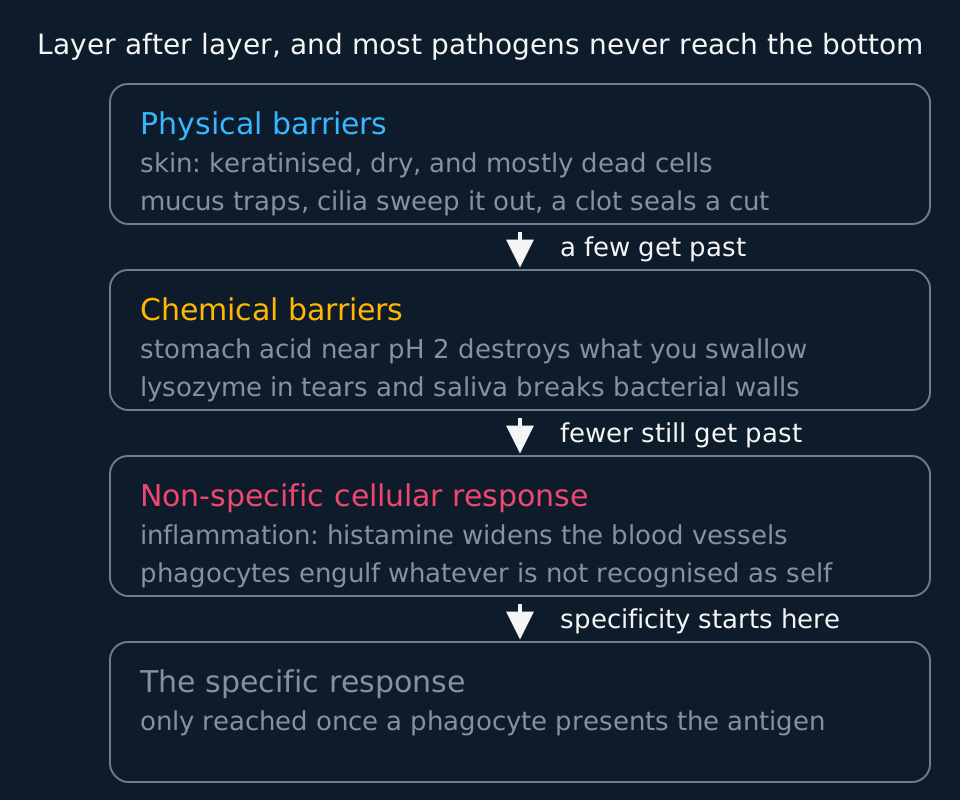
Before the specific immune response is involved, a pathogen must cross defences that act on all pathogens equally. They are classified as physical, chemical and biological.
Describe the drawing
Four stacked bands with arrows running down between them. The top band is physical barriers: skin, mucus and cilia, and clotting. The second is chemical barriers: stomach acid near pH 2 and lysozyme in tears and saliva. The third is the non-specific cellular response of inflammation and phagocytosis. The bottom band, reached only after a phagocyte presents an antigen, is the specific response.
Physical barriers keep pathogens out by being in the way. Skin is the obvious one: its outer layer is dead, dry, and packed with keratin, which almost nothing can digest. Where the body has to be moist and permeable, as in the airways, gut and reproductive tract, it uses mucous membranes instead. Goblet cells secrete sticky mucus that traps particles, and the ciliated epithelium beneath beats the mucus upwards to the throat, where it is swallowed. Damage that cilia layer, as cigarette smoke does, and the trapped material stays put.
A wound is a hole in the barrier, and blood clotting seals it within minutes. Platelets at the damaged vessel release substances that set off a cascade of reactions in the plasma, ending in the formation of insoluble fibrin. The fibrin fibres form a network that traps platelets and blood cells into a clot, which dries into a scab. Under the scab, wound repair begins: cells in the deeper layers of the skin divide to replace the lost tissue, and collagen is laid down to strengthen it. Expulsive reflexes remove what has already got in: coughing and sneezing expel irritants and pathogens from the airways, and vomiting and diarrhoea expel them from the gut.
Chemical barriers destroy pathogens rather than blocking them. Stomach acid holds the gut contents near pH 2, which denatures the enzymes and other proteins of most swallowed bacteria. Mucus cleared from the airways is swallowed, so bacteria trapped in it also reach the stomach: the ciliated epithelium removes them from the airways and the acid destroys them in the stomach. Lysozyme, an enzyme in tears, saliva and mucus, hydrolyses bonds in bacterial cell walls so the cell bursts. Sebum makes the skin surface slightly acidic and inhospitable.
| Defence | Kind | How it works | Where |
|---|---|---|---|
| Skin | Physical | Keratinised, dry, impermeable | Body surface |
| Mucus and cilia | Physical | Traps particles and sweeps them out | Airways, gut |
| Blood clotting | Physical | Seals a break in the barrier | Any wound |
| Stomach acid | Chemical | pH near 2 denatures enzymes and proteins | Stomach |
| Lysozyme | Chemical | Hydrolyses bacterial cell walls | Tears, saliva, mucus |
| Gut and skin microbiota | Biological/competitive | Occupy space and use resources that might otherwise be available to pathogens | Gut, skin |
If a pathogen does get through, the response at the site of damage is inflammation. Mast cells in the damaged tissue release histamine, which dilates arterioles and makes capillary walls more permeable. More blood arrives, so the area is red and warm, and more tissue fluid leaks out, so it swells. That fluid carries antibodies into the infected tissue, and cytokines released at the site attract phagocytes, which squeeze out of the capillaries between the cells of their walls. The swelling is part of the response rather than incidental damage.
A raised body temperature has two effects: most pathogens reproduce more slowly above about 37 °C, and the specific immune response operates faster at the higher temperature. Very high fevers are dangerous because human enzymes also denature.
Interferon: a warning to neighbouring cells
Inflammation, lysozyme and phagocytosis all act on pathogens outside cells. A virus that has entered a cell is out of their reach, and the infected cell's first response is a signal to the cells around it. The cell detects viral nucleic acid in its cytoplasm, in particular double-stranded RNA, which human cells do not normally contain, and responds by transcribing the genes for interferon. Interferons are small proteins, one class of cytokine, and the infected cell secretes them into the tissue fluid.
The infected cell gains little from this; the interferon acts on its neighbours. It diffuses to nearby cells and binds to receptors on their cell-surface membranes, which switches on a set of genes inside each one. The proteins they code for put the cell into an antiviral state. One is an enzyme that stops protein synthesis at the ribosomes as soon as viral double-stranded RNA appears; another activates a ribonuclease that hydrolyses RNA, so viral mRNA is destroyed. A virus that then enters one of those cells cannot be replicated, because it has no ribosomes or enzymes of its own and depends entirely on the host cell's. Interferon also makes cells display more antigen on their surface, which helps T-killer cells find the infected ones, and it activates macrophages and natural killer cells.
- Interferon
- A protein secreted by a virus-infected cell that binds to receptors on neighbouring cells and makes them resistant to viral replication, by inhibiting protein synthesis and destroying viral RNA.
Interferon is non-specific. It is released in response to many different viruses, and the antiviral state it produces works against all of them. It does not bind the virus and is not an antibody: it acts on host cells, not on the pathogen. It appears within hours of infection, days before any antibody, and holds the spread of a virus back while the specific response develops. Several symptoms of a viral infection come from it: the aching, fever and tiredness of influenza are partly the effect of interferon and other cytokines, and patients given interferon as a drug report much the same symptoms.
Interferons were discovered in 1957 by Alick Isaacs and Jean Lindenmann in London, who found that cells exposed to inactivated influenza virus released something that interfered with infection by live virus, which gave the protein its name.
| Non-specific response | What sets it off | What it does |
|---|---|---|
| Inflammation | Tissue damage; histamine from mast cells | Dilates arterioles and makes capillaries leaky, bringing fluid, antibodies and phagocytes to the site |
| Lysozyme | Always present in tears, saliva and mucus | Hydrolyses bacterial cell walls, so the bacteria burst |
| Interferon | Viral double-stranded RNA in an infected cell | Puts neighbouring cells into an antiviral state |
| Phagocytosis | Chemicals from pathogens and damaged tissue | Engulfs and hydrolyses pathogens, then presents antigen |
Phagocytosis, stage by stage
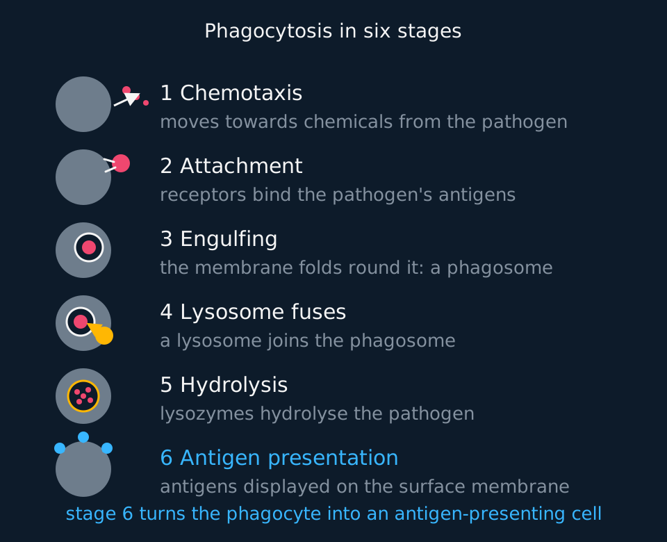
Phagocytes are white blood cells that engulf and digest pathogens. Neutrophils are the short-lived ones that arrive first and die in large numbers, which is what pus is; macrophages are larger, live longer, and present antigen to lymphocytes. The process is the same in both, and is described as an ordered sequence.
Describe the drawing
Six numbered rows, each with a small drawing of a phagocyte on the left. The cell moves along a chemical gradient, binds the pathogen's antigens with its receptors, folds its membrane round the pathogen to form a phagosome, has a lysosome fuse with that phagosome, hydrolyses the pathogen and absorbs the products, and finally displays the pathogen's antigens on its own surface membrane.
- Phagocytosis
- The engulfing and hydrolysis of a pathogen or other foreign material by a phagocyte, forming a phagosome that fuses with a lysosome.
- Phagosome
- The vesicle formed when a phagocyte's cell-surface membrane closes around an engulfed pathogen.
- Lysosome
- An organelle containing hydrolytic enzymes, including lysozymes, kept separate from the cytoplasm.
- Antigen-presenting cell
- A cell displaying antigens from a pathogen or other non-self material on its own cell-surface membrane, such as a phagocyte after phagocytosis, an infected body cell or a B cell that has taken in the antigen.
In words: the phagocyte moves towards the pathogen along a concentration gradient of chemicals released by the pathogen itself or by damaged tissue, which is chemotaxis. Receptors on the phagocyte's membrane attach to the pathogen's surface: some recognise molecular patterns shared by whole classes of microbe, others grip antibodies or complement proteins already coating the pathogen, coatings called opsonins that make the hold much firmer. The membrane then folds around it, and the two edges fuse to enclose the pathogen in a vesicle, the phagosome. Lysosomes move to the phagosome and fuse with it, emptying hydrolytic enzymes inside. Those enzymes hydrolyse the pathogen, and the soluble products are absorbed into the cytoplasm.
Two details matter. The enzymes remain enclosed by membrane throughout, so the phagocyte is not hydrolysed. And engulfing is endocytosis: it requires ATP and a flexible membrane, which a plant cell with a rigid cell wall does not have.
Antigen presentation
After hydrolysis, the phagocyte moves some of the pathogen's antigens to its own cell-surface membrane and displays them there. That is antigen presentation, and it turns the phagocyte into an antigen-presenting cell.
Presentation is required because of how a complementary lymphocyte is found. Millions of different T cells circulate, each with a differently shaped receptor, and the probability of the complementary one encountering free pathogen in an infected tissue is low. Presented antigen, concentrated on the surface of a cell that then travels to a lymph node full of lymphocytes, makes the meeting far more likely. In the A-level account, nothing specific begins until this has happened: no clonal selection, no antibody, no memory cell. (In the fuller picture, dendritic cells are the most effective presenters, and a B cell can also bind free antigen directly with its own receptor; the macrophage route here is the examined one.)
Phagocytosis therefore has two functions. It removes pathogens immediately, at the same rate on every exposure, and it initiates the specific response, which is faster on a second exposure. Antigen presentation is the step that links the non-specific response to the specific one.
TRY IT: Two defences, two speeds
A person is infected with the same strain of a bacterium twice, six months apart. Phagocytosis begins within hours on both occasions, but detectable antibody appears after about eight days the first time and about two days the second. Explain both observations.
Check your answer
Phagocytosis is non-specific. Phagocytes attach to antigens on anything they do not recognise as self, and nothing about that process improves with practice, so it happens just as quickly the second time as the first.
Antibody production is specific. The first time, a phagocyte had to present the antigen, the few lymphocytes with a complementary receptor had to be found and selected, and they had to divide repeatedly before enough cells existed to secrete measurable antibody, hence roughly eight days.
The second time, memory cells specific to that antigen were already present in far greater numbers than the few original lymphocytes. The selection and much of the division had already happened, so antibody appears in about two days.
The distinction is that the non-specific response has no memory and the specific response does.
In the exam
- Name the causative organism rather than repeating the disease named in the question: a bacterium, a virus, a fungus or a protoctist, with the species where known.
- To explain why antibiotics do not treat a viral infection, link the drug target to a structure or process found in bacteria but absent from viruses, such as peptidoglycan cell walls, 70S ribosomes or bacterial metabolic enzymes.
- Give phagocytosis in causal order: engulfing, then lysosome fusion, then hydrolysis, then antigen presentation. The order carries the explanation.
- Use 'hydrolyse' rather than 'break down' or 'destroy' for the action of lysosomal enzymes on a pathogen; it names the reaction.
- Classify the barriers correctly: skin, mucus, cilia and clotting are physical; stomach acid, lysozyme and sebum are chemical; the resident microbiota are a biological, competitive defence.
- An exotoxin is secreted by a living bacterium; an endotoxin is part of the Gram-negative outer membrane, released when the bacterium dies. Say which, and name the genus with it.
- Interferon acts on the neighbouring cells, not on the virus and not on the cell that made it. Write that it makes those cells resistant to viral replication.
Check yourself
A long-term smoker has destroyed much of the ciliated epithelium lining her airways, and she also takes a drug that greatly reduces the secretion of stomach acid. She suffers repeated chest infections, and a sample of her sputum is found to contain very large numbers of dead neutrophils. Explain how each of the two changes makes infection more likely, and explain what the neutrophils show about the defences that are still working.
Answer
Losing the cilia breaks a physical defence in the middle. Goblet cells are still secreting mucus, and that mucus still traps bacteria and virus particles as it always did, so the trapping half of the barrier is intact. What has gone is the removal. Cilia normally beat the mucus upwards to the throat, where it is swallowed; with the ciliated epithelium damaged, the mucus and everything caught in it stays in the airways, warm and undisturbed, which is a good place for bacteria to reproduce.
Reducing stomach acid breaks a chemical defence at the other end of the same chain. Acid normally holds the gut contents near pH 2, which denatures the enzymes and other proteins of most swallowed bacteria and kills them. Raise that pH and swallowed pathogens survive, including the ones in whatever mucus she does still clear from her lungs, which are swallowed rather than coughed out. Both defences are non-specific: they act on any pathogen at all, and they act at the same speed every time, so neither is improved by how often she has been infected before.
The dead neutrophils show that phagocytosis is running hard. Neutrophils are the short-lived phagocytes that arrive first, drawn to the infected tissue by chemotaxis along a gradient of chemicals from the bacteria and the damaged cells. Receptors on the neutrophil attach to the bacterial surface, the membrane folds round the bacterium to enclose it in a phagosome, lysosomes fuse with the phagosome and empty hydrolytic enzymes into it, and the bacterium is hydrolysed. Neutrophils die in the process in very large numbers, which is what pus and that sputum sample are.
So the phagocytes are doing the work the barriers are no longer doing, and they are doing it without needing to know what the pathogen is. They also do what the barriers cannot: macrophages at the site present the bacterial antigens on their own cell-surface membranes, which is the route by which a specific response, meaning clonal selection, antibody production and memory cells, begins. Antigen presentation is the link between the non-specific and specific responses.
Questions
Question 15 marks
Describe the stages of phagocytosis, in order, from the phagocyte approaching a bacterium to the display of its antigens.
Mark scheme
- B1 the phagocyte moves towards the bacterium along a concentration gradient of chemicals released by the pathogen or by damaged tissue, which is chemotaxis
- B1 receptors on the phagocyte's cell-surface membrane attach to antigens on the bacterium's surface
- B1 the membrane folds around the bacterium and the edges fuse, enclosing it in a vesicle called a phagosome
- B1 lysosomes move to the phagosome and fuse with it, emptying hydrolytic enzymes inside, and those enzymes hydrolyse the bacterium
- B1 the phagocyte moves some of the bacterium's antigens to its own cell-surface membrane and displays them, becoming an antigen-presenting cell
Question 25 marks
Describe the transmission, mode of infection and pathogenic effect of the stem rust fungus, Puccinia graminis, on wheat.
Mark scheme
- B1 spores are released from pustules on infected plants and carried by the wind, over long distances, to other wheat plants
- B1 a spore germinates on the stem or leaf in a film of water, and the germ tube grows to a stoma and enters through it
- B1 hyphae grow between the host cells and haustoria push into living cells and absorb sugars and other nutrients
- B1 less sugar reaches the grain, so the grains are small and shrivelled and yield falls, and the infected tissue photosynthesises less
- B1 pustules rupture the epidermis so more water is lost, and weakened stems break or fall over
Question 34 marks
Compare physical barriers with chemical barriers as defences against infection, using a named example of each.
Mark scheme
- B1 both act against any pathogen without recognising which one it is, and both act at the same speed on every exposure
- B1 a physical barrier blocks or removes pathogens, whereas a chemical barrier destroys them
- B1 a named physical barrier with how it works, for example skin, which is dry, dead and keratinised, or mucus which traps particles while cilia sweep it to the throat
- B1 a named chemical barrier with how it works, for example stomach acid near pH 2, which denatures the enzymes and other proteins of swallowed bacteria, or lysozyme, which hydrolyses bacterial cell walls
Question 44 marks
A drug prevents lysosomes from fusing with phagosomes inside a phagocyte. Suggest how this would affect a patient's response to a bacterial infection.
Mark scheme
- B1 bacteria are still engulfed, because chemotaxis, attachment and the forming of the phagosome are unaffected
- B1 hydrolytic enzymes are not emptied into the phagosome, so the bacteria are not hydrolysed and may survive inside the phagocyte
- B1 fewer antigens become available to be moved to the phagocyte's cell-surface membrane, so antigen presentation is reduced
- B1 the specific response is therefore slow to begin, since clonal selection cannot start until antigen is presented, and the infection is likely to be more severe and to last longer
Question 54 marks
Explain how cholera toxin causes the severe diarrhoea seen in cholera.
Mark scheme
- B1 cholera toxin binds to cells lining the small intestine and raises the level of the messenger molecule cyclic AMP inside them
- B1 this holds open a chloride channel, so chloride ions flow out of the cells through it into the lumen of the gut
- B1 this lowers the water potential of the gut lumen below that of the epithelial cells and the tissue fluid behind them
- A1 water then moves out of the cells and into the lumen down a water potential gradient by osmosis, producing large volumes of watery diarrhoea
Question 64 marks
A virus enters a host cell, replicates and is released. Explain how the virus uses the host cell to produce new virus particles, and explain how the release of those particles usually affects the host cell.
Mark scheme
- B1 the virus's nucleic acid is transcribed and translated using the host cell's own ribosomes, enzymes and nucleotides, since a virus has no metabolism of its own
- B1 hundreds of new virus particles are assembled inside the host cell using these host resources
- B1 many viruses are released by bursting the cell open, which is lysis and destroys the host cell
- A1 other viruses, HIV among them, bud out through the cell surface membrane instead, so the infected cell declines more gradually rather than bursting at once
Question 74 marks
A cell lining the airway is infected by influenza virus and secretes interferon. Explain how this reduces the spread of the virus to other cells in the tissue.
Mark scheme
- B1 interferon is a protein that diffuses from the infected cell and binds to receptors on the cell-surface membranes of neighbouring cells
- B1 binding switches on genes in those cells, putting them into an antiviral state
- B1 the proteins made inhibit protein synthesis at the ribosomes, or activate an enzyme that hydrolyses viral RNA
- B1 a virus entering one of those cells cannot be replicated, because it depends on the host cell's ribosomes and enzymes to make new virus particles
Question 84 marks
Staphylococcus species produce exotoxins and Salmonella species produce endotoxins. Compare exotoxins with endotoxins.
Mark scheme
- B1 an exotoxin is a protein, whereas an endotoxin is a lipopolysaccharide that forms part of the outer membrane of the bacterial cell wall
- B1 an exotoxin is secreted by living bacteria, whereas an endotoxin is released mainly when the bacteria die and their walls break down
- B1 exotoxins are made by Gram-positive and Gram-negative bacteria, whereas endotoxins come only from Gram-negative bacteria such as Salmonella
- B1 an exotoxin acts on a specific target and is potent in small amounts, whereas an endotoxin causes general effects such as fever and inflammation through the host's response
Question 93 marks
Explain why an antibiotic that cures tuberculosis has no effect whatever on influenza.
Mark scheme
- B1 tuberculosis is caused by a bacterium while influenza is caused by a virus
- B1 antibiotics work by disrupting structures or processes that bacteria have and human cells do not, such as the peptidoglycan cell wall or the 70S ribosomes
- B1 a virus has no cell wall, no ribosomes and no metabolism of its own, so there is nothing for the drug to disrupt
Question 103 marks
Explain how inflammation at the site of an infection helps the body to clear the pathogen.
Mark scheme
- B1 mast cells in the damaged tissue release histamine
- B1 histamine dilates the arterioles and makes capillary walls more permeable, so more blood arrives and more tissue fluid leaks out into the tissue
- B1 that tissue fluid carries antibodies into the infected tissue, and cytokines attract phagocytes, which leave the capillaries and move to the site
Question 113 marks
Oral rehydration solution, a drink of water, salts and glucose, does nothing whatever to the cholera bacterium, yet it keeps most cholera patients alive. Suggest why.
Mark scheme
- B1 the damage in cholera is done by a toxin rather than by the bacterium itself: the toxin holds open the chloride channels of the cells lining the small intestine, so chloride ions pour into the lumen
- B1 water follows the chloride into the lumen by osmosis, and the diarrhoea can remove several litres a day, so what kills is dehydration, the loss of water and salts, which the solution replaces by mouth
- A1 the bacterium does not destroy the host's cells the way a virus does by lysis, so a patient whose water and salts are kept replaced survives while the body's defences clear the infection
Question 123 marks
Bacteria in a deep wound release tetanus toxin, which travels to the spinal cord. There it stops certain neurones releasing a transmitter that normally inhibits motor neurones, making them less likely to fire. Suggest how the toxin causes the sustained muscle spasms of tetanus.
Mark scheme
- B1 without that inhibition, the motor neurones fire impulses far more often, even continuously
- B1 so acetylcholine is released at the neuromuscular junctions almost without pause, and the muscle fibres are stimulated again and again
- B1 the muscle contracts and cannot relax, producing sustained spasms
Question 133 marks
Much of the lung damage seen in tuberculosis is caused by the body's own inflammatory response rather than by a toxin the bacterium releases. Explain how this happens.
Mark scheme
- B1 Mycobacterium tuberculosis survives inside the very macrophages that engulf it, rather than being destroyed by them
- B1 the host's immune system responds with a sustained inflammatory response that walls the infected cells off into masses called granulomas
- A1 it is this ongoing walling-off response, rather than a released toxin, that causes much of the tissue damage seen in the lungs
Question 143 marks
Compare a neutrophil with a macrophage as phagocytes, referring to how long-lived each is and what each does after it has engulfed a pathogen.
Mark scheme
- B1 neutrophils are short-lived and arrive first at a site of infection, whereas macrophages are larger and live for longer
- B1 large numbers of neutrophils die in the process of phagocytosis, and their dead bodies are what makes up pus, whereas macrophages usually survive engulfing a pathogen
- A1 macrophages, but not neutrophils, present antigens from the pathogen on their own cell-surface membrane, linking the non-specific response to the specific one
Question 153 marks
Antibodies and complement proteins can coat a pathogen before a phagocyte engulfs it. Explain how this coating, called an opsonin, makes phagocytosis more effective.
Mark scheme
- B1 receptors on the phagocyte's cell-surface membrane can grip the antibody or complement coating a pathogen, as well as recognising patterns on the pathogen's own surface
- B1 this gives the phagocyte an additional, firmer hold on the pathogen than binding directly to the pathogen's own surface alone would give
- A1 opsonisation therefore speeds up attachment and increases the chance that the pathogen is engulfed
Question 163 marks
A fever raises body temperature by a few degrees. A bacterium that infects humans grows fastest at 37 °C and more slowly at 40 °C. Suggest why the bacterium grows more slowly at 40 °C, and suggest why a body temperature above about 41 °C is dangerous to the patient.
Mark scheme
- B1 40 °C is above the optimum of some of the bacterium's enzymes, so bonds holding their tertiary structure break and the shape of the active site changes
- B1 fewer enzyme-substrate complexes form, so the bacterium's metabolic reactions slow and it grows and divides more slowly
- B1 the patient's own enzymes and other proteins also begin to denature at such temperatures, disrupting metabolism, especially in the brain
Question 173 marks
A patient prescribed a broad-spectrum antibiotic for a bacterial infection develops digestive upset during the course of treatment. Suggest why.
Mark scheme
- B1 resident microbiota in the gut normally provide a biological, competitive defence by occupying space and using resources that would otherwise be available to pathogens
- B1 a broad-spectrum antibiotic kills bacteria generally, including a good many of the resident gut microbiota, not only the pathogen being treated
- A1 with the resident microbiota reduced, the balance of the gut is disturbed, which is what produces the digestive upset
Question 183 marks
The table shows the usual time between eating contaminated food and the first symptoms for two causes of food poisoning. Suggest an explanation for the difference, in terms of the type of toxin each produces.
| Cause of food poisoning | Usual time from eating to first symptoms |
|---|---|
| Staphylococcus aureus | A few hours |
| Salmonella species | 12 hours to 3 days |
Mark scheme
- B1 Staphylococcus produces an exotoxin, which it secretes into the food while growing in it before the food is eaten
- B1 the toxin is already present when the food is eaten, so it acts at once without the bacteria needing to grow in the gut
- B1 Salmonella produces endotoxin, which is part of its wall, so the bacteria must first multiply and invade the gut lining and be killed before enough endotoxin is released to cause symptoms
Question 193 marks
A race of stem rust called Ug99 was identified in Uganda in 1999. It can infect wheat varieties carrying the resistance gene Sr31, which most other races cannot. Explain how Ug99 could have spread once wheat carrying Sr31 was widely grown.
Mark scheme
- B1 a mutation in the fungus produced a variant that the Sr31 resistance did not recognise or stop
- B1 on fields of Sr31 wheat that variant could infect plants and reproduce while other races could not, so it was selected
- B1 it produced large numbers of spores which were carried by the wind to other fields and regions, so its frequency in the fungus population increased
Question 202 marks
Give one named human disease caused by a bacterium and one named human disease caused by a protoctistan.
Mark scheme
- B1 a bacterial disease named, for example tuberculosis caused by Mycobacterium tuberculosis, or cholera caused by Vibrio cholerae
- B1 a protoctistan disease named, for example malaria caused by Plasmodium
Question 212 marks
Interferon is classed as part of the non-specific response to infection. Explain why.
Mark scheme
- B1 it is released in response to infection by many different viruses, and the antiviral state it produces acts against any virus
- B1 it does not recognise or bind a particular antigen, and it is produced at the same speed on every infection, with no memory
Worth remembering
- Four groups of pathogen: bacteria, viruses, fungi, protoctista. Learn one named disease for each.
- Pathogens damage the host mainly by releasing toxins or by destroying the cells they reproduce inside.
- Physical barriers block; chemical barriers destroy. Both act on any pathogen at all.
- Phagocytosis in order: chemotaxis, attachment, engulfing into a phagosome, lysosome fusion, hydrolysis, antigen presentation.
- Antigen presentation is the handover from the non-specific response to the specific one.
- Exotoxins are secreted proteins (Staphylococcus); endotoxins are wall lipopolysaccharide (Salmonella); M. tuberculosis damages by invasion.
- Interferon from an infected cell puts its neighbours into an antiviral state, against any virus.
- Stem rust: wind-borne spores, entry through stomata, haustoria taking sugars from living cells, shrivelled grain.
CHECK YOUR PROGRESS
Rate how confident you are with each objective for this lesson. Ratings are saved in this browser, on this device, unless you sign in.
- Name the four groups of pathogen and give a disease caused by each.
- Explain how a pathogen damages its host, using toxins and cell lysis as the two standard exam routes.
- Distinguish a physical barrier from a chemical one, with named examples of both.
- Describe phagocytosis as an ordered sequence, ending with antigen presentation.
- Explain why an antibiotic that cures tuberculosis does nothing whatever for influenza.
- Distinguish an exotoxin from an endotoxin, with Staphylococcus and Salmonella as the examples, and set both beside invasion of tissue by Mycobacterium tuberculosis.
- Describe the transmission, mode of infection and pathogenic effect of the stem rust fungus, Puccinia graminis, on wheat.
- Explain how interferon released by a virus-infected cell protects the cells around it.
Open the full revision checklist to see every objective in the curriculum in one place.
Practise this lesson
WORKBOOK
The written questions from this topic, on paper with room to work, and a separate book of mark schemes. The question that is set on a diagram stays on this page, where the diagram can be drawn. Free to use; please do not redistribute or sell.