Biology › Nervous coordination, receptors and muscles › Receptors: transduction and the reflex arc
Receptors: transduction and the reflex arc
A receptor detects a particular form of stimulus and transduces its energy into a change in membrane potential. The resulting generator potential is graded, and if threshold is reached in the associated sensory neurone, action potentials of constant amplitude are produced. The reflex arc, the Pacinian corpuscle, a taste-bud chemoreceptor, and rods against cones are the worked examples.
Before this Membrane structure and channel proteins · Active transport and the use of ATP
COMMON MISCONCEPTION
Your fingertip detects heat and sends the heat up your arm; your eye detects colour and sends colour along the optic nerve.
A receptor transduces stimulus energy into a change in membrane potential. The nervous system distinguishes stimuli through the pathway carrying the impulses and their frequency; the original pressure, light or chemical does not travel along the sensory neurone.
What you should be able to do
- Put the stages from stimulus to response in order, and name the three neurones in a spinal reflex arc.
- Explain what makes a reflex fast, and what it protects you from.
- Describe how pressure on a Pacinian corpuscle produces a generator potential.
- Distinguish a generator potential from an action potential, using the word graded correctly.
- Describe how a taste bud chemoreceptor turns a dissolved chemical into an action potential in the sensory neurone at its base.
- Compare rods and cones on sensitivity, acuity, pigment and distribution, and explain the first two using convergence.
- Distinguish a taxis from a kinesis, and explain how each keeps a mobile organism in a favourable environment.
- Describe the layers of the retina from the ganglion cells to the rods and cones, and explain why there is a blind spot.
- Describe how to use a choice chamber or maze to investigate an animal's response to light or humidity, with its controls, and test the counts with chi-squared.
From stimulus to response
Coordination in an animal runs through five stages in a fixed order: stimulus, receptor, coordinator, effector, response. The coordinator is the central nervous system, and the two neurones on either side of it are the sensory and the motor.
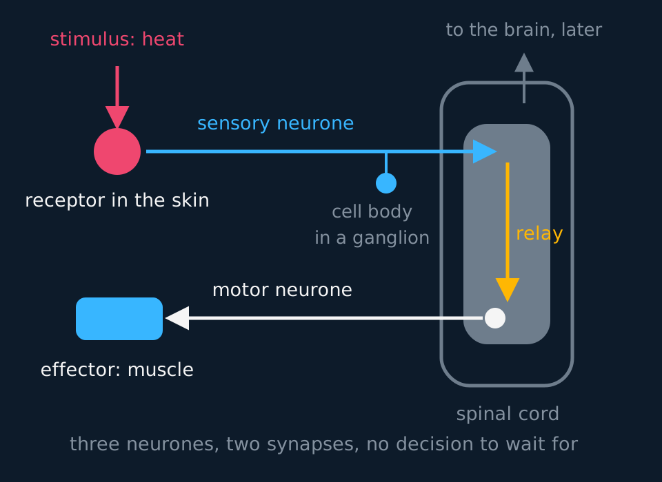
A reflex uses the shortest available pathway, so the response begins before the stimulus has been consciously perceived. Impulses pass from the receptor along a sensory neurone into the spinal cord, across a relay neurone and out along a motor neurone to the biceps, which contracts. The pathway crosses two synapses. That three-neurone withdrawal arc is the standard A-level example rather than the shape of every reflex: the knee-jerk stretch reflex, for one, runs sensory neurone straight onto motor neurone with no relay neurone at all.
Describe the drawing
A reflex arc drawn as a loop: heat reaching a receptor in the skin on the left, a sensory neurone carrying impulses right into the grey matter of a spinal cord shown in section, with its cell body on a short side branch in a ganglion outside the cord, a relay neurone running down inside the cord, and a motor neurone whose cell body lies in the cord carrying impulses back left to a muscle acting as the effector. A faint arrow leaves the cord upwards to show the brain being informed afterwards rather than consulted first.
State the brain's part precisely. Impulses do reach the brain, which is how the stimulus is perceived and how the response is learned from, but the response does not depend on a decision from the brain.
Three features make the pathway fast: a short pathway, only two synapses to cross, and a route through the spinal cord rather than the brain. Each synapse adds about half a millisecond of delay, so the number of synapses has a measurable effect on the response time. Reflexes limit damage in the interval before a conscious response could occur: withdrawal from heat, blinking at a moving object, coughing on an inhaled particle and constriction of the pupil in bright light.
- Stimulus
- A detectable change in the internal or external environment.
- Sensory receptor
- A cell or structure that detects one kind of stimulus and converts it into a change in membrane potential.
- Effector
- A muscle or gland that brings about a response.
- Reflex
- A rapid, automatic response to a stimulus, involving a fixed pathway of neurones and no conscious decision.
- Reflex arc
- The pathway of neurones taken by a reflex. In the examined withdrawal reflex: receptor, sensory neurone, relay neurone, motor neurone, effector.
Simple responses: taxes and kineses
An organism survives better if it stays in conditions that suit it, and a mobile organism can do that with responses far simpler than a reflex arc. AQA names two kinds.
A taxis is a directional movement in response to a directional stimulus. The organism moves towards the stimulus, a positive taxis, or away from it, a negative taxis, and the name of the stimulus goes in front: phototaxis for light, chemotaxis for a chemical. Woodlice show negative phototaxis, moving away from light, which tends to take them into dark, sheltered places where they lose less water and are less easily found by predators. Single-celled photosynthetic organisms such as Euglena show positive phototaxis, swimming towards light, which keeps them where they can photosynthesise. Many bacteria swim up a concentration gradient of a nutrient, which is positive chemotaxis.
A kinesis is a non-directional response. The organism does not move towards or away from the stimulus; instead the intensity of the stimulus changes how fast it moves or how often it turns. Woodlice move faster in dry air than in humid air. A woodlouse that wanders into a dry area therefore tends to leave it quickly, and one in a damp area slows down and stays, so over time the population gathers where it is damp, although no individual ever moved towards the damp. A kinesis suits stimuli such as humidity and temperature, which often have no clear direction.
| Taxis | Kinesis | |
|---|---|---|
| Direction of the stimulus | Matters | Does not matter |
| Response | Moves towards or away from the stimulus | Changes speed of movement or rate of turning |
| What sets the response | The direction of the stimulus | The intensity of the stimulus |
| Example | Woodlice moving away from light | Woodlice moving faster in dry air |
These responses are investigated with a choice chamber or a maze, which is AQA's required practical 10 and is set out below. A headcount at the end shows where the animals gathered but not which kind of response put them there: telling a taxis from a kinesis needs individual paths tracked. Testing the counts from a woodlouse choice chamber with chi-squared is worked through in the lesson on choosing and concluding a statistical test.
- Taxis
- A directional movement in response to a directional stimulus, towards it (positive) or away from it (negative).
- Kinesis
- A non-directional response in which the speed of movement or the rate of turning changes with the intensity of a stimulus.
Required practical 10: the choice chamber and the maze
optional
AQA sets this as required practical 10: Investigation into the effect of an environmental variable on the movement of an animal using either a choice chamber or a maze. Woodlice or blowfly maggots are the usual animals.
A choice chamber is a shallow dish divided into two or four compartments under a gauze floor, with a hole in the centre of the lid. To set humidity, put a drying agent, anhydrous calcium chloride or silica gel, beneath the gauze on one side and a little water on the other. To set light, cover the lid over one half with black card. Change one variable at a time: in a humidity run the whole chamber is lit evenly, and in a light run both sides have the same water underneath.
| Step | What to do | Why |
|---|---|---|
| 1 | Close the chamber for about 10 minutes, with the animals in damp leaf litter beside it | Each side reaches its humidity; the animals settle after handling |
| 2 | Release ten animals together through the central hole and close it | No animal starts nearer one side |
| 3 | Count the animals on each side every 30 s for 10 minutes, or once at a fixed end time, with a rule decided beforehand for an animal on the line. In a light run, lift the card only briefly to count the dark side | A time series shows how fast they redistribute |
| 4 | Repeat with fresh animals, swapping which side holds the stimulus | A bias towards one side cancels out |
Run the chamber once with both sides identical. The animals should then split about evenly; if not, the chamber, a window or a draught is biasing them. Keep it away from a lamp, which warms one side as well as lighting it.
Woodlice and light
These results are idealised. Four runs of ten woodlice, light side swapped each time, gave 27 in the dark and 13 in the light after 10 minutes. The critical value at p = 0.05 with 1 degree of freedom is 3.84.
Show the working
Null hypothesis: there is no significant difference between the observed numbers and those expected if light had no effect.
With no effect the split would be 1:1, so E = 40 ÷ 2 = 20 on each side, above 5. (27 − 20)² ÷ 20 = 2.45 and (13 − 20)² ÷ 20 = 2.45, so chi-squared = 4.90. Degrees of freedom = 2 categories − 1 = 1.
4.90 is greater than 3.84, so the null hypothesis is rejected: significantly more woodlice settled in the dark than chance would give.
To tell a taxis from a kinesis, follow single animals across a gridded arena or film them from above, and measure speed and turns per minute on each side. Paths that head straight away from the light are a taxis; faster movement or more turning in dry air, with no direction to the paths, is a kinesis.
A T-maze gives one choice per trial: the animal walks up the stem and turns left or right, with the stimulus in one arm. Woodlice tend to turn the opposite way to their previous turn, so a forced turn before the junction biases the choice. Approach the junction straight, or alternate the side of the forced turn.
Handle the animals gently with a soft paintbrush, keep runs short, and return them to where they were found. Keep the drying agent under the gauze, and check its CLEAPSS Hazcard and your school's risk assessment before starting.
Limitations: woodlice gather where other woodlice are, so the animals in a run are not independent; humidity is assumed unless a probe measures it; and light leaks under the card's edge.
What a receptor does
A receptor detects a particular form of stimulus and transduces its energy into a change in membrane potential. The resulting generator potential is graded: a stronger stimulus produces a larger change. If threshold is reached in the associated sensory neurone, action potentials are produced. Their amplitude is constant; stimulus intensity is represented mainly by their frequency.
Different receptor types are most sensitive to different forms of stimulus energy. Rod and cone cells transduce light; a Pacinian corpuscle transduces changes in pressure.
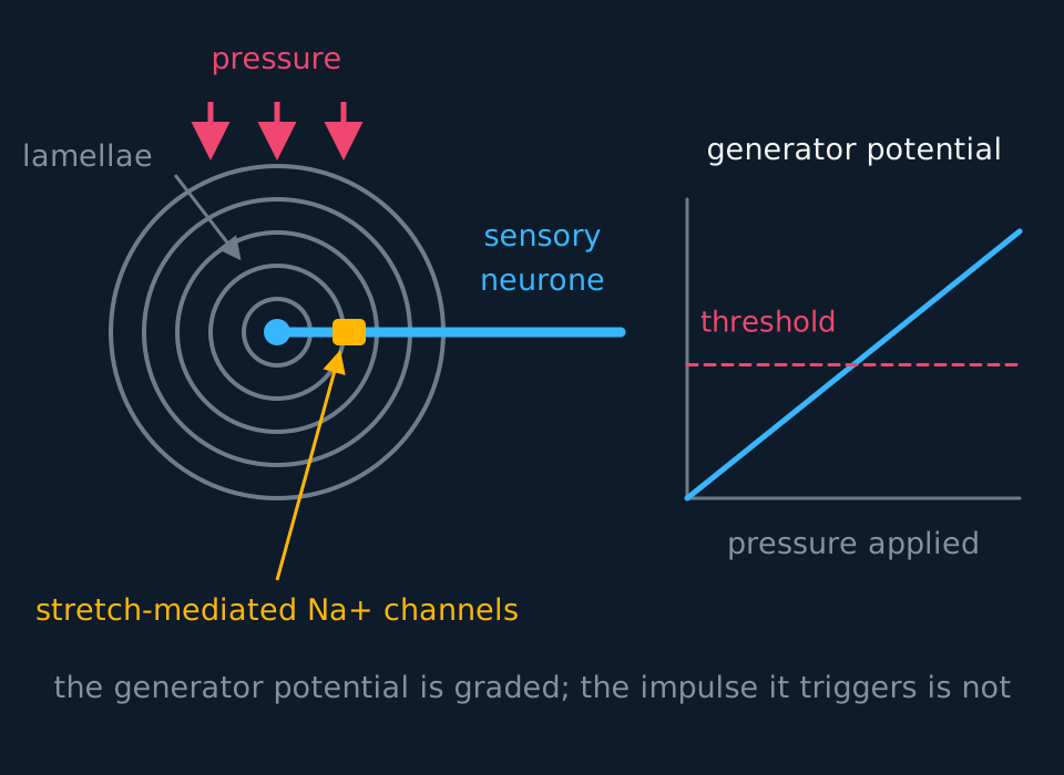
The Pacinian corpuscle is the standard worked example. It sits deep in the skin, in joint capsules, in ligaments and tendons, and it responds to changes in pressure. Under the microscope it looks like an onion about a millimetre long: rings of connective tissue called lamellae, separated by a gel, wrapped round the ending of a single sensory neurone.
Describe the drawing
A Pacinian corpuscle drawn as five concentric rings of lamellae round a single nerve ending, with pressure arrows pressing down on the top, a stretch-mediated sodium channel marked on the membrane of the ending and the sensory neurone leaving to the right. Beside it a graph of generator potential against applied pressure rises as a straight line and crosses a marked threshold, showing the response is graded rather than fixed.
Press on the corpuscle and the lamellae deform, which stretches the membrane of the nerve ending inside them. That membrane contains stretch-mediated sodium channels: channels whose shape, and therefore whether they are open, depends on being physically pulled. Stretched open, they let sodium ions diffuse in down their electrochemical gradient, and the inside of the ending becomes less negative. That depolarisation is the generator potential.
A greater pressure deforms more lamellae, stretches more channels, admits more sodium and gives a bigger generator potential. If the generator potential is large enough to bring the membrane to about −55 mV, which is the threshold, an action potential is triggered in the sensory neurone. If not, the depolarisation simply fades away and nothing is sent. So the receptor's response is graded and the neurone's is all-or-nothing, and the threshold is the step between them.
- Transducer
- Something that converts energy from one form into another; a receptor converts the energy of a stimulus into a change in membrane potential.
- Generator potential
- The graded depolarisation produced in a receptor by a stimulus, whose size depends on the strength of that stimulus.
- Threshold
- The membrane potential, around −55 mV, that must be reached before an action potential is triggered.
Working out what will and will not fire
In one Pacinian corpuscle a pressure of one arbitrary unit produces a generator potential of 6 mV of depolarisation, and the relationship is proportional. The resting potential is −70 mV and the threshold is −55 mV. Will pressures of 2 units and of 3 units each trigger an impulse?
Show the working
First find what is needed. Going from −70 mV to −55 mV is a depolarisation of 15 mV, so that is the size of generator potential required.
Two units gives 2 × 6 = 12 mV, which reaches −58 mV. That is short of threshold, so no action potential is triggered and the depolarisation dies away.
Three units gives 3 × 6 = 18 mV, which takes the membrane past −55 mV. An action potential is triggered, and it will be exactly the same size as one triggered by ten units, because the generator potential is graded but the impulse is not.
A chemoreceptor transduces a different form of stimulus by the same general route. A taste bud on the tongue holds a cluster of receptor cells, each an elongated epithelial cell rather than a neurone, with microvilli poking through a pore into the saliva above. A chemical dissolved in the saliva is the stimulus; sodium chloride is the cleanest case. Sodium ions from the salt diffuse through channels in the microvillus membrane into the receptor cell, and the entering positive charge depolarises it, exactly as stretched-open channels depolarise the Pacinian ending.
If that depolarisation reaches threshold, voltage-gated calcium channels in the receptor cell open, calcium ions enter, and vesicles of neurotransmitter fuse with its basal membrane, because a taste receptor cell does not run its own impulse anywhere: it synapses with the sensory neurone wrapped round its base, and the transmitter it releases is what triggers the action potential that travels to the brain. The sequence to give is four steps, in this order: stimulus, depolarisation of the receptor cell, transmitter across the synapse, action potential in the sensory neurone. The same sequence applies to other receptor types, with a different stimulus and a different channel.
The retina, layer by layer
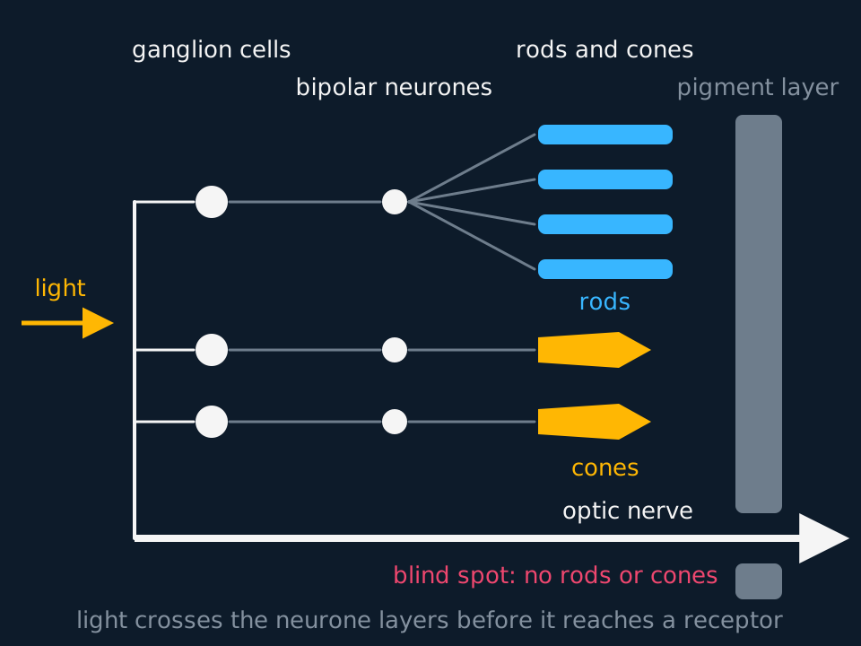
The retina lines the back of the eye, and in section it has three layers of cells, arranged the opposite way round from what might be expected: the light-sensitive cells are at the back. Light entering through the lens passes first through a layer of ganglion cells, then through a layer of bipolar neurones, and only then reaches the rods and cones. Both neurone layers are thin and nearly transparent, so little light is lost crossing them. Behind the receptors is a pigmented layer that absorbs the light the receptors did not, so it is not scattered back across the retina to blur the image.
Describe the drawing
A cross-section of the retina with light arriving from the left. Nearest the light is a column of ganglion cells, then a column of bipolar neurones, then the rods and cones, with a grey pigment layer behind them at the back of the eye. Four rods connect to a single bipolar neurone, while each of two cones connects to a bipolar neurone of its own, and every bipolar neurone connects to one ganglion cell. The ganglion cells' axons run along the inner surface to one point, then pass back through all the layers as the optic nerve. That point has no rods or cones and is labelled the blind spot.
Signals run in the other direction. A rod or cone synapses with a bipolar neurone, and the bipolar neurone synapses with a ganglion cell. The ganglion cells generate the action potentials that are sent to the brain. Their axons run across the inner surface of the retina to one point, where about a million of them gather and leave the eye together as the optic nerve.
Where the optic nerve leaves, its axons pass back through every layer, and there is no room for receptors. This is the blind spot, also called the optic disc: it has no rods or cones, so light falling on it is not detected. The gap goes unnoticed in everyday vision because the blind spots of the two eyes receive light from different parts of the scene and the brain fills in what is missing.
The fovea is the opposite case. It lies close to the centre of the retina, directly behind the lens, and there the ganglion and bipolar layers are pushed aside, so light reaches the tightly packed cones with less tissue in the way. Together with the one-to-one wiring of its cones, this is why detail is seen best in the centre of the visual field.
TRY IT: Finding the blind spot
Draw a cross and, about 8 cm to its right, a dot. Close the left eye, look steadily at the cross with the right eye, and move the page slowly towards your face. At one distance the dot disappears. Explain why, and explain why it reappears when the page is moved closer still.
Check your answer
Looking at the cross keeps its image on the fovea. The dot's image falls on another part of the retina, and its position changes as the page moves.
At one distance the dot's image falls on the blind spot, where the optic nerve leaves the eye. There are no rods or cones there, so no generator potentials are produced, no impulses are sent from that point, and the dot is not seen. The brain fills the gap with the background, so a blank page appears instead.
Moving the page closer still shifts the dot's image further across the retina. Once it is off the blind spot it falls on rods and cones again and is seen.
- Bipolar neurone
- A neurone in the retina that carries the signal from rods or cones to a ganglion cell.
- Ganglion cell
- A neurone in the retina whose axon forms part of the optic nerve, carrying action potentials to the brain.
- Blind spot
- The point where the optic nerve leaves the retina; it has no rods or cones, so light falling there is not detected.
- Fovea
- The small region of the retina directly behind the lens, packed with cones and containing no rods, where acuity is greatest.
Two kinds of photoreceptor
The retina carries two receptor types, and every comparison question about them can be answered from four differences: the pigment, the light needed, the detail resolved and where in the retina they sit.
Rods contain the pigment rhodopsin, which is broken down, or bleached, by relatively little light. There are roughly 120 million of them in a human retina, they respond to all wavelengths together and so give a monochrome image, and they are spread across the peripheral retina with none at all in the fovea.
Cones contain iodopsin, which needs a good deal more light to break down. There are about 6 million, in three types, called long-, middle- and short-wavelength cones, whose pigments absorb best at roughly 560, 530 and 420 nm. 'Red, green and blue cones' is the familiar shorthand, though the long-wavelength peak actually sits in the yellow-green. Colour vision comes from comparing how strongly the three are stimulated. They are concentrated at the fovea, directly behind the centre of the lens, and thin out towards the edges.
Because rhodopsin is broken down in dim light and iodopsin is not, only the rods respond at low light intensities, so vision at night is monochrome. Rhodopsin then has to be resynthesised, and that takes time: the twenty minutes or so your eyes need to adjust when you walk into a dark room is the chemistry of that resynthesis, not a mental adjustment.
A rod responds to light in the opposite direction from the Pacinian corpuscle. In the dark, cation channels in the rod membrane are open, sodium ions diffuse in, and the partly depolarised rod releases a transmitter that inhibits its bipolar neurone. Light splits rhodopsin into retinal and opsin, and the opsin sets off a chain of reactions that closes the cation channels. With sodium ions no longer entering, the rod hyperpolarises and stops releasing transmitter. Freed from inhibition, the bipolar neurone depolarises and passes the signal to a neurone of the optic nerve, which carries action potentials to the brain. Pearson Edexcel asks for this sequence; AQA treats the rod's response more simply as a generator potential, which is how the next section describes it.
| Rods | Cones | |
|---|---|---|
| Pigment | Rhodopsin | Iodopsin, in three types |
| Light needed | Low: bleached by dim light | High: needs bright light |
| Number | About 120 million | About 6 million |
| Distribution | Peripheral retina, none in the fovea | Concentrated in the fovea |
| Image | Monochrome | Colour, from three pigments |
| Connection | Several rods share one bipolar neurone | Close to one cone per bipolar neurone at the fovea |
| Sensitivity | High | Low |
| Visual acuity | Low | High |
- Rhodopsin
- The light-sensitive pigment in rod cells, broken down by low light intensities into retinal and opsin.
- Iodopsin
- The light-sensitive pigment in cone cells, in three types, broken down only by higher light intensities.
Convergence, sensitivity and acuity
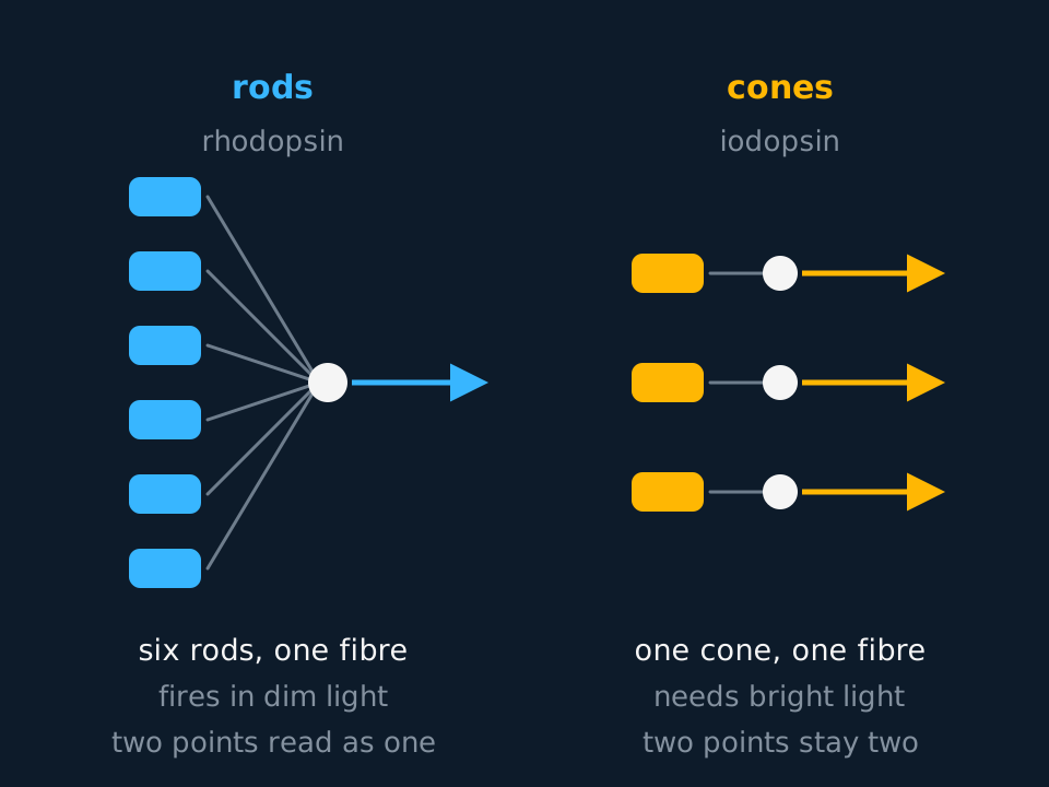
The last two rows of that table both follow from the pattern of connections to the bipolar neurones, which is convergence. Explain rod sensitivity and cone acuity from it.
Describe the drawing
Two wiring diagrams side by side. On the left six rod cells in a column all send connections to a single shared bipolar junction and one nerve fibre leaves it. On the right three cone cells each connect to their own junction and their own separate fibre. Labels give rhodopsin for the rods and iodopsin for the cones, and note that the shared fibre fires in dim light but reports two points of light as one.
Sensitivity first. In dim light a single rod produces only a small generator potential, nowhere near enough on its own. But several rods, in the peripheral retina sometimes a hundred of them, connect to the same bipolar neurone, so their contributions are added together by spatial summation. Enough small potentials arriving at once will reach threshold, and one impulse is sent. At the fovea a cone has a bipolar neurone essentially to itself (cones further out share connections too), so nothing is added to its contribution; it needs enough light to reach threshold single-handed.
Acuity follows from the same arrangement. If two points of light fall on two different rods that share a neurone, the brain receives one impulse from one fibre and cannot tell whether one point or two caused it: the two points are seen as one. Two points falling on two cones send impulses along two separate fibres, and are resolved as two. High sensitivity and high acuity pull in opposite directions, and the retina solves the problem by having both kinds of receptor in different places.
TRY IT: Explaining an observation with the wiring
Astronomers use a technique called averted vision: to see a very faint star, they look slightly to one side of it rather than straight at it. Explain why this works.
Check your answer
Looking straight at something focuses its image on the fovea, and the fovea contains cones and almost no rods. Cones need bright light to break down iodopsin, and a faint star does not provide it, so the cones do not reach threshold and nothing is sent.
Looking slightly to one side moves the image onto the peripheral retina, which is rod territory. Rhodopsin is broken down by much less light, and several rods converge on one bipolar neurone, so their generator potentials sum. Threshold is reached, an action potential is generated, and the star is seen.
State the cost as well. The image formed by the rods has low acuity, so the star is visible but blurred and its position is imprecise. The gain in sensitivity is accompanied by a loss of detail.
- Retinal convergence
- Several rods connecting to one bipolar neurone, so their generator potentials are summed.
- Visual acuity
- The ability to distinguish two points close together as separate.
In the exam
- Name all three neurones rather than referring to a nerve: sensory, relay and motor.
- State that a reflex response does not depend on a decision from the brain, rather than that the brain is not involved: impulses do reach it.
- Use 'graded' for the generator potential and 'all-or-nothing' for the action potential. A stronger stimulus does not produce a larger impulse.
- For the Pacinian corpuscle, give the full sequence: lamellae deformed, membrane stretched, stretch-mediated sodium channels open, sodium ions diffuse in, generator potential, threshold, action potential.
- Explain rod sensitivity and low acuity through convergence and spatial summation. The pigment alone does not account for either.
- A taxis is directional and a kinesis is not. For a kinesis, say that the speed or rate of turning changes with the intensity of the stimulus.
- Give the retinal layers in the order light meets them: ganglion cells, bipolar neurones, rods and cones. The blind spot has no receptors because the optic nerve leaves there.
Check yourself
A student presses gently on the back of their hand and feels nothing; pressing harder in the same place, they feel it clearly. Using what a Pacinian corpuscle does, explain both observations, and explain why the harder press does not produce larger impulses in the sensory neurone.
Answer
Gentle pressure deforms the lamellae only slightly, so few stretch-mediated sodium channels are opened, few sodium ions enter and the generator potential is small. It does not reach the threshold of about −55 mV, no action potential is triggered, and nothing is sent to the central nervous system.
The harder press deforms the lamellae much more, so more channels open, more sodium ions diffuse in and the generator potential is larger. This time the membrane reaches threshold and action potentials are triggered in the sensory neurone.
Their size, though, is fixed. An action potential is all-or-nothing: once threshold is passed, the voltage-gated channels open fully and the same peak is reached every time, whatever the stimulus. What a stronger stimulus changes is the frequency of impulses, and how many neurones are firing at once.
The graded response belongs to the receptor and the all-or-nothing response to the sensory neurone; they are separate stages of the same pathway.
Questions
Question 16 marks
A student covers one half of a choice chamber with black card and releases 50 woodlice at the centre, in five groups of ten. After ten minutes, 33 are in the dark half and 17 in the light half. The critical value of chi-squared at p = 0.05 with 1 degree of freedom is 3.84. Calculate chi-squared and use it to reach a conclusion. In a control run with no card, 29 of 50 woodlice were found in the half that the card had covered. Explain what this control suggests about the conclusion, and how the method should be changed.
Mark scheme
- M1 expected number on each side = 50 ÷ 2 = 25
- M1 (33 − 25)² ÷ 25 = 2.56 and (17 − 25)² ÷ 25 = 2.56
- A1 chi-squared = 2.56 + 2.56 = 5.12
- B1 5.12 is greater than 3.84 at p = 0.05 with 1 degree of freedom, so the null hypothesis is rejected: significantly more woodlice were in the dark half than chance would give
- B1 the control shows a lean towards that half with no difference in light, for example from a window or draught, so part of the excess in the dark may be side bias rather than a response to light
- B1 swap the card to the other half between runs, so any side bias falls equally on the light and dark halves
Question 25 marks
Describe how a firm pressure applied to a Pacinian corpuscle leads to an action potential in the sensory neurone it surrounds.
Mark scheme
- B1 the pressure deforms the lamellae of connective tissue, which stretches the membrane of the nerve ending inside them
- B1 stretch-mediated sodium channels in that membrane are pulled open
- B1 sodium ions diffuse into the ending down their electrochemical gradient, so the inside becomes less negative
- B1 this depolarisation is the generator potential, and it is graded, so a firmer pressure opens more channels and gives a larger one
- A1 the generator potential is large enough to bring the membrane to the threshold of about −55 mV, so an action potential is triggered in the sensory neurone
Question 35 marks
State the five stages, in order, through which a reflex response passes, from the detection of a stimulus to the response itself.
Mark scheme
- B1 stimulus, the detectable change
- B1 receptor, which detects it
- B1 coordinator, the central nervous system
- B1 effector, the muscle or gland
- B1 response, the resulting action
Question 44 marks
A person eats salted food. Describe how the sodium chloride dissolved in the saliva leads to an action potential in the sensory neurone at the base of a taste bud receptor cell.
Mark scheme
- B1 sodium ions from the dissolved salt diffuse through channels in the membrane of the microvilli, which project through the pore of the taste bud into the saliva
- B1 the positive charge entering depolarises the receptor cell, which is the transduction step: the chemical stimulus has become a change in membrane potential
- B1 when the depolarisation reaches threshold, voltage-gated calcium channels in the receptor cell open and calcium ions enter
- A1 the calcium makes vesicles of neurotransmitter fuse with the basal membrane, and the transmitter released across the synapse triggers the action potential in the sensory neurone, because the receptor cell is an epithelial cell and carries no impulse of its own
Question 54 marks
Explain why a withdrawal reflex is faster than a conscious response, and explain why the statement that the brain is not involved in a reflex would not be credited.
Mark scheme
- B1 the pathway is short: the impulses enter the spinal cord and leave it again without travelling up to the brain first
- B1 there are only two synapses to cross, and each synapse adds a delay of about half a millisecond
- B1 the pathway is fixed, so no conscious decision has to be made before the effector is stimulated
- A1 impulses do reach the brain along a branch of the pathway, which is how the pain is felt a moment later; the point is that the response does not wait for a decision from it
Question 64 marks
Compare rod cells with cone cells in the human retina, referring to their pigment, the light each needs, their distribution and the image each produces.
Mark scheme
- B1 rods contain rhodopsin, which is broken down by dim light, whereas cones contain iodopsin, which needs much brighter light to break down
- B1 rods are found in the peripheral retina and are absent from the fovea, whereas cones are concentrated at the fovea and thin out towards the edges
- B1 rods respond to all wavelengths together and give a monochrome image, whereas cones come in three types with different pigments, so comparing them gives colour
- B1 there are about 120 million rods and only about 6 million cones, and several rods share one bipolar neurone whereas each cone has one to itself
Question 74 marks
Explain how the wiring of the retina makes rods more sensitive than cones in dim light but gives them lower visual acuity.
Mark scheme
- B1 several rods converge on a single bipolar neurone, whereas each cone has a bipolar neurone to itself
- B1 in dim light one rod produces only a small generator potential, but the contributions of several rods are added together by spatial summation
- B1 the total is enough to reach threshold and an impulse is sent, while a cone must reach threshold on its own and so needs far more light
- A1 two points of light falling on two rods that share a neurone send one impulse along one fibre and are seen as one point, whereas two cones send impulses along separate fibres and the points are resolved
Question 84 marks
A choice chamber shows that woodlice gather on the humid side. Describe how a student could find out whether this response to humidity is a kinesis or a taxis.
Mark scheme
- B1 follow individual woodlice, by tracing each path on a gridded arena or filming from above
- B1 measure the speed of each woodlouse, as distance moved per unit time, in dry air and in humid air
- B1 count the rate of turning, as turns per minute, in dry air and in humid air
- B1 a kinesis if speed or rate of turning is greater in dry air but the paths have no direction; a taxis if the paths lead directly towards the humid side
Question 94 marks
Describe the structure of the human retina, in the order in which light entering the eye passes through it.
Mark scheme
- B1 light passes first through a layer of ganglion cells
- B1 it then passes through a layer of bipolar neurones, which connect the rods and cones to the ganglion cells
- B1 it then reaches the rods and cones, the light-sensitive receptors, at the back of the retina, with a pigmented layer behind them that absorbs light
- B1 the axons of the ganglion cells run across the inner surface of the retina and leave it as the optic nerve at the blind spot, where there are no rods or cones (the fovea, packed with cones and with no rods, also accepted)
Question 104 marks
A Pacinian corpuscle is the ending of a sensory neurone, wrapped in layers of connective tissue, and pressure opens stretch-mediated sodium channels in the neurone's own membrane. A taste receptor cell is an epithelial cell, not a neurone: sodium ions in saliva enter it through channels in its microvilli, and it passes the signal across a synapse to a sensory neurone. Using this information, explain why the depolarisation in the Pacinian corpuscle can itself start an action potential in the sensory neurone while the depolarisation of the taste receptor cell cannot, and explain one consequence of this difference for the signal.
Mark scheme
- B1 in both, sodium ions entering through the opened channels depolarise the membrane, giving a generator potential whose size depends on the strength of the stimulus
- B1 in the Pacinian corpuscle the depolarised membrane belongs to the sensory neurone itself, so once it reaches threshold, voltage-gated sodium channels open and the action potential starts in that same neurone
- B1 the taste receptor cell is a separate cell, so its depolarisation can affect the sensory neurone only by causing transmitter release, and the transmitter must cross the synapse and bring the neurone to threshold
- B1 any one consequence: the synapse adds a delay; the signal can pass only from receptor cell to neurone; the frequency of impulses in the neurone depends on how much transmitter the receptor cell releases
Question 114 marks
Explain what is meant by saying that a receptor is a transducer, and explain why a Pacinian corpuscle would not respond to light shone directly onto it.
Mark scheme
- B1 a transducer is something that converts energy from one form into another, and a receptor converts the energy of its stimulus into a change in membrane potential
- B1 the Pacinian corpuscle contains stretch-mediated sodium channels, which open only when the membrane they sit in is physically deformed
- B1 light falling on the corpuscle does not deform the lamellae or stretch the membrane of the nerve ending inside them, so those channels are not opened
- B1 without the channels opening, no sodium ions enter and no generator potential is produced, so light alone cannot trigger an action potential in this receptor
Question 123 marks
In a rare condition the stretch-mediated sodium channels in a person's Pacinian corpuscles are locked permanently open. Suggest what this does to the impulses sent along the sensory neurone when no pressure is applied, and suggest why the person would find it hard to judge how firmly they are being pressed.
Mark scheme
- B1 sodium ions would diffuse into the nerve ending continuously even with no pressure applied, so the membrane would be permanently depolarised
- B1 the depolarisation would already be at or beyond threshold, so action potentials would be sent to the central nervous system when nothing was touching the skin
- B1 the generator potential could no longer be graded by the pressure, because the channels cannot open any further, so impulse frequency would no longer report how firm the pressure was
Question 133 marks
Compare a taxis with a kinesis as responses that keep a mobile organism in a favourable environment.
Mark scheme
- B1 a taxis is a directional response, in which the organism moves towards or away from the stimulus, whereas a kinesis is non-directional
- B1 in a taxis the direction of movement is set by the direction of the stimulus, whereas in a kinesis the speed of movement or the rate of turning changes with the intensity of the stimulus
- B1 both keep the organism in favourable conditions: a taxis by moving it towards them, such as woodlice moving away from light into the dark, and a kinesis by making it leave unfavourable areas quickly and stay longer in favourable ones
Question 143 marks
Twenty woodlice are released in the middle of a choice chamber with dry air on one side and humid air on the other, and no difference in light. After ten minutes most of them are on the humid side, although none was seen to head straight for it. Explain how a kinesis could produce this result.
Mark scheme
- B1 a kinesis is non-directional: the woodlice do not move towards the humid air, but their speed of movement or rate of turning depends on how dry the air is
- B1 woodlice in the dry air move faster, so they spend little time there and soon leave
- B1 woodlice in the humid air slow down, so they remain there longer and the number on the humid side builds up, which keeps them where they lose less water
Question 153 marks
Explain why light falling on one small region of the retina, the blind spot, is not detected.
Mark scheme
- B1 the axons of the ganglion cells run across the surface of the retina and leave the eye together at this point as the optic nerve
- B1 there are no rods or cones at the blind spot, because the nerve fibres pass through all the layers of the retina there
- B1 with no receptors, the light is not absorbed by any pigment, no generator potential is produced and no action potentials are sent to the brain from that point
Question 163 marks
Explain why the generator potential produced by a receptor is described as graded, while the action potential it may trigger is described as all-or-nothing.
Mark scheme
- B1 the generator potential varies in size with the strength of the stimulus, because a firmer stimulus opens more of the receptor's channels and admits more ions
- B1 the action potential it triggers is always the same size, reaching the same peak of about +40 mV whatever the size of the generator potential that set it off
- B1 once threshold is reached the action potential is produced by positive feedback that runs to completion on its own, so nothing about the stimulus can make it any larger
Question 173 marks
In one Pacinian corpuscle, each arbitrary unit of pressure produces 4 mV of depolarisation, and the relationship is proportional. The resting potential is −70 mV and the threshold is −55 mV. Calculate the minimum whole number of pressure units needed to trigger an action potential.
Mark scheme
- M1 the depolarisation needed to reach threshold is −55 − (−70) = 15 mV
- M1 dividing by the depolarisation per unit gives 15 ÷ 4 = 3.75 units
- A1 4 units, because 3 units gives only 12 mV of depolarisation, which does not reach threshold
Question 183 marks
A genetic condition reduces the number of sodium channels in the microvilli of a person's taste bud receptor cells. Suggest the effect this has on the depolarisation produced by a given concentration of dissolved salt, and suggest what it does to how salty the food tastes.
Mark scheme
- B1 with fewer channels available, less sodium enters the receptor cell for a given salt concentration, so the depolarisation produced is smaller
- B1 a smaller depolarisation is less likely to reach the threshold at which voltage-gated calcium channels open and neurotransmitter is released onto the sensory neurone
- B1 fewer action potentials are triggered in the sensory neurone at a given salt concentration, and since intensity is coded by frequency, the food would taste less salty than it would to someone with a normal number of channels
Question 193 marks
Describe how the arrangement of neurones in the knee-jerk stretch reflex differs from the arrangement in a withdrawal reflex.
Mark scheme
- B1 a withdrawal reflex arc has three neurones, sensory, relay and motor, connected by two synapses
- B1 the knee-jerk stretch reflex arc has only two neurones, sensory and motor, with no relay neurone at all
- B1 with one fewer synapse to cross, the knee-jerk reflex is faster than a reflex arc that includes a relay neurone
Question 202 marks
Name the type of neurone that carries impulses from a receptor into the spinal cord in a reflex arc, and name the type that carries impulses out of the spinal cord to the effector.
Mark scheme
- B1 the sensory neurone carries impulses from the receptor into the spinal cord
- B1 the motor neurone carries impulses from the spinal cord to the effector
Question 212 marks
State what is meant by a stimulus, and state what is meant by an effector.
Mark scheme
- B1 a stimulus is a detectable change in the internal or external environment
- B1 an effector is a muscle or a gland that brings about a response
Worth remembering
- Stimulus, receptor, coordinator, effector, response: in that order, every time.
- A withdrawal reflex arc has three neurones and two synapses, and the response does not wait for the brain.
- A receptor is a transducer: it converts stimulus energy into a change in membrane potential.
- The generator potential is graded; the action potential it may trigger is not.
- Rods converge on shared bipolar neurones, giving high sensitivity and low acuity; cones converge little at the fovea, so they need more light and resolve more detail.
CHECK YOUR PROGRESS
Rate how confident you are with each objective for this lesson. Ratings are saved in this browser, on this device, unless you sign in.
- Put the stages from stimulus to response in order, and name the three neurones in a spinal reflex arc.
- Explain what makes a reflex fast, and what it protects you from.
- Describe how pressure on a Pacinian corpuscle produces a generator potential.
- Distinguish a generator potential from an action potential, using the word graded correctly.
- Describe how a taste bud chemoreceptor turns a dissolved chemical into an action potential in the sensory neurone at its base.
- Compare rods and cones on sensitivity, acuity, pigment and distribution, and explain the first two using convergence.
- Distinguish a taxis from a kinesis, and explain how each keeps a mobile organism in a favourable environment.
- Describe the layers of the retina from the ganglion cells to the rods and cones, and explain why there is a blind spot.
- Describe how to use a choice chamber or maze to investigate an animal's response to light or humidity, with its controls, and test the counts with chi-squared.
Open the full revision checklist to see every objective in the curriculum in one place.
Practise this lesson
WORKBOOK
The questions from this topic, on paper with room to work, and a separate book of mark schemes. Free to use; please do not redistribute or sell.
Used later in Hormonal communication and negative feedback