Biology › Nervous coordination, receptors, muscles and behaviour › Organisation of the nervous system
Organisation of the nervous system
The central nervous system consists of the brain and spinal cord. The peripheral nervous system contains sensory pathways carrying impulses towards the CNS and motor pathways carrying impulses to effectors. Motor pathways are divided into the somatic system, which supplies skeletal muscle, and the autonomic system, which supplies cardiac muscle, smooth muscle and glands.
Before this The reflex arc and the three neurones in it · Hormonal coordination and the pituitary gland
COMMON MISCONCEPTION
Sensory, relay and motor neurones are the same kind of cell; the only difference is which way the impulse happens to be travelling.
Sensory, relay and motor neurones differ in structure. A sensory neurone usually has its cell body in a ganglion beside the conducting fibre; a motor neurone has its cell body in the CNS and a long axon; a relay neurone is short and lies within the CNS.
What you should be able to do
- Divide the nervous system into central and peripheral, and the peripheral system into somatic and autonomic.
- Set sympathetic against parasympathetic, with the effects of each on the same named organs.
- Locate the cerebrum, cerebellum, medulla oblongata, hypothalamus and pituitary, and give one function of each.
- Distinguish sensory, relay and motor neurones by their structure: where the cell body sits, and dendron against axon.
- Compare skeletal, involuntary and cardiac muscle as tissues, including which division of the nervous system controls each.
Two divisions of the nervous system
The nervous system contains of the order of 86 billion neurones and is classified by two divisions, each made on a single criterion.
The first division is anatomical. The central nervous system, or CNS, is the brain and the spinal cord: the coordinating region, containing the relay neurones. The peripheral nervous system, or PNS, is everything else: the sensory neurones carrying impulses from receptors into the CNS, and the motor neurones carrying them out to effectors. A nerve is not a neurone: it is a bundle of the axons and dendrons of many neurones wrapped in connective tissue.
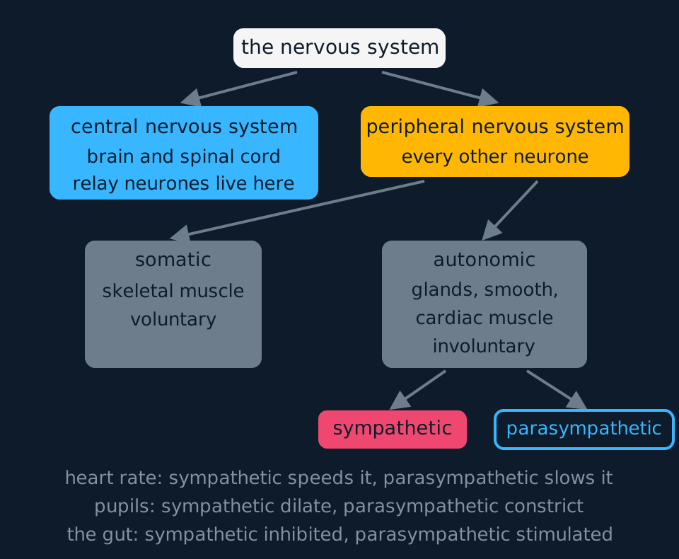
The second division separates the motor side of the PNS by whether its responses are under conscious control. The somatic nervous system carries impulses to skeletal muscle, and its responses are voluntary or reflex: walking, writing, pulling your hand off a pan. The autonomic nervous system carries impulses to glands, to the smooth muscle of the gut and blood vessels and to cardiac muscle, and its responses are not under conscious control. The secretion of gastric juice, for example, is regulated without any voluntary input.
The autonomic system then splits once more, into two systems that innervate the same organs and pull in opposite directions. The sympathetic system readies the body for action, and the parasympathetic system runs it at rest, which the old tags 'fight or flight' and 'rest and digest' summarise fairly. Most of the time both are active at once, and the state of an organ reflects the balance between them, which is the same antagonistic logic as insulin and glucagon.
| Sympathetic | Parasympathetic | |
|---|---|---|
| Overall effect | Prepares the body for activity | Returns the body to rest |
| Heart rate | Speeds it up | Slows it down |
| Pupils | Dilate | Constrict |
| Gut movement and secretion | Inhibited | Stimulated |
| Bronchioles | Dilate | Constrict |
| Usual transmitter at the effector | Noradrenaline | Acetylcholine |
- Central nervous system
- The brain and the spinal cord: the coordinating part of the nervous system, containing the relay neurones.
- Peripheral nervous system
- The neurones outside the brain and spinal cord, carrying impulses into and out of the CNS.
- Somatic nervous system
- The part of the peripheral system carrying impulses to skeletal muscle, under voluntary control.
- Autonomic nervous system
- The part of the peripheral system carrying impulses to glands, smooth muscle and cardiac muscle, not under conscious control.
The brain, region by region
Gross structure means what is visible on a cut brain without a microscope. Five regions are named at A level, each with a distinct function.
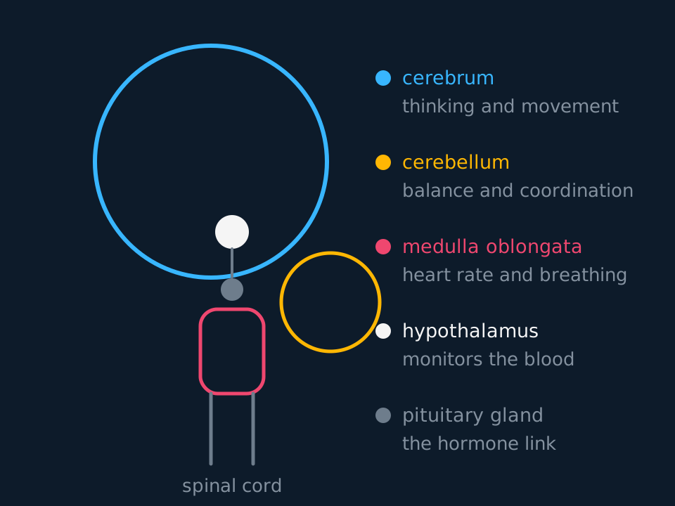
The cerebrum is the great folded pair of hemispheres that makes up most of the brain's mass. Its outer few millimetres, the cerebral cortex, handle conscious thought, language, memory, learning and the initiation of voluntary movement: everything the somatic system does on purpose starts here.
The cerebellum sits below and behind it, and coordinates rather than commands. It takes in a stream of information from the organs of balance in the inner ear and from stretch receptors in muscles and tendons, and adjusts the timing and strength of contractions so that movement is smooth. None of this is conscious: the cerebrum initiates a movement and the cerebellum coordinates its execution.
The medulla oblongata is the swollen top of the spinal cord, and it holds the regulatory centres of the autonomic system: the cardiovascular centre that sets heart rate and blood vessel diameter, and the ventilation centre that sets breathing rhythm. Damage to the medulla is rapidly fatal because these centres are not duplicated elsewhere.
The hypothalamus monitors the composition of the blood. It holds the osmoreceptors used in osmoregulation and the thermoregulatory centre, and it manufactures ADH. Next topic for both control systems: osmoregulation and thermoregulation in the hormones unit. The pituitary gland hangs beneath it on a stalk and is its link to the endocrine system: the posterior lobe stores and releases the hormones the hypothalamus makes, and the anterior lobe secretes its own under hypothalamic control. Together these two structures link nervous coordination to endocrine coordination.
- Cerebrum
- The paired, folded hemispheres responsible for conscious thought, learning, memory and the initiation of voluntary movement.
- Cerebellum
- The region below and behind the cerebrum that coordinates balance and the timing of movement, without conscious involvement.
- Medulla oblongata
- The base of the brainstem, containing the autonomic centres controlling heart rate, vessel diameter and breathing.
- Hypothalamus
- The region beneath the thalamus that monitors the blood, controls thermoregulation and osmoregulation, and directs the pituitary.
- Pituitary gland
- The endocrine gland hanging beneath the hypothalamus, whose two lobes release hormones under its control.
Three neurones, told apart by structure
Sensory, relay and motor neurones are usually named by their function in a reflex arc. They also differ in structure, and OCR A requires each type to be identified from a diagram. Two terms carry the distinction. A dendron is a fibre carrying impulses towards a cell body, and an axon carries them away from it; dendrites are the fine branches that feed a dendron or the cell body itself.
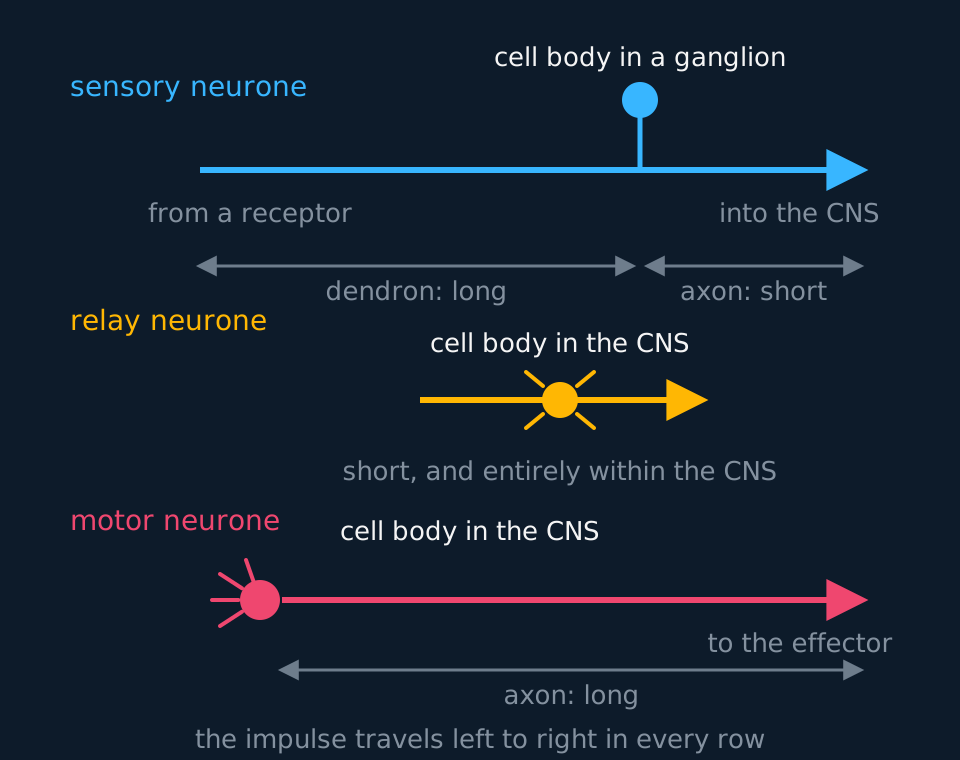
A motor neurone has its cell body in the CNS, wearing many short dendrites that collect input from other neurones. One long axon then runs all the way out to the effector, a metre of it if the effector is in the foot. Cell body first and long axon after is the identifying arrangement.
A sensory neurone is arranged differently. Its cell body sits neither at the start nor at the end but on a short side branch part-way along, housed with thousands of its neighbours in a ganglion just outside the spinal cord. The long fibre from the receptor is therefore a dendron, because the impulse in it is travelling towards the cell body, and only the short fibre from the cell body into the CNS is an axon. A neurone drawn with its cell body on a side branch of the fibre is therefore sensory.
A relay neurone, also called an intermediate neurone, lies entirely within the CNS, and it is small: a cell body with many short processes and no long fibre at all, because nothing it connects to is far away. It links sensory pathways to motor pathways within the CNS, which is its position in a reflex arc.
| Sensory | Relay | Motor | |
|---|---|---|---|
| Cell body | On a side branch, in a ganglion outside the CNS | In the CNS | In the CNS |
| Dendron | One, long: from the receptor | Many short processes | None; short dendrites on the cell body |
| Axon | One, short: into the CNS | Short | One, long: to the effector |
| Where it runs | Receptor to CNS | Within the CNS | CNS to effector |
- Dendron
- A fibre of a neurone that carries impulses towards the cell body.
- Axon
- A fibre of a neurone that carries impulses away from the cell body.
- Ganglion
- A swelling containing the cell bodies of many neurones, outside the central nervous system.
Three kinds of muscle and the division that controls each
An effector is a muscle or a gland, and the muscle category covers three distinct tissues. Next topic for the first of them in detail: muscles and movement.
Skeletal muscle is the tissue attached to bone: long multinucleate fibres with the regular striations the sliding filament lesson explains, contracting quickly and powerfully, fatiguing with use, and taking orders from the somatic system. Involuntary muscle, also called smooth muscle, lines the gut, the blood vessels, the airways and the iris: small spindle-shaped cells, one nucleus each, no striations because the filaments are not in register, contracting slowly and steadily without tiring, under autonomic control. Peristalsis, the wave of contraction that moves food along the gut, is carried out by this tissue.
Cardiac muscle differs from both. Its cells are striated like skeletal muscle but short, branched and joined end to end at intercalated discs, so the tissue contracts as a connected sheet. It is myogenic: the beat starts in the muscle itself, at the sinoatrial node, and the autonomic system only adjusts the rate, sympathetic impulses raising it and parasympathetic impulses lowering it, under the control of the medulla. Cardiac muscle does not fatigue, which allows contraction to continue throughout life.
| Skeletal | Involuntary (smooth) | Cardiac | |
|---|---|---|---|
| Appearance | Striated | No striations | Striated, cells branched |
| Cells | Long multinucleate fibres | Spindle-shaped, one nucleus | Branched, one nucleus, joined at intercalated discs |
| Control | Voluntary: somatic system | Involuntary: autonomic system | Myogenic; rate adjusted by the autonomic system |
| Contraction | Rapid and powerful; fatigues | Slow and sustained; does not fatigue | Rhythmic and continuous; does not fatigue |
| Found | Attached to the skeleton | Gut, vessels, airways, iris | The heart, and nowhere else |
TRY IT: Placing a symptom in the diagram
After a stroke, a patient can understand speech and decide to reach for a cup, but the movement is clumsy and overshoots, and their heart rate and breathing are normal. Suggest which region of the brain has been damaged, and justify the answer by eliminating the others.
Check your answer
The clumsy, overshooting movement points to the cerebellum. Its job is coordinating the timing and strength of contractions using information from balance organs and stretch receptors, so damage leaves movement possible but poorly controlled.
The cerebrum can be ruled out because the conscious functions it handles are intact: the patient understands language, forms the intention to move, and initiates the movement. The medulla oblongata can be ruled out because heart rate and ventilation, which its centres control, are normal.
The hypothalamus and pituitary govern homeostasis and hormone release rather than movement, and none of the symptoms involves either. The argument works by naming the function of each region and testing it against the symptoms.
In the exam
- Central against peripheral is a division by anatomy; somatic against autonomic is a division by control. The autonomic system is part of the peripheral nervous system, not of the brain.
- Give sympathetic and parasympathetic effects as a pair: name the organ and give both effects, as in 'the sympathetic system increases heart rate and the parasympathetic system decreases it'.
- Tie each brain region to a function, not a vague theme: the medulla controls heart rate and breathing; the cerebellum coordinates balance and movement; the hypothalamus monitors temperature and water potential.
- A neurone whose cell body sits on a side branch of the fibre is sensory; a cell body in the CNS with one long axon leaving it is motor. These questions are set on diagrams, so identify the type from the structure.
- Dendron and axon are defined by direction relative to the cell body, towards and away. The long fibre of a sensory neurone is a dendron.
- Cardiac muscle is myogenic. The autonomic system changes the rate of the heartbeat; it does not cause it.
Check yourself
A textbook drawing shows a neurone with a long fibre, a cell body on a short stalk part-way along that fibre, and a short fibre continuing beyond it. Name the type of neurone, name the two fibres, and state where in a reflex pathway this neurone runs. Then explain why the muscle this pathway finally reaches is under somatic rather than autonomic control.
Answer
The cell body on a side branch identifies it as a sensory neurone, with the cell body sitting in a ganglion just outside the spinal cord.
The long fibre from the receptor is a dendron, because the impulses in it travel towards the cell body; the short fibre carrying them onwards into the CNS is an axon, because they travel away from it.
In a reflex pathway this neurone runs from the receptor into the spinal cord, where it synapses with a relay neurone; a motor neurone then carries the impulses out to the effector.
The effector in a withdrawal reflex is skeletal muscle, and skeletal muscle is served by the somatic nervous system: the system whose motor pathways can also be driven voluntarily. The autonomic system serves glands, smooth muscle and cardiac muscle, which are not attached to the skeleton and are not supplied by somatic motor pathways.
Questions
Question 15 marks
A person treads on a sharp stone, pulls the foot away without thinking, and then notices that their heart is beating faster. Explain which divisions of the nervous system bring about each of these two responses, and explain why only the first of them could also have been performed on purpose.
Mark scheme
- B1 the withdrawal is a reflex carried by the peripheral nervous system: a sensory neurone carries impulses from the receptor into the spinal cord, a relay neurone in the CNS connects the pathway, and a motor neurone carries impulses out to the effector
- B1 the effector is skeletal muscle, which is supplied by the somatic nervous system, and the same motor pathways can be driven voluntarily from the cerebrum, which initiates movement, so the action could also have been performed on purpose
- B1 the faster heartbeat is the work of the autonomic nervous system, whose sympathetic division prepares the body for activity, acting on instruction from the cardiovascular centre in the medulla oblongata
- B1 cardiac muscle is myogenic, so the beat itself originates at the sinoatrial node and the sympathetic impulses only raise its rate, with parasympathetic impulses lowering it again once the danger has passed
- B1 the autonomic system supplies glands, smooth muscle and cardiac muscle, and its pathways are not driven from the conscious cortex, so the change in heart rate happens whether or not the person notices it and cannot be decided on
Question 24 marks
Describe the effects of the sympathetic and the parasympathetic systems on the heart, the pupils, the gut and the bronchioles.
Mark scheme
- B1 the sympathetic system speeds the heart up and the parasympathetic system slows it down
- B1 the sympathetic system dilates the pupils and the parasympathetic system constricts them
- B1 the sympathetic system inhibits movement and secretion in the gut, whereas the parasympathetic system stimulates them
- B1 the sympathetic system dilates the bronchioles and the parasympathetic system constricts them; overall the first prepares the body for activity and the second returns it to rest
Question 34 marks
Compare skeletal muscle, involuntary muscle and cardiac muscle as tissues.
Mark scheme
- B1 skeletal muscle is made of long multinucleate striated fibres, involuntary muscle of small spindle-shaped cells with one nucleus and no striations, and cardiac muscle of short branched striated cells joined end to end at intercalated discs so the tissue contracts as a connected sheet
- B1 skeletal muscle takes its orders from the somatic system and involuntary muscle from the autonomic system, whereas cardiac muscle is myogenic: the beat begins in the muscle itself at the sinoatrial node
- B1 the autonomic system only adjusts the heart's rate, sympathetic impulses raising it and parasympathetic impulses lowering it on instruction from the medulla, rather than causing the beat as it causes contraction in the other two tissues
- B1 skeletal muscle contracts rapidly and powerfully and fatigues, whereas involuntary muscle contracts slowly and steadily and cardiac muscle rhythmically and continuously, and neither of these fatigues
Question 44 marks
After an injury to the brainstem a patient's heart rate and breathing become irregular, while their speech, memory and balance are unaffected. Suggest which region of the brain has been damaged, and suggest how the other regions can be ruled out.
Mark scheme
- B1 the medulla oblongata, which holds the cardiovascular centre that sets heart rate and blood vessel diameter and the ventilation centre that sets breathing rhythm
- B1 the cerebrum can be ruled out because the conscious functions of its cortex, language and memory, are intact
- B1 the cerebellum can be ruled out because balance and the coordination of movement, which it handles, are unaffected
- B1 the hypothalamus and pituitary govern thermoregulation, osmoregulation and hormone release rather than heart rate and breathing, and nothing in the symptoms involves them
Question 54 marks
Compare the somatic nervous system with the autonomic nervous system, referring to what each supplies, whether its responses are voluntary, and an effector each system controls.
Mark scheme
- B1 the somatic system supplies skeletal muscle, whereas the autonomic system supplies glands, smooth muscle and cardiac muscle
- B1 somatic responses are voluntary or reflex, whereas autonomic responses are not under conscious control
- B1 the somatic system's effector is exemplified by a skeletal muscle such as the biceps, whereas the autonomic system's effector is exemplified by cardiac muscle or the smooth muscle of the gut
- B1 both are motor divisions of the peripheral nervous system, carrying impulses out from the central nervous system to their own effectors
Question 64 marks
Name the two lobes of the pituitary gland, and name what each one does with the hormones of the hypothalamus.
Mark scheme
- B1 the posterior lobe of the pituitary
- B1 stores and releases hormones made by the hypothalamus
- B1 the anterior lobe of the pituitary
- B1 secretes its own hormones, under the control of the hypothalamus
Question 74 marks
A patient loses the ability to regulate body temperature and water balance after a head injury, but their movement, speech and heart rate are normal. Suggest which region of the brain has been damaged, and suggest how the other regions can be ruled out.
Mark scheme
- B1 the hypothalamus, because it holds the osmoreceptors that monitor water potential and the centre that regulates temperature
- B1 the cerebrum can be ruled out because the conscious functions of its cortex, speech and voluntary movement, are unaffected
- B1 the medulla oblongata can be ruled out because heart rate and breathing rhythm, which its centres control, are normal
- B1 the cerebellum can be ruled out because balance and the coordination of movement, which it handles, are unaffected
Question 83 marks
Name the region of the brain that sets heart rate and breathing rhythm, the region that coordinates balance and the timing of movement, and the region that monitors the water potential of the blood.
Mark scheme
- B1 the medulla oblongata contains the cardiovascular centre and the ventilation centre
- B1 the cerebellum coordinates balance and the timing and strength of contractions
- B1 the hypothalamus holds the osmoreceptors that monitor the water potential of the blood
Question 93 marks
Explain how a sensory neurone can be recognised in an unlabelled drawing, and explain why its long fibre is called a dendron rather than an axon.
Mark scheme
- B1 its cell body lies neither at the start nor at the end of the fibre but on a short side branch part-way along it, housed in a ganglion just outside the spinal cord
- B1 a dendron carries impulses towards a cell body and an axon carries them away from it
- B1 the impulse in the long fibre is travelling from the receptor towards the cell body, so that fibre is a dendron, and only the short fibre running on into the CNS is an axon
Question 103 marks
Describe the structure of a relay neurone, and describe its position within a reflex arc.
Mark scheme
- B1 a relay neurone has a cell body with many short processes and no long fibre, because nothing it connects to is far away
- B1 it lies entirely within the central nervous system
- B1 within a reflex arc it links the sensory neurone to the motor neurone
Question 113 marks
Explain why both the sympathetic and parasympathetic systems are usually active in an organ at the same time, rather than only one being switched on.
Mark scheme
- B1 the two systems act antagonistically on the same organs, one increasing and the other decreasing its activity
- B1 the state of the organ at any moment reflects the balance between the two opposing sets of impulses arriving at it, rather than either system being fully off
- B1 this lets the balance shift smoothly in either direction, for example by raising sympathetic activity, lowering parasympathetic activity, or both together, which a simple on/off switch could not achieve
Question 123 marks
Explain why damage to the medulla oblongata is more rapidly life-threatening than similar damage to the cerebellum.
Mark scheme
- B1 the medulla oblongata contains the cardiovascular centre, which sets heart rate and blood vessel diameter, and the ventilation centre, which sets the rhythm of breathing
- B1 these centres are not duplicated anywhere else in the nervous system, so damage to them can stop the heart or breathing directly
- B1 the cerebellum coordinates the timing and strength of movement and balance, so damage there causes clumsy, poorly coordinated movement rather than removing a function needed to stay alive from moment to moment
Question 132 marks
State which parts of the body make up the central nervous system, and state what the somatic nervous system supplies.
Mark scheme
- B1 the central nervous system is the brain and the spinal cord
- B1 the somatic nervous system carries impulses to skeletal muscle, and its responses are voluntary or reflex
Question 142 marks
State what a nerve is, and state how it differs from a single neurone.
Mark scheme
- B1 a nerve is a bundle of the axons and dendrons of many neurones, wrapped together in connective tissue
- B1 a neurone is a single cell, so a nerve is not a neurone but contains the fibres of many of them
Question 152 marks
Name the term for a swelling containing the cell bodies of many neurones outside the central nervous system, and name the type of neurone whose cell body is found there.
Mark scheme
- B1 a ganglion, outside the spinal cord
- B1 a sensory neurone
Worth remembering
- CNS is brain plus spinal cord; the PNS is every neurone outside them.
- Somatic serves skeletal muscle and is voluntary; autonomic serves glands, smooth muscle and cardiac muscle and is not.
- Sympathetic readies the body and parasympathetic rests it, acting antagonistically on the same organs.
- The cerebrum handles conscious function, the cerebellum coordinates movement, the medulla controls heart rate and breathing, the hypothalamus monitors the blood, and the pituitary releases hormones under its control.
- Sensory neurones carry a cell body on a side branch and a long dendron; motor neurones keep the cell body in the CNS and run one long axon out.
CHECK YOUR PROGRESS
Rate how confident you are with each objective for this lesson. Ratings are saved in this browser, on this device, unless you sign in.
- Divide the nervous system into central and peripheral, and the peripheral system into somatic and autonomic.
- Set sympathetic against parasympathetic, with the effects of each on the same named organs.
- Locate the cerebrum, cerebellum, medulla oblongata, hypothalamus and pituitary, and give one function of each.
- Distinguish sensory, relay and motor neurones by their structure: where the cell body sits, and dendron against axon.
- Compare skeletal, involuntary and cardiac muscle as tissues, including which division of the nervous system controls each.
Open the full revision checklist to see every objective in the curriculum in one place.
Practise this lesson
WORKBOOK
The same questions as the player, on paper with room to work, and a separate book of mark schemes. Free to use; please do not redistribute or sell.