Chemistry › Practicals › Thin-layer chromatography: separating amino acids
Required practical 12: separating amino acids by TLC
Run an amino acid mixture beside known standards on one TLC plate, show the spots with ninhydrin, and compare Rf values.
This is the method section of Chromatography: TLC, column and gas on a page of its own, with its variables, sample results, an evaluation and questions set on it. The chemistry behind it is in that lesson.
The capability this develops
This investigation is one instance of capability P09: Thin-layer chromatography, which sets out the techniques it belongs to, the calculations it carries and the errors that cost marks in all of them. The notes behind the chemistry are in Organic analysis.
Where this sits on the specifications
Each qualification is mapped to its own specification one numbered heading at a time, against a document whose sha256 is recorded. These are the headings that list the lesson this method is taught in, on the four qualifications mapped. Each links to the route map the row belongs to.
| Qualification | Heading |
|---|---|
| AQA 7404/7405 | 3.3.13.2 Proteins |
| AQA 7404/7405 | 3.3.16 Chromatography |
| OCR Chemistry A H032/H432 | 6.3.1 Chromatography and qualitative analysis |
| Pearson Edexcel Chemistry 8CH0/9CH0 | 19.6 mobile and stationary phases |
| Pearson Edexcel Chemistry 8CH0/9CH0 | 19.7 Rf values |
| Pearson Edexcel Chemistry 8CH0/9CH0 | 19.8 HPLC and GC, with mass spectrometry |
| Cambridge International 9701 | 37.1 Thin-layer chromatography |
| Cambridge International 9701 | 37.2 Gas / liquid chromatography |
Three boards number their practical activities, and those lists were read here from the same specifications as the headings above. This investigation is filed on them as:
- AQA 7404/7405 required practical 12: Thin-layer chromatography
Use your awarding organisation’s current specification and its practical handbook as the authority on which activities your course requires and how they are assessed.
Apparatus
A silica TLC plate held by its edges in gloves, a pencil and ruler, capillary tubes, a beaker with a watch-glass lid as the tank, the solvent (butan-1-ol, ethanoic acid and water in the ratio 12:3:5 is a common choice for amino acids), solutions of the unknown and of known amino acids, ninhydrin spray and an oven or hairdryer.
Safety
Risk assessment. The solvent mixture is flammable and ethanoic acid is corrosive; keep it away from flames and use it in a well-ventilated room. Ninhydrin is harmful and is sprayed only in a fume cupboard. Ultraviolet light damages eyes, so never look into the lamp. Wear eye protection and gloves. Check the CLEAPSS Hazcards and the school's risk assessment; where they differ from the advice here, follow them.
Variables
Independent variable
What is spotted on each lane of the plate: the unknown mixture and each known amino acid.
Dependent variable
The distance each spot travels from the baseline, as a fraction of the distance the solvent travels: the Rf value.
Control variables
- The plate, so that every lane runs on the same stationary phase, in the same solvent, at the same temperature and for the same time.
- The depth of solvent in the tank, below the baseline, and the lid, which keeps the air in the tank saturated with vapour.
- The size of each spot: small, concentrated and dry before the plate goes in, built up by spotting and re-spotting.
- The handling: the plate held by its edges in gloves, pencil for every mark, and the solvent front marked the moment the plate comes out.
Rf is the ratio of two distances on one plate, so everything that affects how far the solvent and the solutes travel is held the same by running every lane on the same plate at the same time. A standard run on another day is not a fair comparison, which is why the standards go beside the unknown rather than in a table.
Method
| Step | What to do | Why |
|---|---|---|
| 1 | Draw a pencil line 1 cm from the bottom of the plate and mark evenly spaced crosses on it | Pencil is graphite and does not dissolve or run; ink would |
| 2 | Spot the unknown and each standard on its own cross with a capillary tube; let it dry and re-spot to keep each spot small | Small, concentrated spots give compact, separate spots on the chromatogram |
| 3 | Pour solvent into the tank to a depth below 1 cm, cover it and leave it for a few minutes | The baseline must be above the solvent, or the spots dissolve into it; the lid saturates the air with vapour |
| 4 | Stand the plate in the tank, replace the lid and leave it until the solvent is about 1 cm from the top | The lid stops solvent evaporating from the plate as it rises |
| 5 | Remove the plate and at once mark the solvent front in pencil | The front is invisible once the solvent evaporates |
| 6 | Dry the plate in a fume cupboard, spray with ninhydrin and warm; circle each purple spot | Amino acids are colourless until ninhydrin reacts with them |
| 7 | Measure from the baseline to the centre of each spot and to the front, and calculate each Rf | Rf values from one plate are directly comparable |
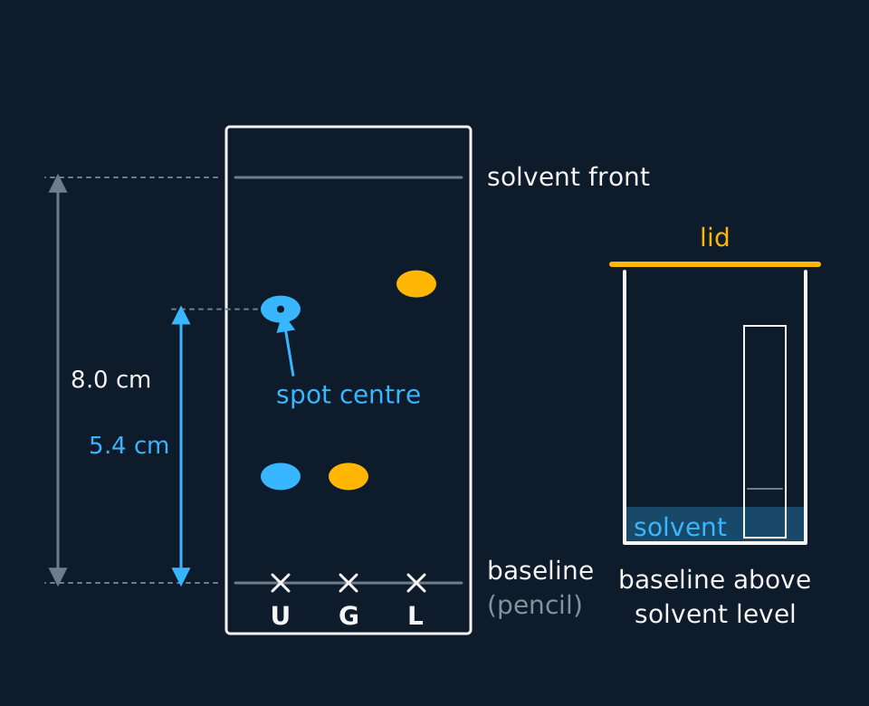
Describe the drawing
A developed thin-layer plate with a pencil baseline near the bottom and the solvent front 8.0 cm above it. Three lanes are marked U, G and L. The unknown U has two spots, at 2.1 and 5.4 cm; glycine has one spot at 2.1 cm level with U's lower spot; leucine has one at 5.9 cm, above U's upper spot. Dimension arrows measure 5.4 cm from the baseline to the centre of U's upper spot and 8.0 cm to the front. An inset shows the plate in a lidded beaker with the solvent below the baseline.
What is measured
Each component is described by its Rf value, the distance the spot travels as a fraction of the distance the solvent travels. It has no unit and always lies between 0 and 1. It is constant for one compound only on the same stationary phase, with the same solvent and at the same temperature.
Many organic compounds are colourless, so the spots have to be made visible. A plate with a fluorescent additive glows under ultraviolet light and the compounds show as dark spots, which are circled in pencil. Iodine vapour in a closed jar stains most organic spots brown. Ninhydrin sprayed on and warmed turns amino acids purple.
Sample results
These readings are idealised: they are the distances in the lesson's worked example, measured from the baseline on one plate.
| Spot | Distance from the baseline / cm | Rf |
|---|---|---|
| Solvent front | 8.0 | |
| Unknown, lower spot | 2.1 | 0.26 |
| Unknown, upper spot | 5.4 | 0.68 |
| Glycine standard | 2.1 | 0.26 |
| Leucine standard | 5.9 | 0.74 |
The lower spot matches glycine run on the same plate, so the mixture probably contains glycine. The upper spot does not match leucine, so it is some other amino acid, which needs another standard to name.
Evaluation
The limitations of the method, what each does to the result and in which direction, and the improvement a mark scheme credits.
| Limitation | What it does to the result | Improvement |
|---|---|---|
| A spot has a width, and Rf is measured to its centre | A spot 3 mm across on a 2.1 cm run puts ±1.5 mm, about 7%, on the smaller distance; in the sample results the lower spot and glycine agree within that, while the upper spot at 5.4 cm and leucine at 5.9 cm differ by 0.5 cm, well outside it | Use a longer plate so the spots spread further apart, measure to the densest part of each spot with a ruler reading to 1 mm, and quote Rf to two decimal places only |
| Two amino acids can run together in one solvent | Compounds with similar polarity have similar Rf values in the butan-1-ol, ethanoic acid and water mixture, so one spot can be two compounds | Run the plate again in a second solvent of different polarity, or turn the dried plate through 90° and run it in the second solvent for a two-dimensional separation |
| A matching Rf is evidence, not proof | The lower spot matches glycine at 0.26, but any compound with that Rf in this solvent on this plate would match it too | Spot the unknown and glycine on top of each other in a third lane: a single spot after the run supports the match, a doubled spot refutes it; confirm by mass spectrometry if the identity matters |
| Ninhydrin shows that an amino acid is there, not which one | Almost every amino acid gives the same purple; proline shows yellow, and the colour fades within hours | Circle every spot in pencil as soon as it appears and measure at once; identification rests on Rf against standards, not on the colour |
Errors in carrying it out
The errors that matter: spots that are too large or too concentrated streak and overlap, so the distance to the centre is uncertain. Touching the face of the plate leaves amino acids from the skin, which ninhydrin shows up as extra spots. A tank without a lid lets the solvent evaporate from the plate, so the front runs unevenly. Running the standards on the same plate as the unknown removes any difference in solvent, temperature or plate between runs.
Questions
Question 1 (6 marks)
Describe how thin-layer chromatography is used to identify the amino acids in a mixture, using standards of known amino acids.
Mark scheme
Indicative content
- a pencil baseline is drawn about 1 cm from the bottom of a silica plate, handled only by its edges
- small spots of the mixture and of each standard are placed on the baseline with capillary tubes and allowed to dry
- the plate stands in a covered tank of solvent whose level is below the baseline, until the solvent nears the top
- the solvent front is marked in pencil as soon as the plate is removed, and the plate is dried
- the plate is sprayed with ninhydrin in a fume cupboard and warmed, so the amino acids show as purple spots
- the Rf of each spot is calculated and compared with the standards on the same plate; a match identifies the amino acid
Levels
- Level 3 (5–6 marks) all stages are present in order: the pencil baseline, small spots of the mixture and each standard, the covered tank with the solvent below the baseline, marking the solvent front, developing with ninhydrin in a fume cupboard, and the Rf comparison with standards on the same plate
- Level 2 (3–4 marks) the plate is spotted, run and compared with the standards; the solvent level below the baseline, the marked solvent front, the ninhydrin step or the Rf values are not there
- Level 1 (1–2 marks) the mixture is spotted and run, often with the baseline drawn in ink or the solvent above the spots, and the colourless amino acids are never made visible
Question 2 (2 marks)
Explain why the baseline on a TLC plate is drawn in pencil and placed above the level of the solvent.
Mark scheme
- B1 pencil is graphite, which does not dissolve in the solvent or move up the plate, whereas ink would
- B1 above the solvent level the spots are not dissolved off the plate into the solvent in the tank
Question 3 (2 marks)
Explain why a lid is placed on the tank while a TLC plate is being developed.
Mark scheme
- B1 the lid keeps the air in the tank saturated with solvent vapour
- B1 so the solvent does not evaporate from the plate as it rises and the front moves evenly
Question 4 (4 marks)
Evaluate a student's identification of a spot with Rf 0.45, on a plate run on Monday, as leucine, because a leucine standard gave Rf 0.45 on a plate run on Friday.
Mark scheme
- B1 the equal Rf values are consistent with the spot being leucine
- B1 Rf depends on the solvent, the plate and the temperature, which may have differed between the two days
- B1 other amino acids can have the same Rf as leucine in a given solvent
- B1 a sound identification runs the standard on the same plate as the unknown, and confirms it in a second solvent or by another technique
Question 5 (2 marks)
Explain why the solvent front is marked as soon as the plate is taken out of the tank.
Mark scheme
The solvent evaporates quickly, and once it has the front cannot be seen.
The distance to the front is the denominator of every Rf value on the plate.
Question 6 (3 marks)
A spot lies 3.6 cm from the baseline and the solvent front 7.5 cm from it. Calculate the Rf value and explain why it has no unit.
Mark scheme
Rf = distance moved by the spot ÷ distance moved by the solvent.
= 3.6 ÷ 7.5 = 0.48.
It is a ratio of two distances in the same unit, so the units cancel, and it always lies between 0 and 1.
Question 7 (3 marks)
Explain, in terms of the two phases, why amino acids separate on a silica plate.
Mark scheme
Silica is polar and is the stationary phase; the solvent is the mobile phase that carries the compounds up the plate.
Each amino acid spends part of its time adsorbed on the silica and part dissolved in the solvent, in a balance set by its attractions to each.
The more strongly a compound is adsorbed, or the less soluble it is in the solvent, the less far it travels and the lower its Rf, so compounds with different balances separate.
Question 8 (2 marks)
The upper spot of the unknown has Rf 0.68 and the leucine standard 0.74 on the same plate. Explain why they are judged to be different compounds, and suggest how the identity of the upper spot could be found.
Mark scheme
On one plate under the same conditions a difference of 0.06, 5 mm, is larger than the uncertainty in locating the centre of a spot, so the compounds differ.
Run further standards beside the unknown, co-spot with a candidate, or identify the spot by mass spectrometry.
Question 9 (3 marks)
Two spots lie 1.8 cm and 2.0 cm from the baseline, with the solvent front at 8.0 cm. Calculate their Rf values and suggest how they could be separated more clearly.
Mark scheme
Rf = 1.8 ÷ 8.0 = 0.23 and 2.0 ÷ 8.0 = 0.25.
Spots this close are likely to overlap, so neither distance can be measured to its centre with confidence.
Run a longer plate, or a solvent of different polarity, or a second solvent at 90° for a two-dimensional separation.
Question 10 (2 marks)
State why the plate is handled by its edges in gloves and why it is sprayed with ninhydrin in a fume cupboard.
Mark scheme
Skin carries amino acids, which would appear as extra spots.
Ninhydrin is harmful and its spray must not be breathed in.
Coverage maps are available for AQA 7404/7405, OCR Chemistry A H032/H432, Pearson Edexcel Chemistry 8CH0/9CH0 and Cambridge International 9701. Use the relevant route map to check what InkChemistry covers, and your awarding organisation’s current specification as the final authority on examinable content.