Physics › Medical physics › X-ray detectors and CT scanning
X-ray detectors and CT scanning
An intensifying screen absorbs far more of the beam than bare film and turns each X-ray into thousands of light photons, so the dose falls. A flat-panel detector reads the transmitted beam out as numbers instead. A CT scanner records attenuation along rays from many angles and reconstructs a cross-sectional slice.
Choose your exam board to show the notes and questions relevant to your specification. You can change this setting at any time, and content for other boards remains available.
X-rays and CT scanning, part 2 of 2. Part 1 is The physics of diagnostic X-rays.
Builds on The physics of diagnostic X-rays and The photoelectric effect and the photon.
IN THIS TOPIC
- Say how an intensifying screen and a flat-panel detector record the beam, and why each can cut the dose.
- Explain how a CT scanner builds cross-sectional images, what its narrow beam and detector array are for, and weigh it against a plain X-ray.
COMMON MISCONCEPTION
A CT scanner is just a sharper X-ray photograph.
A CT scan is not a single sharper image: the scanner records many X-ray projections from different angles and reconstructs a cross-sectional slice, locating structures in depth as one shadow image cannot.
Recording the shadow
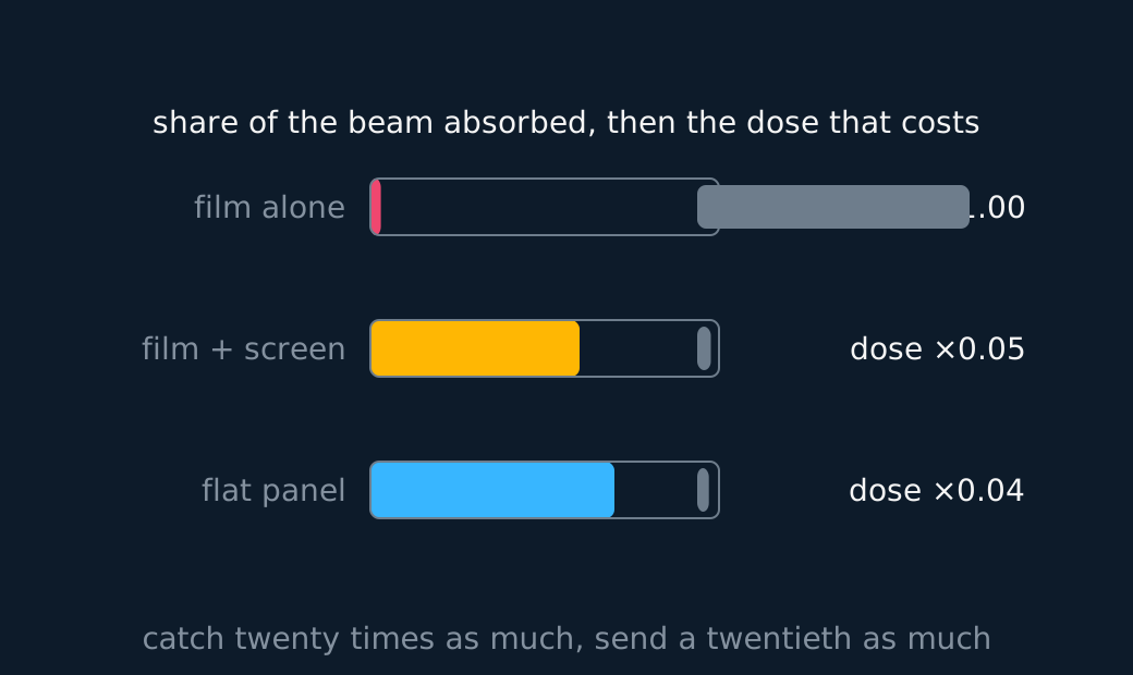
The beam that survives the patient has to be turned into an image, and the receptor chosen sets how much beam the patient had to be given in the first place. Photographic film on its own is a poor X-ray detector. The emulsion is thin and made of light elements, so only a per cent or two of the arriving photons interact with it at all. The rest cross the patient, deliver their dose and blacken nothing.
An intensifying screen is the fix. It is a layer of fluorescent material, calcium tungstate in the older ones and a rare-earth phosphor in the newer, held in contact with the film and usually one on each side of it. Its atoms have high atomic numbers, so it absorbs X-rays far more readily than the emulsion does, and each X-ray it absorbs releases thousands of visible-light photons, which the film is highly sensitive to. Almost all the blackening is then done by light rather than by X-rays directly.
Follow that through to the patient and the reason for the screen appears. The same darkening now needs perhaps a fiftieth of the photons through the body, so the exposure, and with it the dose, falls by that factor. What is lost is sharpness, because the light spreads sideways from the point where the X-ray was absorbed before it reaches the emulsion, so each photon blackens a small patch instead of a point. A thicker screen absorbs more and saves more dose, and blurs more. Dose against detail is the trade-off in every receptor.
Digital flat-panel detectors have largely replaced film and screen, and they begin the same way. A scintillator layer converts each X-ray to light, and in the better panels the caesium iodide is grown as fine parallel needles so the light is channelled down its own column rather than spreading sideways, which recovers much of the sharpness lost with a screen. Under the scintillator sits a matrix of photodiodes, one per pixel, each with its own thin-film transistor. Light frees charge in the diode, the charge is stored, and the array is read out row by row as numbers.
Reading numbers instead of developing a film changes what can be done next. The response stays proportional to the intensity over a far wider range, so one exposure covers bone and soft tissue where a film would be over-exposed in one and under-exposed in the other; the contrast can then be stretched electronically to suit whatever is being looked for; and the image exists at once and can be sent anywhere. The panel also absorbs a larger fraction of the arriving photons than film, so the same image can be made at a lower dose, provided the exposure settings are actually turned down to take the benefit.
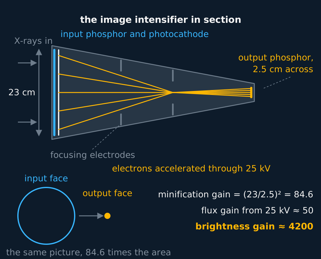
Live screening needs a different instrument, and the image intensifier supplies it. An input phosphor converts the X-rays to light, a photocathode converts that light to electrons, and those electrons are accelerated through tens of kilovolts and focused down onto a small output phosphor. Accelerating them is what supplies the gain: each arriving X-ray ends as a far brighter flash than it began, so a surgeon can watch a catheter move in real time on a beam weak enough to be run continuously.
From shadows to slices
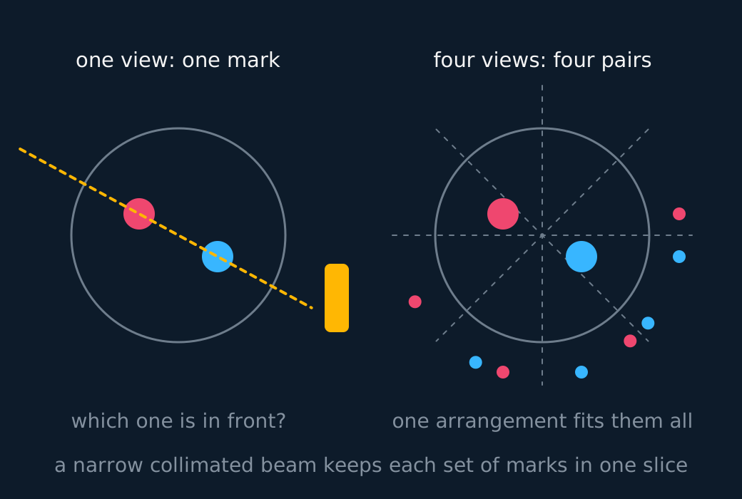
A CT scanner records X-ray attenuation from many angles and uses these projections to reconstruct a cross-sectional image. A plain X-ray superposes everything along each ray into a single value of transmitted intensity; a CT scanner's tube and detectors rotate around the patient, recording attenuation along thousands of directions, and a computer then solves for the map of μ across the slice that is consistent with all of those measurements at once.
Two pieces of hardware make that possible, and questions ask what each is for. The first is the shape of the beam. A collimator at the tube narrows the output to a thin fan, wide enough to cross the patient but only a few millimetres thick, and that narrowness does two jobs at once. It confines the dose to the slice being imaged, leaving the tissue above and below it alone, and it defines how thick that slice is, which sets the resolution along the length of the body.
The narrow beam also improves the reconstruction. Reconstruction assumes each reading is the attenuation along one straight ray, and a Compton-scattered photon breaks that assumption, since it arrives from the wrong direction but is counted as though it had travelled straight. Fewer photons are set scattering in the first place when only a thin slab is irradiated, and each detector carries its own collimator facing the tube, so what reaches it has come along its own ray or not at all.
The second is the detector array facing the tube across the patient, an arc of hundreds of small scintillator and photodiode elements in place of a sheet of film. Each element measures the intensity transmitted along one ray and reports it as a number, and the whole arc is read hundreds of times per rotation. That is what the computation needs, a table of transmitted intensities indexed by ray and by angle rather than a picture. The elements are small, which sets the detail across the slice, and they respond in microseconds, so a full set of angles is gathered while the patient holds one breath.
The answer is a cross-sectional image on which soft tissues are distinguishable, and stacking slices gives a three-dimensional reconstruction. Set against that, there are drawbacks. Many exposures mean a far larger radiation dose than a single plain film, and the machine is expensive, so the sharper picture has to be clinically justified. Detail against dose is the recurring theme of every ionising technique.
ASSESSMENT FOCUS
- Answer receptor questions in terms of dose. An intensifying screen absorbs X-rays far better than the emulsion and turns each one into thousands of light photons, so far fewer photons need to cross the patient, and the drawback is a blurrier image because the light spreads sideways.
- CT against plain film needs both halves. Cross-sectional detail with no superposition of structures, but a much larger dose. If the question asks about the hardware, the narrow collimated beam fixes the slice and keeps scatter out, and the detector array reads each ray as a number the computer can reconstruct from.
- Name the receptor the question is about before explaining anything. Film alone, film behind an intensifying screen, an image intensifier and a flat panel are four different answers, and the last two are the ones a modern department actually uses.
- Say what the collimation is for when a CT question mentions a narrow beam. It fixes which slice is being imaged and keeps scattered photons from other planes out of the detectors, and scatter is what would otherwise blur the reconstruction.
CHECK YOURSELF
A radiographer replaces bare film with a cassette carrying an intensifying screen. Explain how that lowers the dose to the patient and what it costs, then say what a CT scan of the same region would add and what it would cost.
Show a hint
For the screen, follow the photons: how many are absorbed, and what each absorbed one becomes. For CT, one axis is detail and the other is dose.
Show the answer
Film alone absorbs only a small percentage of the X-rays reaching it, so most of the beam sent through the patient records nothing.
The screen absorbs a far larger share and converts each absorbed X-ray into thousands of light photons, which expose the film. A much smaller number of X-rays therefore has to cross the patient for the same exposure, so the dose falls.
The cost is sharpness: the light spreads sideways in the screen before reaching the film, so each X-ray marks a small patch rather than a point.
A CT scan would add cross-sectional images with no superposition of structures, so soft-tissue detail and depth that a single shadow cannot give.
It costs a much larger dose, because the region is imaged from many angles rather than once, and it costs money and time.
An intensifying screen lowers the dose at the expense of a little sharpness, and a flat panel reads the beam out as numbers.
A single projection superposes everything along the ray; CT reconstructs the slice from projections recorded at many angles, which is what removes the superposition.
Or read them with their mark schemes on the physics of diagnostic x-rays questions page.
CHECK YOUR PROGRESS
Rate how confident you feel with each objective for this lesson. Ratings are saved in this browser, on this device, unless you sign in.
- Say how an intensifying screen and a flat-panel detector record the beam, and why each can cut the dose.
- Explain how a CT scanner builds cross-sectional images, what its narrow beam and detector array are for, and weigh it against a plain X-ray.
Open the full revision checklist to track your progress across the whole unit.