Biology › Animal transport and cardiovascular biology › Blood vessels, tissue fluid and lymph
Blood vessels, tissue fluid and lymph
Arteries, arterioles, capillaries, venules and veins have structures suited to their pressure and function. Capillary exchange produces tissue fluid, and excess fluid and escaped proteins enter the lymphatic system. Hydrostatic and oncotic pressures are used to explain filtration and oedema.
Before this The cardiac cycle and the pressures the ventricles generate · Diffusion and osmosis: water potential · Proteins as large molecules that cannot cross membranes freely
COMMON MISCONCEPTION
Arteries carry oxygenated blood and veins carry deoxygenated blood.
Artery means carrying blood away from the heart, which is a statement about direction and not about cargo. The pulmonary artery carries deoxygenated blood and the pulmonary vein oxygenated blood, and the umbilical vessels break the pattern the same way round.
What you should be able to do
- Compare an open circulatory system with a closed one, and say what a closed system gives an animal in delivery rate and in control.
- Relate the structure of an artery, arteriole, capillary, venule and vein to its function and to the pressure it carries.
- Distinguish an elastic artery from a muscular artery by the composition of its wall, its distance from the heart and the job it does.
- Explain why a capillary wall is one cell thick and why the blood moves through it slowly.
- Account for the formation of tissue fluid at the arteriole end of a capillary bed using hydrostatic and oncotic pressure.
- Account, in the A-level examination model, for the return of most of that fluid at the venule end using the same two pressures.
- Describe what the lymphatic system does with the fluid that is not returned.
- Explain three different causes of oedema using nothing but hydrostatic and oncotic pressure.
Open and closed circulatory systems
Not every animal with a heart keeps its blood inside vessels. Insects, and most other arthropods and molluscs, have an open circulatory system.
In an insect the pump is a dorsal vessel, a muscular tube running along the back of the animal with a row of valved holes in its wall. It contracts from the rear forwards and pushes haemolymph, the insect's equivalent of blood, towards the head. There it leaves the vessel through an open end and empties into the body cavity, the haemocoel, where it washes directly over the organs. Nothing separates the fluid from the tissues, so haemolymph is blood and tissue fluid at once. It drains slowly back along the body and re-enters the dorsal vessel through the valved holes, and the cycle starts again.
In a closed circulatory system, which is what vertebrates have, the blood never leaves the vessels. It goes from heart to artery to arteriole to capillary to venule to vein and back to the heart, and the only thing that leaves is a filtrate pushed through the capillary wall as tissue fluid.
Two consequences follow. The first is delivery rate. In a haemocoel there is nothing to contain the fluid, so the pressure the pump generates is lost almost at once and the haemolymph moves slowly, at a few millimetres a second. In a closed system the vessel walls hold the pressure, so the ventricle can generate 120 mmHg and blood leaves the aorta at something like half a metre a second. Fast flow means a substance is delivered and carried away quickly, which is what an animal with a high metabolic rate needs.
The second consequence is control of distribution. Blood in vessels can be directed. Constricting the arterioles supplying the gut and dilating those supplying the leg muscles redistributes blood to the tissues using it, which is what happens at the onset of exercise. Haemolymph in a body cavity cannot be directed, because there is no vessel whose radius can be changed. An open system delivers to all tissues at the same rate.
Insects are nevertheless active animals, and the reason is that they do not transport oxygen in haemolymph. Air is delivered to the tissues directly through a branching system of tracheae opening at the body surface, so the slow circulation only has to move nutrients, hormones and waste, for which a low-pressure system is adequate. State the conclusion in those terms: an open system is enough for an insect because its haemolymph does not carry the oxygen, while a large, active animal whose blood does carry it needs the pressure and directed flow of a closed system.
| Open system, as in an insect | Closed system, as in a mammal | |
|---|---|---|
| Where the fluid goes | Into the haemocoel, bathing the organs directly | Stays inside vessels throughout |
| Blood and tissue fluid | The same fluid, haemolymph | Separate; tissue fluid is filtered out of capillaries |
| Pressure | Low, and lost as soon as the fluid leaves the vessel | Held by the vessel walls, so it can be high |
| Speed of delivery | Slow | Fast |
| Can flow be directed? | No | Yes, by vasoconstriction and vasodilation of arterioles |
| Oxygen carried by it? | Usually not; tracheae deliver air directly | Yes, by haemoglobin |
- Open circulatory system
- A circulation in which blood leaves the vessels and bathes the organs directly in a body cavity before draining back to the heart.
- Closed circulatory system
- A circulation in which blood remains inside vessels at all times and is returned to the heart through them.
- Haemocoel
- The body cavity of an insect or other arthropod, in which haemolymph collects and washes over the organs.
- Haemolymph
- The fluid of an open circulatory system, acting as blood and tissue fluid at the same time.
The five vessel types and their walls
Each vessel wall is a compromise between three requirements: withstanding the pressure inside it, offering low resistance to flow, and allowing exchange across the wall. The vessels differ in which of the three predominates, and the wall structure follows from the pressure the vessel carries.
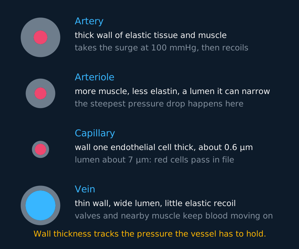
Describe the drawing
Four vessels drawn in cross-section down the left of the figure as rings, with a description of each beside it. The artery has a thick wall around a narrow lumen, the arteriole a smaller version of the same, the capillary a wall one cell thick around a lumen wide enough for a single red blood cell, and the vein a thin wall around a very wide lumen. The pressure each carries falls from about 100 millimetres of mercury in the artery to about 8 in the vein.
The walls of arteries and veins are built in three layers. The inner layer, the tunica intima, is the endothelium: one layer of flattened cells whose smooth surface reduces friction with the blood. The middle layer, the tunica media, holds the elastic tissue and smooth muscle, and it is much thicker in an artery than in a vein. The outer layer, the tunica externa, is mostly collagen, a tough fibrous protein that stops the vessel being overstretched or bursting. A capillary wall is the endothelium alone.
An artery takes the full force of ventricular systole, so its wall is thick, with a lot of elastic tissue and a layer of smooth muscle, and its lumen is narrow relative to that wall. The elastic tissue does more than resist bursting: it stretches during systole, stores energy, then recoils during diastole and drives the blood onwards. That recoil is why arterial pressure does not fall to zero between beats and why the flow becomes continuous rather than intermittent.
That description treats the artery wall as one design, which is sufficient for most questions. Some specifications divide arteries into two kinds, which resolve the same compromise differently at different distances from the heart.
An elastic artery is one of the great vessels near the heart: the aorta, the pulmonary artery, the common carotid and the subclavian. Its middle layer is dominated by thick concentric sheets of elastic tissue, the elastic laminae, with comparatively little smooth muscle between them. These are the vessels that take the full force of ventricular systole, and what they do with it is stretch. The wall balloons outward as blood is ejected, storing energy in the stretched elastic; during diastole it recoils and squeezes the stored blood onwards. That is what turns the intermittent output of a pump into a continuous flow, and it is why arterial pressure is still about 80 mmHg at the moment the ventricle is empty and relaxed.
A muscular artery is one of the named branches further out: the femoral, the brachial, the renal, the splenic. By then the pulse has already been smoothed, so the elastic laminae thin down to little more than a boundary at each face of the middle layer, and smooth muscle takes over the bulk of the wall. These vessels distribute blood between organs. Contracting the muscle constricts the vessel and reduces the supply to the tissue downstream; relaxing it increases the supply. Arterioles make the finer adjustment, described below, while a muscular artery controls the supply to a whole organ.
Elastic arteries therefore absorb the pressure surge and muscular arteries distribute the flow. Both have a thick wall, a narrow lumen and no valves, so the general description of an artery applies to each.
- Elastic artery
- A large artery near the heart, such as the aorta, whose wall is dominated by elastic laminae and which stretches and recoils to smooth out the pulse.
- Muscular artery
- An artery further from the heart, such as the femoral or renal artery, whose wall holds more smooth muscle and which distributes blood to organs by vasoconstriction and vasodilation.
An arteriole is the same design shrunk, but with the proportions shifted towards muscle. Contract that muscle (vasoconstriction) and the lumen narrows, resistance rises and less blood reaches the tissue beyond. Relax it (vasodilation) and more does. This is how blood is redistributed during exercise, and it is why the arterioles are where the pressure drop happens.
A capillary is a tube whose wall is a single layer of endothelium, about 0.6 μm thick, with a lumen around 7 μm, narrow enough that red blood cells go through in single file, pressed against the wall. Three consequences follow. The diffusion distance is tiny. The red cells are held right at the exchange surface. And because the total cross-sectional area of all the capillaries is enormous compared with the aorta, the blood slows right down, giving longer for exchange. Gaps between the endothelial cells let water and small solutes through while keeping the plasma proteins in.
Venules collect blood from capillary beds and merge into veins, which carry it back at about 8 mmHg. At that pressure there is no point in a thick wall, so veins have thin walls and very wide lumens, which lowers resistance and holds a large volume of blood. About 60 per cent of the blood volume is in the veins at any moment. With little pressure behind it, venous return depends on two features: semilunar valves along the veins prevent backflow, and contraction of surrounding skeletal muscle drives blood along. Standing still without using those muscles reduces venous return.
| Artery | Arteriole | Capillary | Vein | |
|---|---|---|---|---|
| Pressure carried | ≈ 100 mmHg | ≈ 60 mmHg | ≈ 25 mmHg | ≈ 8 mmHg |
| Wall | Thick: elastic + muscle | Muscular, adjustable | One cell thick | Thin |
| Lumen | Narrow | Narrow, variable | ≈ 7 μm | Very wide |
| Valves | No | No | No | Yes |
| Main job | Withstand and smooth the surge | Control where blood goes | Exchange | Return blood cheaply |
The pressure profile around the circulation
Blood leaves the left ventricle at about 120 mmHg and returns to the right atrium at close to zero. Where that pressure is lost accounts for the structure of each part of the circulation.
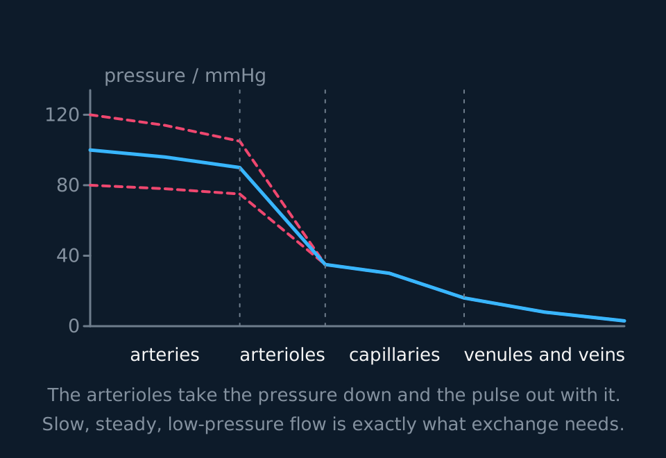
Describe the drawing
A graph of blood pressure against position round the circulation. A solid line for mean pressure falls from about 100 millimetres of mercury in the arteries to near zero in the great veins, with the steepest fall across the arterioles. Two dashed lines above and below it mark the systolic and diastolic swing, and they close onto the mean line at the arterioles, showing the pulse disappearing before the capillaries.
Arterioles are narrow, and resistance to flow rises steeply as radius falls: halving the radius raises the resistance sixteen-fold. Millions of arterioles in parallel take the mean pressure from about 90 mmHg down to about 35 mmHg at the start of a capillary bed. That pressure drop protects the capillaries, whose walls are one cell thick and would not withstand 120 mmHg arriving in pulses.
Blood therefore enters the capillaries with a flow that is steady, slow and at low pressure, which suits an exchange surface: slow flow allows time for exchange, steady flow avoids damaging surges, and low pressure permits a wall thin enough for rapid diffusion.
Formation of tissue fluid
Cells are surrounded by tissue fluid, not by blood. Tissue fluid is plasma forced out of capillaries, without most of the plasma proteins and without the red blood cells, which are too large to leave. Where fluid leaves and where it returns depends on two opposing pressures, of which only one changes along the capillary.
- Hydrostatic pressure
- The physical pressure the blood exerts against the capillary wall, pushing fluid out through the gaps between endothelial cells.
- Oncotic pressure
- The inward pull created by plasma proteins, which stay in the capillary and lower the water potential of the blood relative to the tissue fluid. Also called colloid osmotic pressure.
- Tissue fluid
- The fluid surrounding cells, formed by ultrafiltration from capillaries; it is very poor in plasma protein, though not literally protein-free, and has no red blood cells and no platelets.
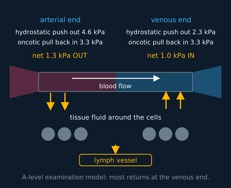
Describe the drawing
A capillary drawn as a horizontal tube running from an arteriole on the left to a venule on the right, with blood flowing left to right. Above the left end the hydrostatic push out of 4.6 kilopascals is set against the oncotic pull in of 3.3, giving a net outward pressure; above the right end the hydrostatic push has fallen to 2.3 while the oncotic pull is unchanged, giving a net inward pressure. Arrows point down out of the capillary at the left end and up back into it at the right end, into and out of tissue fluid surrounding a row of cells, and a further arrow leads down from the tissue into a lymph vessel. A line beneath the drawing labels it as the A-level examination model, in which most of the fluid returns at the venous end.
At the arteriole end the hydrostatic pressure is about 4.6 kPa, pushing outwards. The oncotic pressure pulls inwards at about 3.3 kPa. The net pressure is 1.3 kPa outwards, so water and dissolved small molecules such as glucose, oxygen, amino acids and mineral ions are forced out through the gaps in the wall. This is ultrafiltration: the wall acts as a filter, and almost all the plasma protein is held back from the fluid leaving.
Along the capillary, hydrostatic pressure falls. Some of it is lost to friction against the wall, and some to the fluid that has already left. By the venule end it is down to about 2.3 kPa. The oncotic pressure, meanwhile, has not budged: it is still 3.3 kPa, because the proteins responsible are still inside the capillary and if anything are now slightly more concentrated. The balance therefore reverses: the net pressure is 1.0 kPa inwards, and water moves back into the capillary down the water potential gradient, carrying dissolved waste including carbon dioxide with it.
Working out the direction of movement at each end
At the arteriole end of a capillary the hydrostatic pressure is 4.6 kPa and the oncotic pressure is 3.3 kPa. At the venule end the hydrostatic pressure is 2.3 kPa and the oncotic pressure is unchanged. Calculate the net pressure at each end and state which way fluid moves.
Show the working
Arteriole end: 4.6 − 3.3 = 1.3 kPa net outwards. Fluid leaves the capillary and forms tissue fluid.
Venule end: 3.3 − 2.3 = 1.0 kPa net inwards. Fluid returns to the capillary.
The outward net pressure of 1.3 kPa exceeds the inward net pressure of 1.0 kPa, so in this model more fluid leaves than returns. The figure usually quoted is that about 10 per cent of the fluid that left does not return directly to the capillary; that surplus is returned through the lymphatic system.
State the direction of each pressure and compare their magnitudes. In the classical A-level model, hydrostatic pressure falls along the capillary while the inward oncotic pressure due to plasma proteins is treated as approximately constant.
Two accounts of the same capillary are in use, and they should be labelled rather than blended.
A-level examination model. Hydrostatic pressure favours filtration at the arteriole end; as hydrostatic pressure falls, plasma proteins favour reabsorption towards the venule end; excess fluid enters lymph vessels. This is the classical Starling model, and it is the account the A-level specifications and mark schemes in this topic are written around.
Current physiological model. In most tissues at steady state, sustained venous reabsorption is limited and most net filtered fluid returns through the lymphatic system. This is the revised Starling principle, drawn from measurements made since the 1990s. Check your own specification for the model it requires.
Where the leftover fluid goes
In the examination model, roughly nine tenths of the fluid that left the capillary returns to it at the venule end. The rest is collected by lymph capillaries: blind-ended, very permeable tubes that thread through every tissue. Once inside, the fluid is called lymph.
Lymph has no central pump, but it is not entirely passive either. The larger collecting vessels carry smooth muscle in their walls that contracts rhythmically, working alongside the squeeze of surrounding skeletal muscle and the one-way valves that stop the lymph sliding back. It passes through lymph nodes, where lymphocytes and phagocytes intercept bacteria and other material, and eventually drains back into the bloodstream at the subclavian veins near the collarbone.
One detail explains a lot of medicine: lymph capillaries are permeable enough to take up proteins that have escaped from the blood, and blood capillaries are not. Without the lymphatic system, protein would accumulate in the tissue fluid, its oncotic pressure would climb, and less and less water would be drawn back into the blood.
| Blood | Tissue fluid | Lymph | |
|---|---|---|---|
| Red blood cells and platelets | Present | None | None |
| White blood cells | Present | A few | Many lymphocytes |
| Plasma proteins | High concentration | Very little | More than tissue fluid, because lymph capillaries take them up |
| Lipid | Carried as lipoproteins | Very little | Much more after a meal, in lymph draining the gut |
| Oxygen and glucose | More | Less: used by the cells | Less |
| Carbon dioxide | Less | More: made by the cells | More |
- Lymph
- Tissue fluid that has drained into the lymphatic system; similar to tissue fluid but containing lymphocytes and, after a meal, more lipid, carried in chylomicrons.
- Lymph node
- A swelling in a lymph vessel where lymphocytes collect and pathogens in the lymph are removed.
Oedema and its three causes
Oedema is swelling caused by an accumulation of fluid in a tissue. Explain any case of it using the same two pressures. There are three ways to alter the balance between them.
Raise the hydrostatic pressure. If the left side of the heart is failing, blood backs up in the pulmonary veins, capillary hydrostatic pressure in the lungs rises, more fluid is forced out than can be drawn back, and fluid collects in the alveoli. If the right side is failing, or if venous return from the legs is obstructed, the same thing happens in the ankles. Sitting still on a long flight does a mild version of this by removing the muscle pump.
Lower the oncotic pressure. A diet severely short of protein means the liver cannot make enough plasma proteins, especially albumin. The inward pull at the venule end weakens, less fluid returns, and it accumulates, most visibly in the abdomen, which is the swollen belly of kwashiorkor. Severe liver or kidney disease can do the same, the liver by failing to make albumin and the kidney by losing it in the urine.
Block the drainage. If lymph vessels are blocked, whether by the parasitic worms of filariasis or by lymph nodes removed during cancer surgery, then the tenth of the fluid that relies on that route has nowhere to go, and the affected limb swells.
TRY IT: Explaining a clinical observation
A person with a long-term shortage of protein in their diet develops a swollen abdomen. Explain, in terms of the pressures acting at a capillary bed, why this happens.
Check your answer
Too little dietary protein means too few amino acids for the liver to build plasma proteins, so the concentration of protein in the blood plasma falls.
Plasma proteins are what generate the oncotic pressure, so that inward pull is reduced, perhaps from 3.3 kPa to well under 2 kPa. The hydrostatic pressure is unchanged, so at the arteriole end even more fluid is forced out than usual, and at the venule end the reduced oncotic pressure can no longer overcome the hydrostatic pressure, so far less returns.
Fluid therefore accumulates in the tissues faster than the lymphatic system can drain it, and the tissue swells. In the abdomen this is visible as distension.
Name the pressure that changed and its direction, then follow the consequence to both ends of the capillary. A general reference to osmosis does not answer the question.
In the exam
- Quote pressures with a direction: 'hydrostatic pressure of 4.6 kPa outwards exceeds oncotic pressure of 3.3 kPa inwards, so fluid leaves', rather than 'the pressure is higher so fluid moves'.
- In the classical A-level model, the oncotic pressure is treated as approximately constant along the capillary and the hydrostatic pressure falls. Attribute the reversal to the fall in hydrostatic pressure.
- Tissue fluid contains no red blood cells, no platelets and effectively no plasma proteins, because those are too large to pass through the gaps. Everything small enough goes.
- If asked to relate a vessel's structure to its function, use the structure-then-consequence form: 'elastic tissue in the wall, so it stretches and recoils, so pressure is maintained between beats'.
- Answer oedema questions with the pressures. State whether hydrostatic pressure rose or oncotic pressure fell, then give the effect at each end of the capillary.
- Define an artery by direction of flow, away from the heart, rather than by the oxygenation of the blood it carries.
- For open against closed, give both consequences. Pressure is lost in a haemocoel, so delivery is slow; and there are no vessels to constrict, so flow cannot be directed to one organ rather than another.
- An insect's open system supports an active animal, because oxygen is delivered separately by tracheae and the haemolymph does not transport it.
- Where a question names an elastic or a muscular artery, answer with the wall structure. Elastic laminae stretch and recoil to smooth the pulse near the heart; smooth muscle constricts and dilates to distribute blood further out.
Check yourself
Explain why a capillary wall being one cell thick is useful, and why the same feature makes it essential that arterioles reduce blood pressure before blood arrives.
Answer
A wall one endothelial cell thick, roughly 0.6 μm, gives a very short diffusion pathway. Oxygen, glucose and other small solutes cross quickly, and the rate of diffusion is inversely proportional to the distance travelled, so a thin wall means fast exchange. The gaps between the endothelial cells also allow the bulk flow of fluid out of the capillary that forms tissue fluid.
The cost of that thinness is fragility, and low resistance to being stretched. Blood leaving the left ventricle at 120 mmHg in pulses would rupture such a wall, or at the very least drive far more fluid out of it than could ever be drawn back, producing oedema.
Arterioles are narrow and heavily muscled, and resistance to flow rises steeply as radius falls. Millions of them in parallel drop the mean pressure from around 90 mmHg to around 35 mmHg and damp out the difference between systolic and diastolic entirely, so what reaches the capillary bed is a slow, steady, low-pressure flow.
The two features are complementary: the capillary wall can be that thin only because the arteriole upstream has already reduced the pressure, and the muscular arteriole wall is required because the capillary downstream is that thin.
Questions
Question 14 marks
Compare the open circulatory system of an insect with the closed circulatory system of a mammal, referring to where the fluid travels, the pressure reached and the control of where it goes.
Mark scheme
- B1 in the insect haemolymph is pumped by a dorsal vessel and then leaves it to bathe the organs directly in the haemocoel, whereas in the mammal blood stays inside vessels for the whole journey
- B1 in the insect the haemolymph is blood and tissue fluid at once, whereas in the mammal tissue fluid is a separate compartment filtered out through the capillary walls
- B1 pressure is low in the haemocoel because nothing contains the fluid, so delivery is slow, whereas the vessel walls of a closed system hold the pressure the ventricle generates, so blood moves far faster
- A1 flow cannot be directed in an open system because there is no vessel whose radius could be changed, whereas in a closed system vasoconstriction and vasodilation of arterioles redistribute blood between organs
Question 24 marks
At the arteriole end of a capillary the hydrostatic pressure is 4.6 kPa and the oncotic pressure is 3.3 kPa. At the venule end the hydrostatic pressure has fallen to 2.3 kPa. Calculate the net pressure at each end, and calculate by how much the outward net pressure exceeds the inward one.
Mark scheme
- M1 net pressure is the hydrostatic pressure minus the oncotic pressure, and the oncotic pressure is still 3.3 kPa at the venule end because the proteins never left
- A1 arteriole end: 4.6 − 3.3 = 1.3 kPa net outwards
- A1 venule end: 3.3 − 2.3 = 1.0 kPa net inwards
- A1 the outward net pressure exceeds the inward one by 0.3 kPa, so more fluid leaves than returns directly
Question 34 marks
Using the A-level examination model of capillary exchange, explain how tissue fluid is formed at the arteriole end of a capillary bed and how most of it is returned at the venule end.
Mark scheme
- B1 at the arteriole end the hydrostatic pressure of about 4.6 kPa pushing outwards exceeds the oncotic pressure of about 3.3 kPa pulling inwards
- B1 water and small solutes are forced out through the gaps between the endothelial cells by ultrafiltration, while plasma proteins and red blood cells are too large to leave
- B1 hydrostatic pressure falls along the capillary to about 2.3 kPa, lost to friction against the wall and to the fluid that has already left
- A1 the oncotic pressure has not changed, because the proteins responsible are still inside, so it now exceeds the hydrostatic pressure and water returns down the water potential gradient
Question 44 marks
Explain how the elastic tissue in an artery wall keeps blood flowing between beats, and explain why it is the arterioles rather than the arteries that bring the mean blood pressure down.
Mark scheme
- B1 elastic tissue in the artery wall stretches during ventricular systole and stores energy as it does so
- B1 during diastole it recoils and pushes the blood on, so arterial pressure never falls to zero between beats and the flow is smoothed rather than arriving in slugs
- B1 arterioles are narrow and heavily muscled, and resistance to flow rises steeply as the radius falls, so halving the radius raises the resistance sixteen-fold
- A1 millions of arterioles in parallel take the mean pressure from about 90 mmHg down to about 35 mmHg and damp out the difference between systolic and diastolic
Question 54 marks
A capillary wall is a single layer of endothelium about 0.6 μm thick, with gaps between its cells and a lumen about 7 μm across, so red blood cells pass through pressed against the wall in single file. Together the capillaries of a bed have a far greater cross-sectional area than the artery supplying them. Explain how each of these features suits capillaries to exchange.
Mark scheme
- B1 the thin wall gives a very short diffusion pathway, so oxygen, glucose and other small solutes cross quickly between blood and tissue
- B1 gaps between the endothelial cells allow water and small solutes to filter out of the capillary as tissue fluid, while keeping the larger plasma proteins inside
- B1 the narrow lumen holds red blood cells right against the exchange surface, minimising the diffusion distance from the cell carrying oxygen to the tissue
- A1 the huge total cross-sectional area of all the capillaries together, compared with the aorta, slows the blood right down as it passes through, giving more time for exchange to take place
Question 64 marks
In left-sided heart failure, blood backs up in the pulmonary veins. Suggest how this leads to fluid collecting in the alveoli of the lungs.
Mark scheme
- B1 blood backing up in the pulmonary veins raises the pressure in the capillaries of the lungs, since the blood cannot be moved on as quickly as normal
- B1 this raises the hydrostatic pressure along the whole length of these capillaries, so more fluid than usual is forced out into the surrounding tissue
- B1 the oncotic pressure pulling fluid back in is unchanged, so it can no longer draw back as much of the increased outflow
- A1 more fluid is therefore forced out than can be returned or drained by the lymphatic system, and it accumulates in the alveoli, impairing gas exchange
Question 74 marks
A person on a diet severely short of protein develops a swollen abdomen, a condition called kwashiorkor. Explain, in terms of the pressures acting at a capillary bed, why this happens.
Mark scheme
- B1 too little dietary protein means the liver cannot make enough plasma proteins, such as albumin, so the concentration of protein in the blood plasma falls
- B1 plasma proteins are what generate the oncotic pressure, so this inward pull is reduced
- B1 the hydrostatic pressure pushing fluid out at the arteriole end is unchanged, so as much fluid as usual, or more, is still forced out
- A1 at the venule end the weakened oncotic pressure can no longer draw back as much fluid as normal, so fluid accumulates in the tissues faster than the lymphatic system can drain it, causing the swelling
Question 83 marks
The aorta is classed as an elastic artery and the renal artery as a muscular artery. Explain the difference between the walls of the two vessels and how each wall suits the job that vessel does.
Mark scheme
- B1 the wall of the aorta is dominated by thick concentric elastic laminae with relatively little smooth muscle, whereas the wall of the renal artery has much more smooth muscle and only thin elastic layers
- B1 the aorta lies next to the heart and takes the full force of ventricular systole, so it stretches as blood is ejected and recoils during diastole, which maintains pressure between beats and smooths the pulse out
- A1 the renal artery lies further out where the pulse has already been smoothed, and contracting or relaxing its smooth muscle constricts or dilates the vessel, which adjusts how much blood is distributed to the kidney
Question 93 marks
Explain why blocking the lymph vessels that drain a limb makes that limb swell.
Mark scheme
- B1 in the examination model about a tenth of the fluid leaving the capillary does not return at the venule end, and is normally collected by the blind-ended lymph capillaries
- B1 with the drainage blocked that surplus fluid has nowhere to go, so it accumulates in the tissue as oedema
- A1 lymph capillaries also take up plasma proteins that have escaped from the blood, so protein now builds up in the tissue fluid, raising its oncotic pressure and drawing back still less water
Question 103 marks
Standing still for an hour on a hot day often makes a person's ankles swell. Suggest why.
Mark scheme
- B1 blood in the veins of the leg is returned largely by contraction of the surrounding skeletal muscle squeezing the veins, and standing still removes that muscle pump; or, in the heat, arterioles supplying the skin dilate, so more blood reaches the capillaries at a higher hydrostatic pressure
- B1 blood therefore pools in the leg veins and the hydrostatic pressure in the capillaries of the ankle rises
- A1 more fluid is forced out at the arteriole end than can be drawn back at the venule end or drained by the lymphatics, so fluid collects in the tissue
Question 113 marks
Explain why a vein has a thin wall and a wide lumen, and explain how blood is kept moving back towards the heart despite the low pressure in it.
Mark scheme
- B1 blood in a vein is at a low pressure of only about 8 mmHg, so there is no need for a thick wall to withstand the pressure, and a thin wall and wide lumen offer low resistance to flow and hold a large volume of blood
- B1 semilunar valves along the length of the vein prevent blood flowing backwards
- A1 contraction of the surrounding skeletal muscle squeezes the vein and drives blood onwards towards the heart, which is why standing still without moving reduces venous return
Question 123 marks
During exercise, blood flow to the gut falls while blood flow to the leg muscles rises. Suggest how arterioles bring this about.
Mark scheme
- B1 arterioles supplying the gut constrict (vasoconstriction), narrowing their lumen and raising resistance, which reduces the blood reaching the gut
- B1 arterioles supplying the leg muscles dilate (vasodilation), widening their lumen and lowering resistance, which increases the blood reaching them
- A1 because arterioles, unlike capillaries, have smooth muscle in their walls that can contract or relax, blood is redistributed to the tissues that need it most at that moment
Question 133 marks
Describe how lymph is moved through the lymphatic system back to the bloodstream, given that it has no central pump.
Mark scheme
- B1 smooth muscle in the walls of the larger lymph vessels contracts rhythmically, helping to move the lymph along
- B1 contraction of surrounding skeletal muscle also squeezes the lymph vessels, and one-way valves stop the lymph sliding back
- A1 lymph eventually drains back into the bloodstream at the subclavian veins near the collarbone
Question 143 marks
State what the classical A-level model of capillary exchange says happens at the venule end and what the current physiological model says instead, and state what each implies for the fluid returned by the lymphatic system.
Mark scheme
- B1 the classical A-level model says that hydrostatic pressure falls enough along the capillary that oncotic pressure exceeds it at the venule end, so most of the fluid that left is directly reabsorbed there
- B1 the current physiological model says that in most tissues at steady state, sustained venous reabsorption at the venule end is limited
- A1 so the lymphatic system returns only a surplus of about a tenth in the classical model, but most of the net filtered fluid in the current one
Question 152 marks
A blood vessel has a thin wall, a very wide lumen and valves along its length. Identify the type of vessel, and give the pressure the blood in it typically carries.
Mark scheme
- A1 it is a vein, or a venule merging into one
- A1 a low pressure of about 8 mmHg, accepting any value from 5 to 15 mmHg
Worth remembering
- Open circulation: haemolymph leaves the dorsal vessel and bathes the organs in the haemocoel, so pressure is low, delivery is slow and flow cannot be directed.
- Closed circulation: blood stays in vessels, so pressure is maintained, delivery is fast and arterioles can direct flow to particular tissues.
- Elastic arteries near the heart stretch and recoil to smooth the pulse; muscular arteries further out constrict and dilate to distribute blood.
- Wall thickness increases with the pressure a vessel carries; the lumen occupies the remainder of the cross-section.
- Mean pressure falls steeply across the arterioles, and the pulse is lost there.
- Tissue fluid forms where hydrostatic pressure exceeds oncotic pressure, at the arteriole end, and returns where the balance reverses, at the venule end.
- In the classical A-level model, hydrostatic pressure falls along a capillary while oncotic pressure is treated as approximately constant, because the plasma proteins remain inside the capillary.
- In that model about a tenth of the fluid leaving does not return directly to the capillary; it is returned through the lymphatic system.
CHECK YOUR PROGRESS
Rate how confident you are with each objective for this lesson. Ratings are saved in this browser, on this device, unless you sign in.
- Compare an open circulatory system with a closed one, and say what a closed system gives an animal in delivery rate and in control.
- Relate the structure of an artery, arteriole, capillary, venule and vein to its function and to the pressure it carries.
- Distinguish an elastic artery from a muscular artery by the composition of its wall, its distance from the heart and the job it does.
- Explain why a capillary wall is one cell thick and why the blood moves through it slowly.
- Account for the formation of tissue fluid at the arteriole end of a capillary bed using hydrostatic and oncotic pressure.
- Account, in the A-level examination model, for the return of most of that fluid at the venule end using the same two pressures.
- Describe what the lymphatic system does with the fluid that is not returned.
- Explain three different causes of oedema using nothing but hydrostatic and oncotic pressure.
Open the full revision checklist to see every objective in the curriculum in one place.
Practise this lesson
WORKBOOK
The questions from this topic, on paper with room to work, and a separate book of mark schemes. Free to use; please do not redistribute or sell.
Used later in Cholesterol, atheroma and what a risk factor means · Thermoregulation in endotherms and ectotherms