Biology › Nutrition, digestion and health evidence › Cholesterol, atheroma and what a risk factor means
Cholesterol, atheroma and what a risk factor means
Cholesterol is essential for membranes and as a precursor of steroid hormones, bile acids and vitamin D. Cardiovascular risk rises when LDL particles enter and are retained in the artery wall.
Before this Cholesterol in the phospholipid bilayer · Receptor-mediated endocytosis · Vessels and tissue fluid: the structure of arteries · Designing an investigation: correlation, confounding and study design
COMMON MISCONCEPTION
HDL and LDL are two kinds of cholesterol, one good and one bad.
There is one cholesterol molecule, and the cholesterol in an HDL particle is chemically identical to the cholesterol in an LDL particle. HDL and LDL are the particles that carry it, differing in density, size, surface proteins and destination.
What you should be able to do
- Describe cholesterol as a sterol and state four things the body needs it for.
- Explain what a lipoprotein is and distinguish LDL from HDL by composition and function.
- Describe the formation of an atheroma in sequence, from endothelial damage to a fibrous plaque.
- Explain how a plaque leads to angina, to a myocardial infarction and to an aneurysm.
- State precisely what the term 'risk factor' licenses you to claim, and what it does not.
- Account for why the evidence on LDL is regarded as causal while the evidence on HDL is not.
Cholesterol is not a fat, and it is not optional
Cholesterol is a lipid but not a triglyceride. It is a sterol: four carbon rings fused together, with a short hydrocarbon tail at one end and a single hydroxyl group at the other. That hydroxyl is the only polar part of an otherwise hydrophobic molecule, which is exactly the arrangement needed to sit in a membrane with its hydroxyl among the phosphate heads and its rings among the fatty acid tails.
Four jobs are worth naming. Cholesterol regulates the fluidity of every cell surface membrane you have, restricting movement of the phospholipids at higher temperatures and preventing them packing too closely at lower ones. It is the precursor of the steroid hormones, such as testosterone, oestrogen, cortisol and aldosterone, and of the bile salts that emulsify lipids in the duodenum. And it is the starting material for vitamin D.
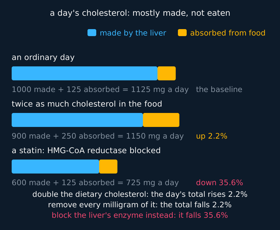
Most of it is not eaten. The liver synthesises on the order of a gram a day, while a typical diet supplies two or three hundred milligrams, and the liver adjusts its synthesis downwards when dietary intake rises. The rate-limiting enzyme of that synthetic pathway is HMG-CoA reductase, and inhibiting it is what a statin does, which is why statins lower blood cholesterol far more effectively than removing cholesterol from the diet does.
Describe the drawing
Three horizontal bars drawn to one scale, each one a day's supply of cholesterol split into a long cyan part made by the liver and a short amber part absorbed from food. The first bar is an ordinary day, a thousand milligrams made and a hundred and twenty-five absorbed. In the second the food has twice as much cholesterol in it, so the amber part doubles, but the cyan part shrinks by most of that increase and the bar as a whole is barely longer. The third bar has a statin blocking the liver's own synthesis, and it is visibly the shortest of the three. The figures beneath each bar are computed from a typical compensation, which varies a good deal between people, and the closing lines put the three percentage changes side by side.
That regulation is why the long-standing advice to limit dietary cholesterol was dropped from several national guidelines around 2015: in most people, eating more of it produces only a small rise in blood cholesterol, because synthesis falls to compensate.
Lipoproteins and how they are classified
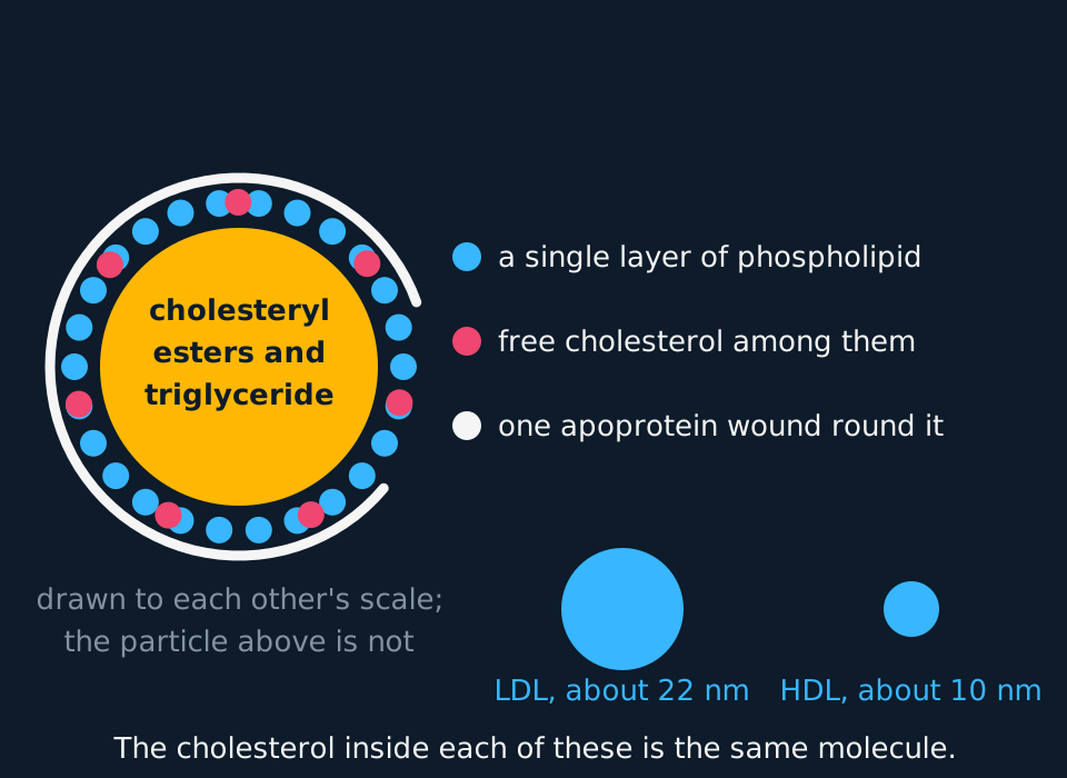
Cholesterol and triglyceride are insoluble in plasma, which is mostly water, so they cannot simply dissolve and travel. They are carried inside particles built for the purpose.
Describe the drawing
A large lipoprotein particle in cross-section: an amber core labelled as cholesteryl esters and triglyceride, ringed by a single layer of cyan phospholipid circles with seven coral free cholesterol molecules among them, and a thick ink arc representing one apoprotein wrapped most of the way round. A key beside it names each part. Below, two cyan discs drawn to the same scale as each other compare a low-density particle of about twenty-two nanometres with a high-density particle of about ten.
A lipoprotein has a core of cholesteryl esters and triglyceride, wrapped in a single layer of phospholipid with the hydrophilic heads facing outwards, with free cholesterol among them and one or more apoproteins threaded through the surface. The apoprotein is the address label: it determines which receptors the particle can bind to and therefore where it is unloaded.
| Particle | Made by | Carries mainly | Delivers to |
|---|---|---|---|
| Chylomicron | Ileum epithelium | Triglyceride from the meal just eaten | Muscle and adipose tissue, via the lymph |
| VLDL | Liver | Triglyceride made in the liver | Muscle and adipose tissue |
| LDL | Formed from VLDL in the blood | Cholesterol, and little triglyceride | Any cell with LDL receptors |
| HDL | Liver and intestine | Protein, and cholesterol collected from tissues | Back to the liver |
LDL is the delivery service. A cell needing cholesterol makes LDL receptors and places them in its surface membrane; an LDL particle binds and the whole particle is taken in by receptor-mediated endocytosis and digested in a lysosome. A cell with plenty of cholesterol makes fewer receptors, so uptake is regulated by need, and LDL not taken up stays in the plasma.
HDL runs the other way. Nascent HDL particles are mostly protein and accept cholesterol from cell membranes, including from cells inside artery walls, and carry it back to the liver, which can excrete it in bile. That is reverse cholesterol transport, and it is where HDL's reputation came from.
The examinable comparison is not 'good and bad'. It is that LDL particles are larger, cholesterol-rich and delivering, while HDL particles are smaller, protein-rich and collecting, and that a high LDL concentration means more particles available to enter an artery wall.
How an atheroma is built
Atherosclerosis takes decades and happens in a fixed order. Fatty streaks are present in the aortas of many teenagers; the first symptom typically arrives in the sixth decade or later. Nothing in this sequence happens quickly except the last step, which happens in minutes.

Describe the drawing
An artery in longitudinal section, animated through five stages. First a coral jagged line marks damage to the smooth endothelium lining the upper wall, with high blood pressure, smoking and high blood glucose named as causes. Second, cyan circles representing LDL particles move from the lumen up into the wall. Third, coral macrophages appear inside the wall and a small amber swelling bulges into the lumen as a fatty streak. Fourth, the swelling is much larger and capped by a dark line, with double-headed arrows comparing the full width of the lumen elsewhere against the narrowed channel at the plaque. Fifth, the cap has torn and a dark clot fills most of the remaining lumen.
It starts with damage to the endothelium, the single layer of cells lining the vessel. High blood pressure provides mechanical stress, particularly where arteries branch; chemicals in tobacco smoke, high blood glucose and a high LDL concentration all injure the lining as well. An intact endothelium is smooth and does not admit much.
Next, LDL particles pass into the wall beneath the endothelium and are retained there, where they are chemically modified, or oxidised. Modified LDL is treated as damage rather than as cargo. White blood cells, monocytes, adhere to the activated endothelium and migrate into the wall, becoming macrophages, and take up the modified LDL through receptors that are not regulated by how much cholesterol the cell already holds. So they keep eating, fill with lipid, and become foam cells. A collection of foam cells is a fatty streak, and this is an inflammatory process from beginning to end.
Smooth muscle cells then migrate from the deeper layers of the artery wall, multiply, and deposit collagen over the accumulating lipid, forming a fibrous cap. The result is an atheroma or plaque: a lipid and cellular core under a fibrous cap, bulging into the lumen and reducing the space for blood to flow. Narrowed, stiffened arteries raise the blood pressure, and the higher pressure damages more endothelium, so the process feeds itself.
- Atheroma
- A plaque in the wall of an artery, consisting of lipid, foam cells and cell debris beneath a fibrous cap, which narrows the lumen and stiffens the wall.
- Thrombosis
- The formation of a blood clot inside a blood vessel, most often on a ruptured atheroma, which may block the vessel completely.
- Aneurysm
- A localised bulge in an artery wall that has been weakened, which may rupture and cause severe internal bleeding.
Two consequences follow, one gradual and one sudden. Gradually, a narrowed coronary artery delivers too little blood when demand rises, so the muscle it supplies runs short of oxygen during exertion and the person feels the pain of angina, which stops on resting.
Suddenly, the fibrous cap tears. Collagen beneath it is exposed to the blood, platelets adhere and release thromboplastin, thromboplastin with calcium ions catalyses the conversion of prothrombin to thrombin, and thrombin catalyses the conversion of soluble fibrinogen into insoluble fibrin, which forms a mesh trapping cells. That clot can occlude the artery within minutes. In a coronary artery the muscle downstream is deprived of oxygen, cannot respire aerobically, and dies: a myocardial infarction. In an artery supplying the brain the result is a stroke. A fragment breaking away and lodging elsewhere is an embolus.
A plaque also weakens the wall it sits in. An artery carries blood at high pressure, and a weakened section stretches, which thins it further and lets it stretch more: an aneurysm. Rupture of a large one, in the abdominal aorta, is usually fatal, which is why the NHS (under its screening programme as it stands in 2025) invites men to a single ultrasound scan in the year they turn 65, the age and sex at which the condition is common enough for screening to do more good than harm.
Risk factors and what the term supports
A risk factor is a variable associated with an increased probability of a disease in a population.
It supports prediction. Age, sex, blood pressure, smoking status, cholesterol concentration and an area-based measure of deprivation together allow a reasonable estimate of a person's probability of a cardiovascular event in the next ten years, and the tools used in general practice do exactly that. In the UK, under the NICE guidance current in 2025, a ten-year risk of 10% or more is the point at which a statin is normally offered and discussed; thresholds are reviewed from time to time.
It does not license three things. It does not establish a mechanism, because an association can exist where the factor causes the disease, where something else causes both, or where early disease causes the factor. It does not predict an individual: a risk of 12% means about twelve of a hundred similar people will have an event, and says nothing about which twelve. And it does not promise that removing the factor removes the risk, which is a separate question needing a trial or something as good as one.
| Cannot be changed | Can be changed |
|---|---|
| Age | Smoking |
| Sex: men develop it earlier | High blood pressure |
| Family history and specific inherited conditions | High LDL concentration |
| Ancestry: risk differs between populations | Diabetes and blood glucose control |
| Physical inactivity, diet and central obesity |
A case-control study published in 2004, covering around 15,000 first heart attacks in 52 countries, found that nine measurable factors accounted for the great majority of the risk of a first myocardial infarction in every region studied: smoking, blood lipids, blood pressure, diabetes, abdominal obesity, psychosocial factors, fruit and vegetable intake, alcohol and physical activity. Notice two things together: consistency across very different populations is strong evidence, and it was still a case-control study, with everything that implies about recall and about choosing controls.
Four classes of drug are used against cardiovascular disease, and each has a benefit and a risk. A statin lowers LDL but can cause muscle pain and, rarely, liver damage. Antihypertensives, such as ACE inhibitors, calcium channel blockers and diuretics, lower blood pressure and so the stress on the endothelium, but can cause dizziness, a dry cough with ACE inhibitors and disturbed ion balance with diuretics. Anticoagulants such as warfarin and heparin slow the clotting cascade, and platelet inhibitors such as aspirin and clopidogrel reduce platelet aggregation; both make a clot on a torn cap less likely, and both raise the risk of bleeding.
How the evidence was gathered, and what is still argued about
optional
The claim that LDL causes atherosclerosis is about as secure as any causal claim in medicine, and the way it was established is worth knowing, because it is the model for what strong evidence looks like when a randomised trial of a lifetime exposure is impossible.
The first strand is a natural experiment. Familial hypercholesterolaemia is caused by a fault in the gene for the LDL receptor, so cells take up LDL poorly and the blood concentration is roughly doubled from birth. It affects around 1 in 250 people and, untreated, produces heart attacks decades early. The raised LDL cannot be a consequence of an unhealthy life, because it was set at conception.
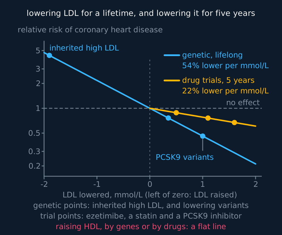
The second strand runs the same logic the other way. Some people carry inactive versions of a gene called PCSK9 and have LDL concentrations around a quarter lower than average for their whole lives. Their rate of coronary heart disease is lower by far more than a quarter. Because the genetic variant is allocated at conception and is not associated with income, smoking or diet, this kind of analysis behaves like a randomised trial run by nature, and it is called Mendelian randomisation.
The third strand is intervention. Trials of drugs that lower LDL by unrelated mechanisms, among them statins, ezetimibe and PCSK9 inhibitors, lower cardiovascular events in proportion to how much LDL they lower. Genetics, mechanism and trials agree, which is the pattern that makes a causal claim safe.
Describe the drawing
A graph with how far LDL has been lowered, in millimoles per litre, along the bottom, and relative risk of coronary heart disease up a logarithmic scale, so that a constant proportional change plots as a straight line. Both lines cross relative risk 1.0 at zero lowering, because that is where nothing has changed. The cyan line, for lifelong genetic differences in LDL, is much the steeper: it runs from a relative risk above four at the left, where inherited high LDL is plotted, down through the no-effect point and on below. The amber line, for drug trials lasting about five years, starts at the same point and falls at roughly a third of the gradient. The percentage reduction per millimole per litre is printed beside each, fitted from the plotted points rather than stated.
Now the contested part, and it is instructive precisely because the same field produced both. HDL concentration predicts cardiovascular risk reliably: people with more of it have fewer events. But raising it has repeatedly failed to help. Drugs that raised HDL substantially did not reduce events, and one had to be abandoned when the treated group did worse. Mendelian randomisation tells the same story: genetic variants that raise HDL are not associated with lower heart disease, while variants that raise LDL are associated with more of it. The plain summary is that HDL is a marker of something protective rather than established as protective itself, so the term 'good cholesterol' is not supported by the evidence.
Dietary fat is contested at a different level. Controlled feeding studies, in which every meal is prepared and measured, showed decades ago that replacing saturated fat with polyunsaturated fat lowers blood LDL, and that part is not seriously disputed. What happens to heart attacks and deaths is less settled: the Cochrane systematic review of the randomised trials, in its 2020 update, found that reducing saturated fat intake cut combined cardiovascular events by about 17%, with no clear effect on death from any cause. What it is replaced with matters as much: unsaturated fat looks beneficial, refined carbohydrate does not.
Two episodes show how an evidence base itself can be distorted. Data from two large dietary trials run in the 1960s and 1970s were only fully published after being recovered from the investigators' records forty years later, and showed that lowering blood cholesterol by changing dietary fat did not translate into fewer deaths in those trials. And documents recovered in 2016 showed that a 1967 review of sugar and heart disease in a leading journal had been funded, undisclosed, by the sugar industry, which chose the papers discussed. Neither overturns the biology. Both are reasons to ask who funded a study, whether it was registered before it began, and whether the result would have been published had it come out the other way.
TRY IT: Two claims, different footings
A student writes: 'HDL is good cholesterol and LDL is bad cholesterol, so raising your HDL and lowering your LDL will both reduce your risk of a heart attack.' Rewrite this as an accurate statement, and explain why the two halves of the original claim are not equally well supported.
Check your answer
Neither HDL nor LDL is a kind of cholesterol. They are lipoprotein particles distinguished by density and by the proteins on their surface, and the cholesterol carried in each is the same molecule.
An accurate version: a high concentration of cholesterol carried in LDL particles raises the risk of atherosclerosis, and lowering it lowers that risk. A high concentration of cholesterol carried in HDL particles is associated with lower risk, but raising it has not been shown to reduce risk.
The two halves rest on different evidence. For LDL, three independent lines agree: an inherited condition raising LDL from birth causes early heart disease, genetic variants that lower LDL for life are associated with much less of it, and drugs working by several different mechanisms lower event rates in proportion to how much LDL they remove.
For HDL, only the observational association exists. Trials of drugs that raise HDL have not reduced events, and variants that raise it are not associated with lower risk, which together suggest HDL marks something protective rather than being it.
In the exam
- Use 'cholesterol carried in HDL' and 'cholesterol carried in LDL' rather than 'good' and 'bad' cholesterol; the longer form names the particle doing the transport.
- For atheroma, give the sequence in order: endothelial damage, LDL entering and being modified, macrophages becoming foam cells, smooth muscle and collagen forming a cap, narrowing of the lumen.
- For a heart attack, the required chain is: cap ruptures, platelets adhere, clot forms, coronary artery blocked, cardiac muscle deprived of oxygen, respiration and therefore ATP supply fails, muscle dies.
- 'Risk factor' means associated with an increased probability in a population. Where a question asks whether a factor causes the disease, state what further evidence would be needed to support a causal claim.
Check yourself
A man of 58 has a total blood cholesterol concentration in the highest fifth for his age. His doctor tells him this is a risk factor for cardiovascular disease, and he replies that his father had the same reading and lived to 92. Explain, in terms of what a risk factor is, why both statements can be true, and what additional information would help.
Answer
A risk factor describes a population, not a person. Saying that a high cholesterol concentration is a risk factor means that among many people with that reading, more will have a cardiovascular event over a given period than among people with a lower one. It predicts nothing about any individual.
His father is one observation. A single person with a high reading and a long life is exactly what a probabilistic claim predicts will sometimes happen, so it is not evidence against the association.
Total cholesterol is also the wrong measurement to argue about, because it lumps together cholesterol carried in LDL particles, which is associated with disease, and cholesterol carried in HDL particles, which is not associated with it in the same direction. Two people with identical totals can carry very different LDL concentrations.
What would help is his absolute risk rather than one measurement: LDL specifically, plus blood pressure, smoking status, diabetes, family history and age, combined into an estimated probability of an event over ten years. That turns a risk factor into a number he can weigh against the benefits and drawbacks of treatment.
Questions
Question 15 marks
Describe the formation of an atheroma, in order, from the first damage to the artery wall through to a fibrous plaque.
Mark scheme
- B1 the endothelium lining the vessel is damaged by mechanical stress from high blood pressure, particularly where arteries branch, by chemicals in tobacco smoke, by high blood glucose or by a high LDL concentration
- B1 LDL particles pass into the wall beneath the endothelium, are retained there and are chemically modified by oxidation, so they are treated as damage rather than as cargo
- B1 monocytes adhere to the activated endothelium and migrate into the wall, where they become macrophages
- B1 the macrophages take up modified LDL through receptors that are not regulated by how much cholesterol the cell already holds, so they keep eating, fill with lipid and become foam cells, and a collection of foam cells is a fatty streak
- B1 smooth muscle cells migrate from the deeper layers, multiply and deposit collagen over the accumulating lipid, forming a fibrous cap, so the plaque bulges into the lumen and narrows it
Question 25 marks
The table gives results from a case-control study of first heart attacks, constructed for this question rather than measured. A health campaign concludes from it that reducing diabetes would prevent more heart attacks in this population than reducing high blood pressure, because diabetes has the larger odds ratio. Evaluate that conclusion.
| Risk factor | Odds ratio for a first heart attack | Percentage of adults in this population with the factor |
|---|---|---|
| Abnormal blood lipids | 3.3 | 45 |
| Current smoking | 2.9 | 26 |
| Diabetes | 2.4 | 8 |
| Abdominal obesity | 2.2 | 33 |
| High blood pressure | 1.9 | 30 |
Mark scheme
- B1 in support: the odds ratio does compare the strength of the two associations, and 2.4 against 1.9 means diabetes is the more strongly associated of the two with a first heart attack, each compared with people who lack that factor
- B1 against: how many cases a factor accounts for in a population depends on how common it is as well as on how strongly it acts, and high blood pressure is present in 30 per cent of these adults against 8 per cent with diabetes
- B1 against: an odds ratio is not a risk, because the investigators chose how many cases and how many controls to recruit, so neither group's risk can be read off this study at all, and for a common outcome an odds ratio sits further from 1 than the relative risk does
- B1 against: a risk factor is a variable associated with an increased probability of the disease in a population, which does not establish that it causes the disease or that removing it removes the risk, and the factors listed travel together, so their contributions cannot simply be added
- B1 judgement: the conclusion is not supported by the table. On these figures high blood pressure is the more promising target for a population despite its smaller odds ratio, and what either change would actually achieve needs a trial rather than a case-control study
Question 35 marks
A systematic review of randomised trials found that reducing saturated fat intake cut combined cardiovascular events by about 17%, with no clear effect on death from any cause. A magazine reports the headline 'Cutting saturated fat doesn't save lives'. Evaluate this headline.
Mark scheme
- B1 in support: the review did find no clear effect on death from any cause, so the headline's specific factual claim about that particular outcome is not unreasonable on its own
- B1 in support: because the review is based on randomised trials rather than observational studies, its findings on both outcomes carry more weight against confounding than an association from a cohort study would
- B1 against: the same review found a real reduction, about 17 per cent, in combined cardiovascular events, which the headline omits entirely, giving a one-sided impression of the evidence
- B1 against: what a food is replaced with matters, since unsaturated fat looks beneficial while refined carbohydrate does not, and a headline about saturated fat alone says nothing about that choice
- B1 judgement: the headline is misleading rather than false; it reports the absence of one specific finding, an effect on overall mortality, as though it were the whole of the evidence, when the same trials also show a genuine benefit for cardiovascular events specifically
Question 44 marks
Explain how the rupture of an atheroma in a coronary artery leads to the death of cardiac muscle.
Mark scheme
- B1 the fibrous cap tears, so collagen beneath it is exposed to the blood and platelets adhere and release thromboplastin
- B1 thromboplastin with calcium ions catalyses the conversion of prothrombin to thrombin, and thrombin catalyses the conversion of soluble fibrinogen into insoluble fibrin, which forms a mesh trapping cells
- B1 that clot can occlude the artery within minutes, so blood no longer reaches the muscle downstream of the blockage
- B1 the muscle is therefore deprived of oxygen and cannot respire aerobically, so its supply of ATP fails and the cells die, which is a myocardial infarction
Question 54 marks
Compare LDL particles with HDL particles, referring to their composition, what each carries and where each takes it.
Mark scheme
- B1 both are lipoproteins, with a core of cholesteryl esters and triglyceride wrapped in a single layer of phospholipid carrying apoproteins, and the cholesterol inside each is chemically the same molecule
- B1 an LDL particle is larger and cholesterol-rich with relatively little protein, whereas an HDL particle is smaller and protein-rich, and since protein is dense that is why HDL is the denser class
- B1 LDL delivers cholesterol to any cell carrying LDL receptors, which take the whole particle in by receptor-mediated endocytosis, whereas HDL accepts cholesterol from cell membranes, including from cells inside artery walls
- B1 HDL carries what it collects back to the liver, which can excrete it in bile, so the two classes of particle run in opposite directions
Question 64 marks
Explain why the evidence that a high LDL concentration causes atherosclerosis is regarded as much stronger than the evidence that a high HDL concentration protects against it.
Mark scheme
- B1 familial hypercholesterolaemia is caused by a fault in the gene for the LDL receptor and roughly doubles the blood LDL concentration from birth, producing heart attacks decades early, and that raised concentration was set at conception, so it cannot be a consequence of how the person lived
- B1 people carrying inactive versions of PCSK9 have lifelong LDL concentrations about a quarter lower and far less coronary heart disease, and because the variant is allocated at conception and is unrelated to income, smoking or diet the comparison behaves like a randomised trial
- B1 trials of drugs lowering LDL by unrelated mechanisms lower cardiovascular events in proportion to how much LDL they remove, so genetics, mechanism and trials all agree
- B1 for HDL only the observational association exists: drugs that raised HDL substantially did not reduce events, and genetic variants raising it are not associated with lower risk, so HDL appears to mark something protective rather than to be the protective thing itself
Question 74 marks
A man's total blood cholesterol concentration is 6.4 mmol dm⁻³ and the cholesterol carried in his HDL particles is 1.2 mmol dm⁻³. Calculate his non-HDL cholesterol concentration, and calculate his total cholesterol concentration in milligrams per 100 cm³, taking the relative molecular mass of cholesterol as 386.7.
Mark scheme
- M1 the total counts the cholesterol carried in every class of particle, so the non-HDL concentration is the total minus the concentration carried in HDL
- A1 6.4 − 1.2 = 5.2 mmol dm⁻³
- M1 mass concentration = concentration in mol dm⁻³ × relative molecular mass, so 6.4 × 10⁻³ × 386.7 = 2.47488 g dm⁻³
- A1 1 dm³ is 1000 cm³, so 100 cm³ holds one tenth of that: 0.247488 g, which is 247 mg per 100 cm³ to three significant figures
Question 84 marks
A man of 64 who smokes gets a cramping pain in his calf muscles after walking about a hundred metres, which goes when he stands still, and the pulse in that foot is weak. Suggest an explanation for these two observations, and suggest why he is also at raised risk of a stroke.
Mark scheme
- B1 an atheroma has formed in an artery supplying the leg, so the lumen is narrowed and the wall stiffened, less blood reaches the muscle each minute, and the pulse felt beyond the narrowing is weak
- B1 at rest the reduced supply is enough, but walking raises the muscle's demand for oxygen and the flow cannot rise to match it, so the muscle respires anaerobically and lactate accumulates and causes the pain
- B1 standing still lowers the demand again until the reduced supply can meet it, which is why the pain goes without anything about the artery having changed
- B1 atherosclerosis is not confined to one artery, because the same causes act on endothelium throughout the arterial system and the chemicals in tobacco smoke reach all of it, so a plaque in an artery supplying the brain is likely too; if its cap tears, a clot forms on it or a fragment breaks away as an embolus, brain tissue downstream is deprived of oxygen and dies, and that is a stroke
Question 94 marks
Explain how an atheroma can lead to an aneurysm, and explain why the rupture of a large aneurysm in the abdominal aorta is usually fatal.
Mark scheme
- B1 a plaque weakens the section of artery wall it sits in, and the wall is carrying blood at high pressure
- B1 the weakened section stretches under that pressure, which thins it further and allows it to stretch even more, forming a localised bulge called an aneurysm
- B1 the abdominal aorta is one of the widest arteries in the body and carries a very large flow of blood at high pressure
- B1 if a large aneurysm there ruptures, blood is lost from the circulation catastrophically fast into the abdomen, faster than the body can compensate for, causing severe internal bleeding that is usually fatal
Question 104 marks
A person's liver synthesises 950 mg of cholesterol a day and their gut absorbs a further 250 mg a day from food. Calculate the total amount of cholesterol entering their pool of cholesterol each day, and calculate what percentage of that total comes from the diet.
Mark scheme
- M1 total = liver synthesis + dietary intake = 950 + 250
- A1 1200 mg per day
- M1 percentage from diet = dietary intake ÷ total × 100 = 250 ÷ 1200 × 100
- A1 21 per cent, to two significant figures
Question 114 marks
Compare cardiovascular risk factors that cannot be changed with those that can, giving two named examples of each and explaining why the distinction matters for using the term 'risk factor' responsibly.
Mark scheme
- B1 age, sex and family history cannot be changed by anything a person does, whereas smoking, high blood pressure and physical inactivity can be changed by treatment or by a change in behaviour
- B1 identifying a changeable risk factor, unlike an unchangeable one, opens the possibility of reducing an individual's risk by acting on it, such as by stopping smoking or by taking a drug that lowers blood pressure
- B1 an unchangeable risk factor such as age is still useful for prediction, contributing to an estimate of a person's ten-year risk, even though nothing can be done about the factor itself
- B1 because 'risk factor' only means 'associated with', not 'caused by' or 'fixable', calling something a risk factor does not by itself guarantee that changing it, where that is possible, will actually reduce the risk; that link needs separate evidence, ideally from a trial
Question 123 marks
State what kind of lipid cholesterol is, and state what the body needs it for.
Mark scheme
- B1 it is a sterol, not a triglyceride: four fused carbon rings with a short hydrocarbon tail at one end and a single hydroxyl group at the other
- B1 it regulates the fluidity of every cell-surface membrane, restricting movement of the phospholipids at higher temperatures and preventing them packing too closely at lower ones
- B1 it is the precursor of the steroid hormones and of the bile salts, and the starting material for vitamin D
Question 133 marks
Explain why a statin lowers blood cholesterol concentration far more effectively than simply eating less cholesterol does.
Mark scheme
- B1 the liver synthesises roughly a gram of cholesterol a day, several times more than a typical diet supplies, and reduces its own synthesis when dietary intake rises, so eating less dietary cholesterol is partly offset by the liver making more
- B1 a statin instead inhibits HMG-CoA reductase, the rate-limiting enzyme of the liver's own cholesterol synthesis pathway, directly reducing the much larger internal source
- B1 because the liver's own synthesis is the dominant source of blood cholesterol and a statin acts on it directly, while diet acts on the smaller source the liver partly compensates for anyway, the drug produces a far bigger fall in blood cholesterol concentration
Question 143 marks
Triglyceride absorbed from a meal leaves the ileum in particles called chylomicrons, and triglyceride made in the liver travels in particles called VLDL. Each particle has a core of triglyceride and cholesterol surrounded by a single layer of phospholipid molecules, with proteins in its surface. Explain why triglyceride has to be carried in such particles, and explain the arrangement of the phospholipids in the outer layer.
Mark scheme
- B1 triglyceride is non-polar and insoluble in water, so it cannot dissolve in plasma, which is mostly water
- B1 the hydrophilic phosphate heads of the phospholipids face outwards into the watery plasma and their hydrophobic fatty acid tails face inwards towards the lipid core
- B1 one layer is enough because the inside of the particle is lipid, not water: unlike a membrane bilayer, which has water on both sides, only its outer surface faces water
Question 153 marks
A man is diagnosed with familial hypercholesterolaemia after a heart attack at 42. Suggest why his doctor recommends that his children should also have their blood cholesterol tested, even though they currently have no symptoms.
Mark scheme
- B1 familial hypercholesterolaemia is caused by a fault in the gene for the LDL receptor, so his children each have a chance of having inherited the same faulty gene from him
- B1 a raised LDL concentration caused by this condition is present from birth and produces no symptoms for decades before a heart attack occurs, so waiting for symptoms would mean missing the chance to act early
- B1 testing while young and without symptoms allows any child who has inherited the condition to be identified and treated, for example with a statin, years before the arteries have accumulated the decades of damage that led to their father's early heart attack
Worth remembering
- Cholesterol is a sterol, the liver makes most of it, and membranes, steroid hormones, bile salts and vitamin D all depend on it.
- HDL and LDL are particles, not kinds of cholesterol: LDL delivers, HDL collects, and the density comes from the protein-to-lipid ratio.
- Atheroma begins with damage to the endothelium and ends with a fibrous cap; what proves fatal is usually a clot on a torn cap, not the narrowing.
- A risk factor licenses prediction in a population, not a mechanism and not a prophecy about a person.
- LDL is regarded as causal because genetics, mechanism and drug trials agree; HDL is not, because only the observational association holds.
CHECK YOUR PROGRESS
Rate how confident you are with each objective for this lesson. Ratings are saved in this browser, on this device, unless you sign in.
- Describe cholesterol as a sterol and state four things the body needs it for.
- Explain what a lipoprotein is and distinguish LDL from HDL by composition and function.
- Describe the formation of an atheroma in sequence, from endothelial damage to a fibrous plaque.
- Explain how a plaque leads to angina, to a myocardial infarction and to an aneurysm.
- State precisely what the term 'risk factor' licenses you to claim, and what it does not.
- Account for why the evidence on LDL is regarded as causal while the evidence on HDL is not.
Open the full revision checklist to see every objective in the curriculum in one place.
Practise this lesson
WORKBOOK
The written questions from this topic, on paper with room to work, and a separate book of mark schemes. The question that is set on a diagram stays on this page, where the diagram can be drawn. Free to use; please do not redistribute or sell.
Used later in Exercise, sports medicine and performance-enhancing drugs