Biology › Nutrition, digestion and health evidence › Digestion and absorption in the human gut
Digestion and absorption in the human gut
The carbohydrates, proteins and lipids in food are molecules too large for cells to absorb or use. Digestion hydrolyses each of them, by named enzymes acting in a set order in compartments whose pH differs by a factor of a million, and the products are then absorbed across the ileum epithelium by co-transport and by micelle formation.
Before this Condensation and hydrolysis of carbohydrates, proteins and lipids · Enzyme specificity and the effect of pH on an active site · Facilitated diffusion, active transport and the sodium-potassium pump
COMMON MISCONCEPTION
Bile is the enzyme that digests fat.
Bile contains no enzymes at all. Bile salts emulsify, breaking large lipid droplets into small ones, which changes no chemistry but multiplies the surface available for it. The enzyme is pancreatic lipase.
What you should be able to do
- Describe digestion as hydrolysis, and say what physical digestion contributes to it.
- Name the enzymes acting on carbohydrate, protein and lipid, and state where each works.
- Explain why endopeptidases are needed before exopeptidases can work quickly.
- Explain emulsification as an argument about surface area, and calculate the change.
- Describe the role of micelles in lipid absorption, and say what is and is not absorbed.
- Explain the co-transport of glucose and amino acids with sodium ions, and say where the ATP is spent.
Digestion as hydrolysis
Digestion is the hydrolysis of large biological molecules into small, soluble ones that can be absorbed across a cell membrane. Every reaction below is a hydrolysis, the reverse of the condensation reactions that formed the molecules: a water molecule is added across a bond and the bond breaks. Starch to maltose, maltose to glucose, protein to amino acids, triglyceride to monoglyceride and fatty acids. All of it is hydrolysis, all of it enzyme-catalysed, and all of it happening outside your cells in a tube that runs through you rather than in you.
One precision worth carrying: not everything digested is a polymer. Starch and proteins are. A triglyceride is a large molecule built by condensation but not a polymer, because it is not a chain of repeating monomers, and a disaccharide is two units rather than many. 'Large biological molecules' is the safe phrase.
Alongside it runs physical digestion, which breaks nothing chemical and matters enormously. Teeth cut and grind, the stomach churns, bile salts split fat droplets. Each increases the surface area available to the enzymes, and since those enzymes work on the outside of solid or oily material rather than on molecules free in solution, area sets the rate.
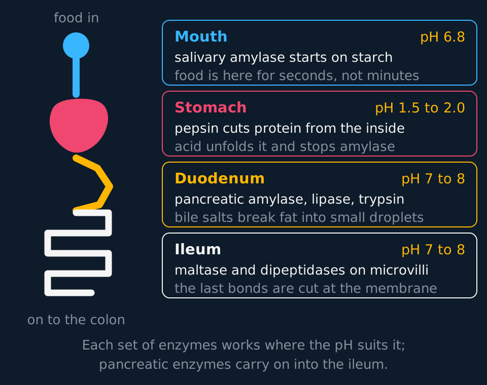
Describe the drawing
A schematic gut down the left of the figure, from the mouth through a coral stomach, an amber duodenum and a coiled ileum to the colon, with four cards beside it in the same colours. Each card names a compartment, gives the pH it holds, names the enzymes working there and says what that compartment achieves: mouth at pH 6.8 with salivary amylase, stomach at pH 1.5 to 2.0 with pepsin, duodenum at pH 7 to 8 with pancreatic enzymes and bile salts, and ileum at pH 7 to 8 with the membrane-bound maltase and dipeptidases.
The stomach holds pH 1.5 to 2.0. The duodenum holds pH 7 to 8. A difference of six pH units is a factor of a million in hydrogen ion concentration, and the gut manages it across a sphincter a few centimetres long, using hydrogencarbonate ions secreted in pancreatic juice and in bile to neutralise the acid arriving from the stomach. That neutralisation is essential. Pancreatic amylase, trypsin and lipase all have optima between about pH 7 and 8 and would be denatured at pH 2, so the pancreas has to fix the pH of its own workplace before its enzymes are of any use.
Two consequences follow. Salivary amylase stops working in the stomach, so starch digestion pauses and resumes in the duodenum with a different enzyme doing the same job. And the acid is doing chemistry although it is not an enzyme: at pH 2 enough of the ionic and hydrogen bonds stabilising a protein's tertiary structure are disrupted (which ones depends on the protein) that the chain unfolds, and peptide bonds buried in the tertiary structure become reachable. Acid denaturation is what makes pepsin efficient.
- Digestion
- The hydrolysis of large biological molecules into small soluble molecules that can be absorbed across cell membranes.
- Physical digestion
- The mechanical breakdown of food into smaller pieces, increasing the surface area available to digestive enzymes without hydrolysing anything.
- Absorption
- The movement of the products of digestion from the lumen of the gut, across the epithelium, into the blood or the lymph.
Exam-board note. AQA sets digestion and absorption out as a section of its own and names the enzymes, the micelles and the co-transport. Cambridge 9700 has no digestion topic, and OCR A does not teach mammalian digestion as a topic either; on those courses the hydrolysis and the membrane transport are examinable and the anatomy is context. Check the current specification for the qualification being taken.
Carbohydrases, and where the last bond is cut
Starch is a polymer of alpha glucose held by glycosidic bonds, in two forms: amylose, an unbranched helix joined only by 1,4 bonds, and amylopectin, which carries a 1,6 bond at each branch point. Amylase hydrolyses the 1,4 bonds and cannot touch the 1,6 ones, so its product is maltose plus short branched fragments needing a separate enzyme.
Amylase appears twice: salivary amylase starts work in the mouth, where food stays for seconds, and pancreatic amylase is secreted into the duodenum and does most of the actual work. Both hydrolyse starch to maltose at a similar, near-neutral optimum pH, and they differ in where they are made and where they work.
The last bond is cut somewhere unexpected. The disaccharidases, maltase, sucrase and lactase, are not released into the lumen at all. They are built into the cell surface membrane of the epithelial cells lining the ileum, with their active sites facing the gut contents. Maltase hydrolyses maltose to two glucose molecules, sucrase gives glucose and fructose, lactase gives glucose and galactose. Being membrane-bound means each monosaccharide is released a few nanometres from the transport protein that will carry it in, so it is absorbed before it can diffuse away or be taken by gut bacteria.
| Enzyme | Where it works | Substrate | Products |
|---|---|---|---|
| Salivary amylase | Mouth | Starch | Maltose |
| Pancreatic amylase | Duodenum | Starch | Maltose |
| Maltase | Membrane of ileum epithelium | Maltose | Two glucose |
| Sucrase | Membrane of ileum epithelium | Sucrose | Glucose and fructose |
| Lactase | Membrane of ileum epithelium | Lactose | Glucose and galactose |
Lactase differs from the others in its expression. Most mammals switch the gene off after weaning, and most adults worldwide make little of it; a change in the DNA controlling that gene keeps it on into adulthood, and is common in northern Europe and in several East African herding populations. Where lactase is absent, lactose reaches the large intestine undigested, lowers the water potential of the gut contents and is fermented by bacteria, which accounts for the watery faeces and the bloating of lactose intolerance, with no allergy involved.
Proteases and the order in which they act
Protein digestion uses three classes of enzyme, and the order in which they act follows from what each does. An endopeptidase hydrolyses peptide bonds in the interior of a polypeptide. An exopeptidase removes amino acids one at a time from an end. A dipeptidase splits the last two-residue fragment into single amino acids.
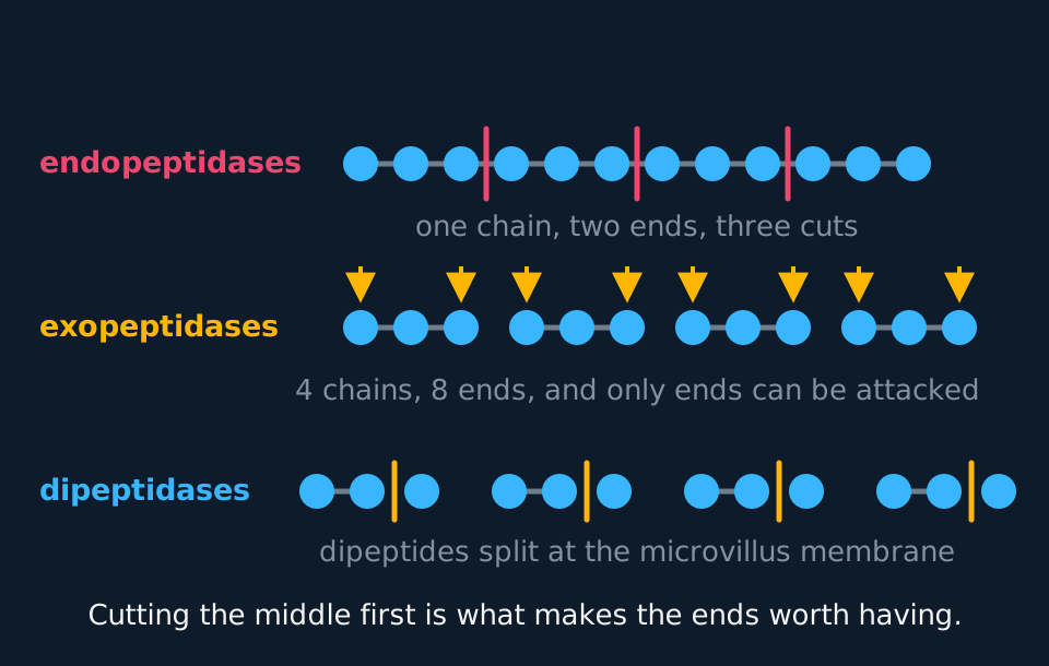
Describe the drawing
Three rows showing one polypeptide taken apart in stages. The top row is a chain of twelve joined circles with three coral cut marks between internal residues. The middle row is the four three-residue fragments those cuts produce, with an amber arrow pointing at each of the eight exposed ends. The bottom row is four dipeptides, each beside the single amino acid an exopeptidase has freed, waiting to be split at the membrane.
An exopeptidase can only work where there is a free end. A chain of three hundred amino acids has two of them, so exopeptidases on their own would have very few places to work. Endopeptidases multiply the ends: each internal cut adds two more places for the exopeptidases to start. That is a rate argument of exactly the same shape as the surface-area argument for bile.
- Endopeptidase
- An enzyme that hydrolyses peptide bonds within a polypeptide chain, producing shorter chains and so more ends. Pepsin, trypsin and chymotrypsin.
- Exopeptidase
- An enzyme that hydrolyses the peptide bond at an end of a chain, removing a single amino acid. Carboxypeptidases work from the end carrying the free carboxyl group, aminopeptidases from the amine end.
- Dipeptidase
- An enzyme, bound to the membrane of the ileum epithelium, that hydrolyses a dipeptide into two amino acids.
The named examples matter. Pepsin is an endopeptidase secreted by the stomach with an optimum near pH 2, and it is the only digestive enzyme named here that works in acid conditions. Trypsin and chymotrypsin are endopeptidases from the pancreas, working near pH 8. The carboxypeptidases are pancreatic as well, while the aminopeptidases and dipeptidases sit in the membrane alongside the disaccharidases.
A gland that makes protein-digesting enzymes must avoid hydrolysing its own proteins. Pepsin is secreted as inactive pepsinogen and converted by the stomach acid once safely out of the cell; trypsin is secreted as trypsinogen and activated by an enzyme anchored in the duodenal wall, after which trypsin activates the rest. The pancreas therefore never contains working protease. When that fails, it digests itself, which is what acute pancreatitis is.
Bile salts, lipase and the micelle
Lipids arrive in the duodenum as droplets, because they are insoluble in the watery gut contents and coalesce. Pancreatic lipase is water-soluble and can only reach the ester bonds at a droplet's surface, so the rate of lipid digestion is set by the total surface area of droplets rather than by how much fat is present.
Bile salts solve that. They are amphipathic: one face is hydrophilic and one hydrophobic, so they coat a lipid surface with their hydrophobic face inwards and leave a hydrophilic outside. Droplets so coated repel each other instead of merging, and the churning of the gut can break them into much smaller ones that stay broken. That is emulsification, and its whole effect is geometric.
What emulsification is worth, in numbers
A single spherical droplet of diameter 1.0 mm is emulsified into droplets of diameter 1.0 micrometre. Assuming the volume is conserved and every droplet is a sphere, calculate the factor by which the total surface area increases. The numbers are idealised: real emulsions give a range of droplet sizes.
Show the working
Volume of a sphere is proportional to the cube of the radius, so if the radius falls by a factor of 1000, each new droplet has one thousand-millionth of the original volume and there must be 1000³ = 1 × 10⁹ of them.
Surface area is proportional to the square of the radius, so each droplet has one millionth of the original area. Total area is therefore 10⁹ × 10⁻⁶ = 1000 times the area of the single droplet.
The general result is worth carrying: dividing a droplet into smaller ones of the same total volume multiplies the surface area by exactly the factor the radius shrank by. Nothing has been digested; what changed is the number of places lipase can work.

Describe the drawing
Lipid absorption animated in four steps. First one large amber lipid droplet is shown beside a cluster of thirteen small ones, with the note that emulsification raises the surface area a thousandfold for droplets a thousandth of the diameter. Second, coral lipase molecules ring the surface of a droplet and an arrow leads to monoglyceride and fatty acids. Third, three micelles, each an amber core of lipid products inside a ring of cyan bile salts, sit above a strip of microvilli, with an arrow under each showing the products passing into the cell while the micelle itself stays outside. Fourth, inside the cell, triglyceride is re-formed, packaged into a larger chylomicron and passed out into a lacteal.
Pancreatic lipase hydrolyses two of the three ester bonds in a triglyceride, giving a monoglyceride and two fatty acids. Those products, together with bile salts, cholesterol and any fat-soluble vitamins present, assemble into micelles: aggregates a few nanometres across with the hydrophobic parts inside and the hydrophilic parts facing the water.
State clearly that a micelle is not itself absorbed. Micelles form and break up continuously, and they carry monoglycerides and fatty acids through the watery layer next to the epithelium and release them there, keeping the concentration of free products high right at the membrane. Because monoglycerides and fatty acids are non-polar and small, they then diffuse straight through the phospholipid bilayer: simple diffusion, no carrier, no ATP.
Inside the epithelial cell the products are rebuilt. The smooth endoplasmic reticulum re-forms triglycerides, and the Golgi apparatus wraps triglyceride and cholesterol in a coat of phospholipid and protein to make a chylomicron. Chylomicrons leave by exocytosis into the lacteal, the blind-ended lymph vessel at the centre of each villus, because they are far too large to cross into a blood capillary. The lymph rejoins the blood near the left shoulder, so absorbed fat reaches the circulation without passing through the liver first.
TRY IT: A blocked bile duct
A gallstone lodges in a patient's bile duct, so no bile reaches the duodenum. The patient passes pale, greasy faeces and, after several months, is found to be deficient in vitamin D. Explain both observations.
Check your answer
Without bile salts the lipid is not emulsified. It stays as a few large droplets, so the surface area available to pancreatic lipase is small and only a fraction of the triglyceride is hydrolysed. Undigested triglyceride cannot be absorbed, so it passes on to the faeces and makes them greasy. They are pale as well because the bile pigments that normally colour faeces brown no longer reach the gut.
Vitamin D is fat-soluble and is normally carried to the epithelium inside micelles, which cannot form without bile salts, so it stays in the lumen and leaves with the fat. Vitamins A, E and K are lost for the same reason.
A good answer names two separate failures: no emulsification, so poor hydrolysis; and no micelles, so poor delivery. Answers that say only 'bile digests fat' cannot explain the vitamin at all.
The ileum, and why glucose needs sodium
The ileum is built for one job. Its wall is folded, the folds carry villi about half a millimetre to a millimetre long, and each epithelial cell on a villus carries several thousand microvilli around a micrometre long. Careful measurements published in 2014 put the mucosal surface area of an adult small intestine at roughly 30 m², closer to the floor of a living room than to the tennis court of older textbooks, which would be about 260 m². Earlier estimates counted folds that are not there in a living, moving gut.
Surface area is only one of the adaptations, and a question asking how the ileum is adapted for absorption wants several. The epithelium is one cell thick, so the diffusion path is short. A dense capillary network carries absorbed products away, maintaining the gradient. Muscle in the wall and in each villus keeps the contents moving, so the fluid next to the membrane is not depleted. The cells are packed with mitochondria, because much of what happens next costs ATP. And the final digestive enzymes are built into the membrane itself.
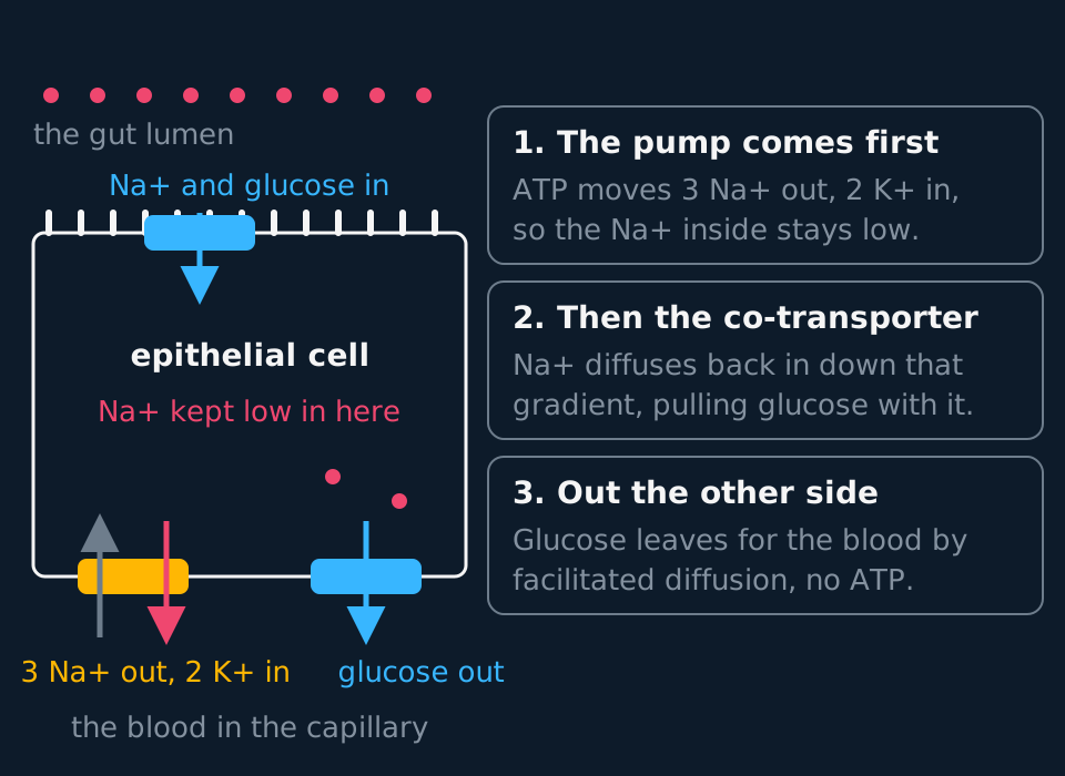
Describe the drawing
One epithelial cell of the ileum, drawn with microvilli along its upper surface facing the gut lumen and the blood below it. Coral dots representing sodium ions crowd the lumen and are sparse inside the cell. A co-transporter protein sits in the upper membrane with an arrow carrying sodium and glucose in together, while a sodium-potassium pump and a separate glucose carrier sit in the lower membrane with arrows leading out to the blood. Three numbered cards beside the drawing give the order of events, starting with the pump.
Glucose in the gut is often at a lower concentration than glucose inside the epithelial cell, so diffusion alone would carry glucose out of the cell into the lumen, and a meal would be left half-absorbed. The solution is co-transport, and it works in three moves.
First, a sodium-potassium pump in the basal membrane, the face towards the blood, hydrolyses ATP and moves three sodium ions out of the cell for every two potassium ions in. The cell's internal sodium concentration falls well below the concentration in the gut lumen.
Second, a co-transporter protein in the apical membrane binds one glucose molecule and two sodium ions together. Sodium moves into the cell down the electrochemical gradient the pump created, and the protein will only let it through if glucose comes with it. Glucose is therefore dragged in against its own concentration gradient, using no ATP at this membrane at all.
Third, glucose leaves the far side of the cell into the blood by facilitated diffusion through a different carrier, down the gradient that its accumulation has created.
- Co-transport
- The transport of two substances across a membrane through the same carrier protein at the same time, one moving down its concentration gradient and providing the means for the other to move against its own.
- Indirect active transport
- Transport that is driven by a gradient which was itself established using ATP elsewhere in the cell. Glucose uptake in the ileum is the standard example.
Amino acids are absorbed by the same mechanism, with their own co-transporters. Fructose is not: it crosses the apical membrane by facilitated diffusion alone, with no sodium involved and no gradient available to concentrate it, which is why large intakes of fructose can overwhelm its carrier and pass on unabsorbed. Both routes are carrier-mediated, so both saturate; the difference is that the co-transporter can move glucose against its own gradient and the fructose carrier cannot.
Glucose is not directly actively transported into the epithelial cell. Its uptake moves glucose against its concentration gradient, so it is active, but only in the indirect, secondary sense defined above: no ATP is hydrolysed at the apical membrane, and a cell whose sodium-potassium pump is inhibited absorbs glucose for a short time and then stops, because the sodium gradient runs down. State where the ATP is spent.
In the exam
- Digestion is hydrolysis, so an answer about how a polymer is broken down should state that water is added across the bond.
- Bile emulsifies and lipase hydrolyses. Bile does not digest, break down or hydrolyse fat.
- Micelles carry the products to the membrane and are not themselves absorbed. Monoglycerides and fatty acids cross by simple diffusion because they are non-polar.
- In a co-transport answer, name the pump, name the gradient and state that the ATP is spent at the basal membrane. Describing glucose uptake as active transport without that chain omits the mechanism.
- Chylomicrons leave in the lacteal rather than the capillary, because of their size.
Check yourself
A drug under development blocks the sodium-potassium pumps in the epithelial cells of the ileum. Predict and explain its effect on the absorption of glucose, of amino acids and of the products of lipid digestion.
Answer
Glucose absorption falls to almost nothing once the existing gradient has run down. The pump is what keeps sodium low inside the cell; without it, sodium no longer moves in down a steep gradient, so the co-transporter has nothing driving it and glucose cannot be taken up against its own gradient.
Amino acid absorption falls by the same mechanism, since amino acids are also carried in on sodium co-transporters.
Absorption of monoglycerides and fatty acids is unaffected. They are non-polar and cross by simple diffusion, using no carrier and no gradient the pump maintains, and the bile salts that deliver them are unaffected too.
Questions
Question 14 marks
Explain why a mixture containing only exopeptidases would digest a protein far more slowly than one that contains endopeptidases as well.
Mark scheme
- B1 an exopeptidase hydrolyses the peptide bond at an end of a chain, removing one amino acid at a time, so it can only work where there is a free end
- B1 a chain of three hundred amino acids has only two ends, so the number of places an exopeptidase can start is very small however much enzyme is present
- B1 an endopeptidase hydrolyses peptide bonds in the interior of the chain, and each internal cut produces two more ends
- B1 far more ends means far more sites at which the exopeptidases can act at the same time, so the overall rate of hydrolysis rises
Question 24 marks
Explain how bile salts increase the rate at which lipid is digested, and explain how micelles help the products of that digestion to be absorbed.
Mark scheme
- B1 bile salts are amphipathic, coating a lipid surface with the hydrophobic face inwards, so the droplets repel one another instead of merging and the churning of the gut can break them into much smaller ones that stay broken
- B1 that emulsification greatly increases the total surface area of lipid, so more ester bonds are accessible to water-soluble pancreatic lipase and the rate of hydrolysis rises, although bile itself breaks no bond
- B1 monoglycerides and fatty acids, with bile salts and any fat-soluble vitamins, assemble into micelles that carry them through the watery layer next to the epithelium and release them at the membrane, keeping the concentration of free products high there
- B1 the micelle itself is not absorbed: monoglycerides and fatty acids are small and non-polar, so they diffuse straight through the phospholipid bilayer by simple diffusion
Question 34 marks
Describe how glucose is absorbed from the lumen of the ileum into the blood, making clear where in the cell ATP is used.
Mark scheme
- B1 a sodium-potassium pump in the basal membrane hydrolyses ATP and moves three sodium ions out of the epithelial cell for every two potassium ions in, so the sodium concentration inside the cell falls below the concentration in the lumen
- B1 a co-transporter protein in the apical membrane binds one glucose molecule together with two sodium ions
- B1 sodium moves into the cell down the electrochemical gradient the pump created and glucose is carried in with it, against its own concentration gradient, with no ATP hydrolysed at this membrane
- B1 glucose then leaves the far side of the cell into the blood by facilitated diffusion through a different carrier protein, down the gradient its accumulation has created
Question 44 marks
Compare pancreatic amylase with maltase, referring to where each enzyme is found, the bond each hydrolyses, the product of each, and one advantage of the difference in position.
Mark scheme
- B1 both are carbohydrases and both catalyse a hydrolysis, adding a water molecule across a glycosidic bond, and neither is absorbed itself
- B1 pancreatic amylase is secreted into the lumen of the duodenum and works free in solution, whereas maltase is built into the cell-surface membrane of the ileum epithelium with its active site facing the gut contents and is never released into the lumen at all
- B1 amylase hydrolyses the 1,4 bonds of starch and cannot touch the 1,6 bonds at a branch point, so it yields maltose and short branched fragments, whereas maltase hydrolyses the single bond of maltose and yields two glucose molecules
- B1 amylase therefore leaves a disaccharide that still cannot be absorbed, whereas maltase releases glucose a few nanometres from the co-transporter that takes it in, so it is absorbed before it can diffuse away or be taken by gut bacteria
Question 54 marks
The table gives idealised dimensions for the folding of the ileum, constructed for this question rather than measured. Treating a villus and a microvillus each as a cylinder, ignoring its tip and the flat wall between neighbouring projections, calculate the factor by which the villi multiply the surface area of a flat gut wall, and calculate the factor by which the microvilli then multiply it again.
| Quantity | Value |
|---|---|
| Length of one villus | 800 µm |
| Diameter of one villus | 100 µm |
| Number of villi on each square millimetre of gut wall | 25 |
| Length of one microvillus | 1.0 µm |
| Diameter of one microvillus | 0.10 µm |
| Number of microvilli on one epithelial cell | 3000 |
| Upper surface of one epithelial cell, unfolded | 100 µm² |
Mark scheme
- M1 curved surface area of a cylinder = π × diameter × length, so one villus offers π × 100 × 800 = 2.5 × 10⁵ µm²
- A1 25 villi stand on each square millimetre, which is 1 × 10⁶ µm² of flat wall, so 25 × 2.5 × 10⁵ = 6.3 × 10⁶ µm² and the villi multiply the area by about 6.3
- M1 one microvillus offers π × 0.10 × 1.0 = 0.314 µm², so 3000 of them offer 3000 × 0.314 = 940 µm² on a cell whose unfolded surface is 100 µm²
- A1 the microvilli multiply the area by about 9.4, and the two factors multiply together, giving 6.3 × 9.4 = about 59 times the area of the same length of unfolded tube
Question 64 marks
An adult who has always drunk milk without difficulty has an infection that destroys much of the epithelium lining her ileum. For several weeks afterwards, milk gives her bloating and watery faeces. Suggest why the infection has this effect, and suggest why the symptoms then disappear.
Mark scheme
- B1 lactase, like the other disaccharidases, is built into the cell-surface membrane of the ileum epithelium rather than secreted into the lumen, so destroying that epithelium destroys the enzyme even though the pancreas is undamaged
- B1 lactose is therefore not hydrolysed to glucose and galactose, and a disaccharide is too large to be absorbed, so it stays in the lumen
- B1 the solute left in the lumen lowers the water potential of the gut contents, so water moves out of the epithelium into the lumen down the water potential gradient by osmosis, and the faeces are watery
- B1 bacteria in the large intestine ferment the lactose and release gas, which distends the gut and causes the bloating; the epithelium is replaced from dividing cells within a few weeks, restoring the membrane-bound enzyme, so the intolerance is temporary rather than the permanent kind set by which alleles a person carries
Question 74 marks
Explain why pepsin is secreted by the stomach as inactive pepsinogen rather than as active pepsin, and explain how it is activated once it is safely in the stomach lumen.
Mark scheme
- B1 pepsin is an endopeptidase, and if it were active while still inside the cells that make it, or in the gastric glands that release it, it would hydrolyse the proteins of those cells and glands themselves
- B1 secreting it as pepsinogen, an inactive form, avoids this: pepsinogen's active site is not exposed or correctly shaped, so it cannot hydrolyse peptide bonds until it is changed
- B1 once pepsinogen reaches the acidic stomach lumen, the low pH converts it into active pepsin
- B1 this activation happening only once the enzyme is outside the cells that made it protects the stomach lining and the secreting cells from being digested by their own product
Question 84 marks
Some people experience bloating after consuming large amounts of fruit juice or foods sweetened with high-fructose syrup, even though they digest lactose and sucrose normally. Suggest why fructose in particular can cause this, given how fructose crosses the ileum epithelium.
Mark scheme
- B1 fructose is absorbed by facilitated diffusion through its own carrier protein in the apical membrane, unlike glucose, which is co-transported with sodium ions
- B1 because facilitated diffusion depends on a concentration gradient rather than being able to move fructose against one, and the carrier itself can be saturated once the fructose concentration is high enough, a very large intake can arrive faster than the carrier can absorb it
- B1 fructose that is not absorbed remains in the lumen and passes on into the large intestine
- B1 there it lowers the water potential of the gut contents, drawing water in by osmosis, and is fermented by gut bacteria, producing gas; both effects together cause the bloating, although here the sugar is failing to be absorbed rather than failing to be digested
Question 94 marks
Describe what happens to monoglycerides and fatty acids once they have diffused into an epithelial cell of the ileum, from arrival at the cell to leaving in the lymph.
Mark scheme
- B1 the smooth endoplasmic reticulum of the epithelial cell re-forms the monoglycerides and fatty acids into triglycerides
- B1 the Golgi apparatus wraps the triglyceride, together with cholesterol, in a coat of phospholipid and protein, forming a chylomicron
- B1 the chylomicron leaves the cell by exocytosis into the lacteal, the blind-ended lymph vessel at the centre of the villus, rather than into a blood capillary, because it is far too large to cross into one
- B1 the lymph eventually rejoins the blood near the left shoulder, so absorbed fat reaches the general circulation without first passing through the liver, unlike glucose and amino acids, which are absorbed straight into the blood capillaries of the villus
Question 104 marks
Compare how glucose is absorbed across the ileum epithelium with how a fatty acid is absorbed, referring to the mechanism used at the apical membrane and the vessel each enters on the other side of the cell.
Mark scheme
- B1 glucose crosses the apical membrane by co-transport with sodium ions, against its own concentration gradient, whereas a fatty acid crosses the same membrane by simple diffusion, needing no carrier and no gradient set up by a pump
- B1 glucose then leaves the far side of the cell into a blood capillary by facilitated diffusion, whereas a fatty acid is first rebuilt into a triglyceride and packaged into a chylomicron before leaving into the lacteal
- B1 glucose absorption therefore depends indirectly on ATP, spent by the sodium-potassium pump maintaining the sodium gradient, whereas the absorption of a fatty acid across the apical membrane uses no ATP at all
- B1 the different vessel each enters follows from size: glucose is small enough to enter a blood capillary directly, while a chylomicron is far too large to do so and must instead enter the wider-gapped lacteal
Question 113 marks
A patient has had a long section of the ileum removed by surgery. Suggest why the patient loses body mass, and suggest why the faeces contain an unusually high proportion of fat.
Mark scheme
- B1 much of the surface area for absorption has gone, with fewer villi and microvilli, so fewer products of digestion are absorbed before the contents pass on to the large intestine
- B1 less glucose and fewer amino acids are absorbed, so less substrate is available for respiration and stored lipid and body protein are respired instead, and mass falls
- B1 monoglycerides and fatty acids are also absorbed across the ileum epithelium, so with less epithelium available a greater proportion of the products of lipid digestion is left in the lumen and leaves in the faeces
Question 123 marks
Explain how the acidity of the stomach makes proteins easier for pepsin to hydrolyse, beyond simply activating the enzyme itself.
Mark scheme
- B1 at pH 2 many of the hydrogen bonds and ionic bonds holding a protein's tertiary structure together are disrupted, so the polypeptide chain unfolds
- B1 peptide bonds that were previously buried inside the folded tertiary structure, and so inaccessible, become exposed on the unfolded chain
- B1 pepsin can therefore hydrolyse those newly exposed bonds
Question 133 marks
Name the two monosaccharides produced when sucrase hydrolyses sucrose, name the two monosaccharides produced when lactase hydrolyses lactose, and name the structure in the ileum that holds both enzymes.
Mark scheme
- B1 sucrase hydrolyses sucrose to give one molecule of glucose and one of fructose
- B1 lactase hydrolyses lactose to give one molecule of glucose and one of galactose
- B1 both enzymes, like maltase, are built into the cell surface membrane of the ileum epithelium rather than released free into the lumen
Question 142 marks
Name the enzyme that hydrolyses maltose, and name the exact place in the ileum at which that enzyme is found.
Mark scheme
- B1 maltase, which hydrolyses maltose to two glucose molecules
- B1 it is built into the cell-surface membrane of the epithelial cells lining the ileum, with its active site facing the gut contents
Question 152 marks
State the approximate pH of the stomach and the approximate pH of the duodenum, and state what would happen to pancreatic enzymes such as amylase if the acid arriving from the stomach were not neutralised.
Mark scheme
- B1 the stomach is at about pH 1.5 to 2.0, and the duodenum is at about pH 7 to 8
- B1 pancreatic enzymes such as amylase, trypsin and lipase have optima between about pH 7 and 8 and would be denatured by the acid arriving from the stomach if it were not neutralised by hydrogencarbonate ions
Worth remembering
- Digestion is hydrolysis, and physical digestion provides surface area for it rather than doing any of it.
- Endopeptidases first, exopeptidases second, dipeptidases at the membrane: cutting the middle multiplies the ends.
- Bile salts emulsify and form micelles; pancreatic lipase does the hydrolysis.
- Micelles deliver, they do not enter. Chylomicrons leave in the lymph.
- In co-transport the ATP is spent by the sodium-potassium pump on the far side of the cell, never at the carrier the glucose uses.
CHECK YOUR PROGRESS
Rate how confident you are with each objective for this lesson. Ratings are saved in this browser, on this device, unless you sign in.
- Describe digestion as hydrolysis, and say what physical digestion contributes to it.
- Name the enzymes acting on carbohydrate, protein and lipid, and state where each works.
- Explain why endopeptidases are needed before exopeptidases can work quickly.
- Explain emulsification as an argument about surface area, and calculate the change.
- Describe the role of micelles in lipid absorption, and say what is and is not absorbed.
- Explain the co-transport of glucose and amino acids with sodium ions, and say where the ATP is spent.
Open the full revision checklist to see every objective in the curriculum in one place.
Practise this lesson
WORKBOOK
The written questions from this topic, on paper with room to work, and a separate book of mark schemes. The question that is set on a diagram stays on this page, where the diagram can be drawn. Free to use; please do not redistribute or sell.
Used later in The liver: detoxification, deamination and the making of urea