Biology › Membranes and transport across cells › Membrane structure and the fluid mosaic model
Membrane structure and the fluid mosaic model
Every cell is surrounded by a bilayer about 7 nm thick, stabilised by the hydrophobic effect and by interactions between the fatty acid tails rather than by covalent bonds between neighbouring phospholipids. Phospholipids, proteins that move laterally within the layer, cholesterol between the tails and glycoproteins on the outer surface make up the fluid mosaic model, which accounts for how a cell controls exchange with its surroundings.
Before this Lipids: triglycerides, phospholipids and ester bonds · Protein structure and function: tertiary structure and denaturation
COMMON MISCONCEPTION
Something must be holding the bilayer together: bonds between the phospholipids, or a frame of protein underneath.
A phospholipid bilayer is not joined by covalent bonds between neighbouring phospholipids. The hydrophobic effect and interactions between fatty-acid tails stabilise the bilayer while phospholipids and many proteins remain able to move laterally.
What you should be able to do
- Explain why a phospholipid bilayer forms without anything assembling it.
- Label a fluid mosaic membrane and give the job of every component on it.
- Distinguish intrinsic from extrinsic proteins, and channel from carrier proteins.
- Outline cell signalling at the membrane: a ligand secreted, transported and bound by a complementary receptor on a target cell.
- Describe what cholesterol does at high temperature and at low temperature, and say why those two effects are not contradictory.
- Explain how temperature and organic solvents change permeability, and describe the beetroot investigation that measures it.
Why a bilayer forms spontaneously
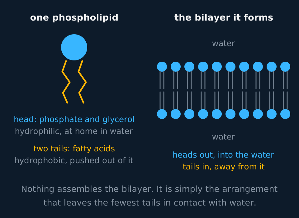
A phospholipid has two chemically different ends. One is a phosphate group attached to glycerol; it carries charge and is soluble in water. The other is two fatty acid chains, uncharged hydrocarbon, which are insoluble in water. A molecule with both properties is amphipathic, and the structure of a membrane follows from that property.
Describe the drawing
On the left, a single phospholipid drawn as a round phosphate head with two long wavy fatty acid tails hanging beneath it, the head marked hydrophilic and the tails hydrophobic. On the right, many of the same molecules arranged as a bilayer: one row of heads facing the water above, a second row facing the water below, and all the tails pointing inwards to meet in a core that water is kept out of.
In water, phospholipids arrange into a double layer with the heads facing the water on both faces and the tails buried between them. No other structure assembles it. The arrangement has the lowest energy, because it shields the greatest number of hydrophobic tails from the greatest volume of water. Fragments of a disrupted membrane reseal into closed vesicles for the same reason: an open edge would leave tails exposed.
The finished sheet is about 7 nm thick, well below the resolving limit of a light microscope, so membrane structure was inferred from permeability and other behaviour before electron microscopy made it visible.
The same bilayer surrounds the organelles of a eukaryotic cell. Inside the cell, membranes are partially permeable barriers between organelles and the cytoplasm, and within organelles such as the chloroplast. They are also sites of reactions: the electron transport chain and ATP synthase are set into the inner mitochondrial membrane, and the proteins of the light-dependent reactions into the thylakoid membranes.
- Phospholipid
- A lipid in which one of the three fatty acids of a triglyceride is replaced by a phosphate group, giving a hydrophilic head and two hydrophobic tails.
- Hydrophilic
- Attracted to water; polar or charged.
- Hydrophobic
- Repelled by water; non-polar.
- Bilayer
- Two layers of phospholipid with the hydrophobic tails facing inwards and the hydrophilic heads facing the watery solutions on each side.
That hydrophobic core is the reason a membrane is selective at all. Small non-polar molecules such as oxygen, carbon dioxide and steroid hormones dissolve in it and pass straight through. Anything charged or large and polar cannot: a sodium ion carries a shell of water molecules around it, and dragging that into a layer of fatty acid tails costs far more energy than it gains. Ions and glucose cross through membrane proteins instead.
The mosaic: what is set into the bilayer
Singer and Nicolson named the fluid mosaic model in 1972, and both halves of the name are accurate. Fluid: the phospholipids and most of the proteins move sideways within their own layer. Mosaic: the proteins are scattered through the phospholipids in an irregular pattern, like tiles set into a floor, rather than forming a continuous layer of their own.
The model replaced an earlier interpretation. In 1935 Davson and Danielli proposed a lipid bilayer coated on both faces by continuous layers of protein. Three kinds of evidence overturned it. Freeze-fracture electron microscopy, which splits a frozen membrane between its two layers, revealed protein particles set into the hydrophobic core. Many membrane proteins turned out to have hydrophobic regions, which could only sit among the tails. When a mouse cell and a human cell with differently labelled surface proteins were fused in 1970, the two labels mixed over the whole surface within about 40 minutes, so the proteins move sideways within the layer.
Describe the drawing
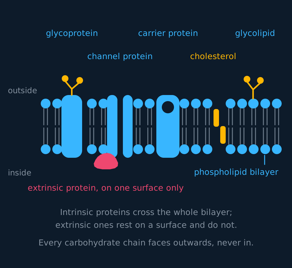
A membrane drawn in cross-section as two rows of round phosphate heads with fatty acid tails meeting in the middle. Set into it, from left to right: a glycoprotein reaching through the whole bilayer with branched sugar chains on the upper surface, a channel protein built as two walls with an open pore between them, a carrier protein with a binding site cut into its upper face, two short cholesterol molecules wedged among the tails, and a glycolipid whose sugar chain also points up. A separate extrinsic protein rests on the lower surface without entering the membrane.
Proteins come in two positions. Intrinsic (or integral) proteins are embedded in the bilayer, and many span it completely; their surfaces that face the core are hydrophobic, which is what holds them there. Extrinsic (or peripheral) proteins sit on one surface, bound to the heads or to an intrinsic protein, and do not enter the core at all. Enzymes and the proteins that anchor the cytoskeleton are often extrinsic.
Two kinds of intrinsic protein carry substances across the bilayer, and they work by different mechanisms.
| Channel protein | Carrier protein | |
|---|---|---|
| Shape | A pore lined with hydrophilic amino acids | A binding site that fits one kind of molecule |
| How it works | The substance passes through the open pore | The protein changes shape to move the substance across |
| Carries | Mostly ions such as Na⁺, K⁺ and Cl⁻; water crosses through channels called aquaporins | Polar molecules such as glucose and amino acids; some carriers move ions |
| Specific? | Yes, usually to one ion | Yes, to one molecule or a small group |
| Used in | Facilitated diffusion only | Facilitated diffusion and active transport |
Many channels are gated: they open in response to a voltage change or to a molecule binding, so a channel is a regulated pore rather than a permanent opening. The gated channels themselves are covered in the action potential and synaptic transmission topics.
On the outer face, some proteins and some lipids carry short branched carbohydrate chains. A protein with sugars attached is a glycoprotein; a lipid with sugars attached is a glycolipid. Both stick out into the surroundings, and much of what they do is recognition: many act as antigens or as receptor sites, some hold cells together in tissues, and they help hold a layer of water at the surface. Not every chain has a known recognition job, but the ABO blood groups are differences in these chains, which is why the wrong transfusion is recognised as foreign within minutes.
- Intrinsic protein
- A protein embedded within the bilayer, often spanning it completely.
- Extrinsic protein
- A protein on one surface of the bilayer that does not enter the hydrophobic core.
- Glycoprotein
- A protein with a carbohydrate chain attached. In the cell surface membrane, glycoproteins act in cell recognition, adhesion or as receptors.
- Glycolipid
- A lipid with a carbohydrate chain attached, acting in cell recognition and adhesion.
Cell signalling at the membrane
Receptor glycoproteins are the basis of communication between the cells of a multicellular organism, examined under the general name cell signalling. The sequence has three steps. One cell secretes a signalling molecule, usually by exocytosis. The molecule is transported to the cells it is meant for: carried in the blood if the target is distant, as a hormone is, or diffusing across a synapse if the target is a neighbouring neurone, or simply drifting between adjacent cells in a tissue. It then binds to a receptor on the target cell's membrane, and the binding changes something inside the cell.
Any molecule that binds specifically to a receptor is called a ligand. Hormones, neurotransmitters and local signalling molecules are all ligands, as are the drugs designed to imitate or block them. The receptor's binding site is complementary to its ligand in exactly the sense an active site is complementary to a substrate: a matter of shape and charge, and specific to one molecule.
That specificity is what makes signalling selective. A hormone circulates in the blood past every cell in the body, and only the cells carrying its receptor respond. Those are its target cells; a cell without the receptor has no binding site for the ligand and does not respond. Insulin binding to receptors on muscle and liver cells, and a neurotransmitter binding to receptors on the postsynaptic membrane, are the usual examples.
A ligand that is small and non-polar, such as a steroid hormone, can cross the bilayer and bind an intracellular receptor instead, so not all receptors are membrane-bound.
- Cell signalling
- Communication between cells in which one cell secretes a molecule that is transported to a target cell and binds to a receptor there.
- Ligand
- A molecule that binds specifically to a receptor, such as a hormone or a neurotransmitter.
- Receptor
- A protein, usually a glycoprotein in the cell surface membrane, whose binding site is complementary to one ligand; only cells carrying the receptor respond to the signal.
Cholesterol at high and at low temperature
Cholesterol is a short, rigid, mostly hydrophobic molecule with one small hydroxyl group at the end. That hydroxyl sits among the phosphate heads and the rest of the molecule wedges between the fatty acid tails. In an animal cell surface membrane, there can be up to one cholesterol molecule for every phospholipid.
Describing cholesterol as making a membrane 'more stable' does not state a mechanism. Its effect depends on the temperature, and both cases should be given.
At high temperature the phospholipids have more kinetic energy and would move apart. Cholesterol binds to the tails around it and restricts that movement, so the membrane stays less fluid than it otherwise would and does not become leaky.
At low temperature the danger is the opposite. The tails would pack tightly together and the membrane would set solid, cracking rather than bending. Cholesterol sits between the tails and stops them packing that closely, so the membrane keeps some fluidity.
The two effects are consistent once cholesterol is described as reducing the change in fluidity as temperature changes. Both follow from the same physical property: a short rigid molecule positioned between the tails restricts their movement at high temperature and prevents close packing at low temperature.
Explaining the contribution of cholesterol
A fish that lives under Antarctic ice at −1.8 °C has membranes with a high proportion of unsaturated fatty acids in its phospholipids. Suggest why, and explain what cholesterol contributes at that temperature.
Show the working
Unsaturated fatty acids contain C=C double bonds, and each double bond puts a kink in the tail. Kinked tails cannot pack closely, so the bilayer stays fluid at temperatures where straight saturated tails would have set.
A membrane that has set solid does not allow its proteins to move or change shape, and it fractures rather than bending, so the cell would leak. Fluidity at −1.8 °C is therefore required for survival.
Cholesterol contributes to the same outcome by a different mechanism. Positioned between the tails, it prevents close packing through its rigid shape rather than through a kink in the hydrocarbon chain.
Factors that increase permeability
Permeability is not fixed. Two factors are examined, and both act on the same two features: the packing of the bilayer and the tertiary structure of the proteins.
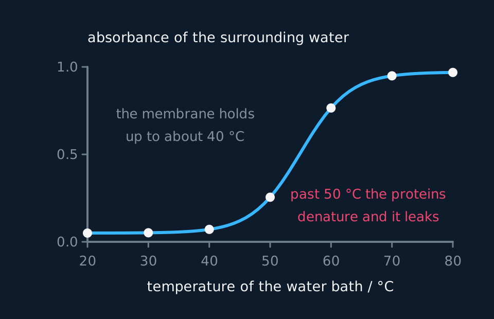
Temperature. Below about 40 °C, warming a membrane raises permeability gently. Phospholipids gain kinetic energy and move more, gaps open and close more often, and the molecules trying to cross are moving faster too. Above roughly 45 to 50 °C the graph turns sharply upward, because the intrinsic proteins begin to denature: hydrogen and ionic bonds break, tertiary structure is lost, and the protein no longer fits the space it occupied or controls what passes through it. The bilayer itself is disordered around each misfolded protein, so gaps open in the membrane and large water-soluble molecules leak through.
Distinguish the two effects. Phospholipids are not proteins and do not denature. State that the phospholipids move more and that the intrinsic proteins denature, rather than that 'the membrane denatures'.
Organic solvents. Ethanol, propanone and methylbenzene dissolve lipids, which is exactly what a bilayer is made of. Increasing the concentration of ethanol around a cell dissolves phospholipids out of the membrane and disrupts what remains, so permeability rises with concentration. The same mechanism accounts for the action of alcohol-based hand gel on the lipid envelope of enveloped viruses.
- Permeability
- How readily a membrane allows a substance to cross it.
- Denaturation
- The loss of a protein's tertiary structure when hydrogen and ionic bonds break, so its shape and therefore its function is lost.
The beetroot investigation
The standard practical uses beetroot because its vacuoles contain betalain, a water-soluble red pigment that is straightforward to measure. The pigment is inside the vacuole, so it must cross two membranes to leave the cell: the tonoplast and the cell-surface membrane. Pigment in the surrounding water is therefore evidence that both membranes have become permeable to it.
Cut discs of equal size with a cork borer, wash them thoroughly to remove pigment released by the cutting itself, then hold each one in its own tube containing a fixed volume of distilled water, in a water bath at a set temperature for a fixed time, such as 15 minutes. Stand each tube of water in the bath for about five minutes before its disc goes in, so that the water starts at the bath temperature. Remove the disc and measure the colour of the water it sat in with a colorimeter using a green filter of around 530 nm. Measure that water, not a fresh volume the disc is moved into afterwards: pigment that leaked at the test temperature is the measurement, and transferring the disc throws it away. Higher absorbance means more pigment escaped, and so more permeable membranes.
Describe the drawing
A line graph rising from left to right. Temperature from twenty to eighty degrees Celsius runs along the horizontal axis and absorbance from zero to one up the vertical axis. The curve stays almost flat and close to zero up to about forty degrees, turns sharply upward between fifty and sixty, then levels off near the top of the scale. Seven plotted points sit on the curve, one for each ten-degree step.
Wash the discs after cutting. Cutting ruptures cells at the surface, and unwashed discs release pigment unrelated to the temperature being tested. Control variables are the disc size and number, the volume of water, the time in the bath, the time before reading, and the age and variety of the beetroot, since older tissue leaks more.
The alcohol-concentration version. Core Practical 3 allows alcohol concentration or temperature as the independent variable, and the method changes only in what is varied. Dilute the stock ethanol with distilled water to make a series such as 0, 20, 40, 60, 80 and 100%, keeping the total volume the same in every tube. Cut and rinse discs of equal size as before, put one in each tube, and leave them all for the same time, such as 15 minutes, in a water bath at one fixed temperature. Remove the discs and read the absorbance of each liquid in the colorimeter with the same green filter, after zeroing it on a blank cuvette of distilled water.
Absorbance rises with concentration because ethanol is an organic solvent: it dissolves phospholipids out of the bilayer and disrupts the packing of those left behind. It also denatures the membrane proteins by disrupting the bonds and hydrophobic interactions that hold their tertiary structure. The 0% tube is the control, showing how much pigment leaks from intact, rinsed discs. Temperature now becomes a control variable, because it changes permeability on its own, alongside disc size, liquid volume, immersion time and the source of the beetroot.
Risk assessment. Ethanol is highly flammable, so keep it away from naked flames. Check the CLEAPSS Hazcard and your school's risk assessment before starting; where they differ from this page, follow them.
TRY IT: Explaining both gradients of the curve
A student reports that absorbance rises slowly from 20 °C to 40 °C and then rises steeply between 50 °C and 60 °C. Explain both parts of that result.
Check your answer
Between 20 °C and 40 °C the rise is caused by the phospholipids gaining kinetic energy. They move more, so the bilayer is slightly more open and slightly more pigment gets through, but the proteins still hold their shape, so the membrane remains largely intact.
Between 50 °C and 60 °C the intrinsic proteins denature. Hydrogen and ionic bonds break, tertiary structure is lost, and the proteins no longer fill the space they occupied or control what crosses. The phospholipids are also moving far more by now. Pigment leaves the vacuole freely and absorbance climbs steeply.
Name the change occurring at each stage rather than describing both as membrane damage. Two distinct processes are involved, which is why the graph has two gradients.
In the exam
- State what a component does, not what it is: 'cholesterol restricts the movement of the phospholipids at high temperature, so the membrane is less fluid and less permeable', rather than 'cholesterol is a lipid'.
- State that the intrinsic proteins denature and that the phospholipids move more. That distinction accounts for the two gradients of the temperature graph.
- Explain both words of 'fluid mosaic'. Fluid because the phospholipids and proteins move laterally within the layer; mosaic because the proteins are scattered irregularly among them.
- Intrinsic against extrinsic is about position, not size. Intrinsic proteins are embedded in the bilayer, often spanning it; extrinsic proteins sit on one surface.
- For the beetroot practical, include washing the discs after cutting when describing the method.
- Carbohydrate chains occur on the outer surface only. Sugars drawn on the cytoplasmic face of a membrane diagram are in the wrong position.
Check yourself
A student places identical discs of beetroot in a range of ethanol concentrations from 0% to 100% at room temperature and measures the absorbance of the surrounding liquid after 30 minutes. Absorbance increases as ethanol concentration increases. Explain this result, and explain why the same student sees almost no pigment released when the discs are left in water at 20 °C.
Answer
Ethanol is an organic solvent and the bilayer is lipid, so ethanol dissolves phospholipids out of the membrane. The more concentrated the ethanol, the more phospholipid is removed and the more disrupted the remaining arrangement is.
Once the bilayer is incomplete, the hydrophobic core no longer forms a continuous barrier. Betalain is water-soluble and would normally be held in the vacuole; with the tonoplast and the cell surface membrane both breached, it diffuses out into the surrounding liquid and the absorbance rises.
In water at 20 °C neither thing has happened. The phospholipids have relatively little kinetic energy and stay closely packed, and the intrinsic proteins are well below the temperature at which they denature, so their tertiary structure is intact and no gaps open around them.
The membrane is therefore still selectively permeable at 20 °C. A large polar molecule like betalain cannot cross the hydrophobic core unaided, and nothing in the membrane transports it, so it stays inside and the water stays clear.
Questions
Question 14 marks
Explain why phospholipids arrange themselves into a bilayer when they are placed in water, and why a torn fragment of membrane reseals itself into a closed vesicle.
Mark scheme
- B1 a phospholipid is amphipathic: the phosphate head is hydrophilic and the two fatty acid tails are hydrophobic
- B1 the heads face the water on both surfaces while the tails are buried between them, facing each other
- B1 no bonds join one phospholipid to the next; this arrangement is simply the one of lowest energy, because it hides the greatest number of tails from water
- B1 an open edge would leave hydrophobic tails exposed to water, so a fragment closes on itself rather than remaining as a sheet
Question 24 marks
Describe what cholesterol does to a cell surface membrane at high temperature and at low temperature, and describe the effect it has on permeability in each case.
Mark scheme
- B1 at high temperature cholesterol binds to the fatty acid tails around it and restricts their movement
- B1 so the membrane is less fluid than it would otherwise be and does not become leaky
- B1 at low temperature cholesterol sits between the tails and stops them packing closely together
- B1 so the membrane keeps some fluidity instead of setting solid and cracking, and its permeability stays close to normal
Question 34 marks
Compare a channel protein with a carrier protein as routes across a cell surface membrane.
Mark scheme
- B1 a channel protein is a pore lined with hydrophilic amino acids, whereas a carrier protein has a binding site shaped to fit one kind of molecule
- B1 a substance passes through the open channel, whereas a carrier protein changes shape to move the substance across
- B1 channels carry water-soluble ions such as sodium, potassium and chloride, whereas carriers take larger polar molecules such as glucose and amino acids
- B1 channels are used in facilitated diffusion only, whereas carrier proteins are used in both facilitated diffusion and active transport
Question 44 marks
A student cuts discs of beetroot with a cork borer and places them directly into water baths at a range of temperatures without washing them first. Suggest how this affects her absorbance readings, and suggest one control variable she must still keep the same.
Mark scheme
- B1 cutting ruptures the cells at the cut surfaces, which releases pigment that has nothing to do with the temperature being tested
- B1 absorbance is therefore raised at every temperature, including those at which the membranes are undamaged
- B1 the readings at low temperature overstate the permeability, so the rise caused by temperature itself is masked
- B1 any one control variable named: size and number of discs, volume of distilled water, time in the water bath, time before the reading is taken, or the age and variety of the beetroot
Question 54 marks
Compare an intrinsic protein with an extrinsic protein in a cell surface membrane, referring to its position and to the kind of job each commonly does.
Mark scheme
- B1 an intrinsic protein is embedded within the bilayer, often spanning it completely, whereas an extrinsic protein sits on one surface and does not enter the hydrophobic core
- B1 intrinsic proteins have hydrophobic surfaces facing the core of the bilayer, which is what holds them there, whereas extrinsic proteins are held by binding to the phospholipid heads or to an intrinsic protein
- B1 intrinsic proteins commonly act as channels or carriers, moving substances across the membrane, whereas extrinsic proteins are often enzymes or proteins that anchor the cytoskeleton
- B1 both kinds are scattered irregularly through the membrane, which is the mosaic of the fluid mosaic model
Question 64 marks
A cell has receptors for insulin on its surface. Suggest what would happen to that cell's response to a rise in blood insulin concentration if a mutation caused the receptor's binding site to no longer be complementary to insulin's shape, and suggest what this shows about the specificity of cell signalling.
Mark scheme
- B1 insulin would no longer be able to bind to the altered receptor, because binding depends on the receptor's site being complementary in shape to the ligand
- B1 without insulin binding, the change inside the cell that binding normally triggers would not happen, so the cell would not respond to a rise in blood insulin concentration even though insulin is present
- B1 this shows that a cell's response to a hormone depends on having a working, complementary receptor and not just on the hormone reaching it, since the hormone passes every cell in the body but only cells with the receptor can respond
- B1 the effects insulin normally has on the cell would not occur: it would not increase its uptake of glucose from the blood or its conversion of glucose to glycogen
Question 73 marks
The table shows results, typical of the beetroot practical, from discs held in water baths for five minutes each. Explain the pattern in the readings, referring to what happens to each membrane component as the temperature rises.
| Temperature / °C | Absorbance of the surrounding water |
|---|---|
| 20 | 0.05 |
| 30 | 0.08 |
| 40 | 0.12 |
| 50 | 0.35 |
| 60 | 0.78 |
Mark scheme
- B1 between 20 and 40 °C absorbance rises gently because the phospholipids gain kinetic energy and move more, so slightly more pigment escapes while the membrane stays largely intact
- B1 above about 45 to 50 °C the intrinsic proteins denature: hydrogen and ionic bonds break and tertiary structure is lost, so the proteins no longer control what crosses, and absorbance climbs steeply
- B1 the phospholipids do not denature, because they are not proteins; the two gradients in the data come from two different changes, one to each component
Question 83 marks
Describe the three steps of cell signalling, from a signalling molecule leaving one cell to a distant target cell responding.
Mark scheme
- B1 one cell secretes a signalling molecule, usually by exocytosis
- B1 the molecule is transported to the cells it is meant for, carried in the blood if the target is distant, as a hormone is
- B1 it binds to a receptor on the target cell's membrane whose binding site is complementary to it, and only cells carrying that receptor respond
Question 93 marks
Explain why a steroid hormone can bind to a receptor inside a target cell, rather than needing a receptor on the cell surface membrane.
Mark scheme
- B1 a steroid hormone is small and non-polar, so it is lipid-soluble
- B1 such a molecule can dissolve in, and diffuse across, the hydrophobic core of the phospholipid bilayer without needing a channel or a carrier protein
- B1 having crossed the membrane, it can bind directly to a receptor inside the cell, so it does not need a complementary receptor on the cell surface
Question 103 marks
A person with blood group A receives blood from a group B donor by mistake. Suggest, referring to glycoproteins and glycolipids, why the wrong blood type is recognised as foreign.
Mark scheme
- B1 the ABO blood groups are set by differences in the short branched carbohydrate chains carried by glycoproteins and glycolipids on the outer surface of red blood cell membranes
- B1 many of these chains act as antigens: molecules the immune system uses to distinguish the body's own cells from foreign ones
- B1 a group B donor carries different chains from the recipient's own cells, so the recipient's immune system does not recognise them as self and responds to them as foreign
Question 113 marks
Explain why increasing the concentration of ethanol around a cell increases the permeability of its membrane.
Mark scheme
- B1 ethanol is an organic solvent, and it dissolves lipids, which is exactly what the phospholipid bilayer is made of
- B1 increasing the concentration of ethanol dissolves more phospholipids out of the membrane and disrupts the packing of what remains
- B1 with the bilayer increasingly disrupted, permeability rises as the ethanol concentration increases
Question 123 marks
Explain why a small non-polar molecule such as oxygen can cross the phospholipid bilayer directly, while a polar molecule such as glucose cannot.
Mark scheme
- B1 the hydrophobic core of the bilayer is made of the fatty acid tails of the phospholipids, and it excludes water and anything charged or strongly polar
- B1 oxygen is small and non-polar, so it can dissolve in this hydrophobic core and diffuse straight through it
- B1 glucose is a large polar molecule, so it cannot dissolve in the hydrophobic core and must cross instead through a membrane protein
Question 132 marks
Name the two kinds of molecule found on the outer surface of a cell surface membrane that carry short branched carbohydrate chains.
Mark scheme
- B1 glycoprotein: a membrane protein with a carbohydrate chain attached
- B1 glycolipid: a lipid with a carbohydrate chain attached
Question 142 marks
State what the word fluid describes in the fluid mosaic model of the membrane, and state what the word mosaic describes.
Mark scheme
- B1 fluid: the phospholipids and most of the proteins move sideways within their own layer
- B1 mosaic: the proteins are scattered through the phospholipids in an irregular pattern, like tiles set into a floor, rather than forming a layer of their own
Question 152 marks
State what is meant by the term ligand, and state one example of a ligand named in this topic.
Mark scheme
- B1 a ligand is a molecule that binds specifically to a receptor
- B1 any one correct example: a hormone, a neurotransmitter, or a drug designed to imitate or block one
Worth remembering
- Heads are hydrophilic and face the water; tails are hydrophobic and face each other.
- Neighbouring phospholipids are not covalently bonded. The bilayer is the arrangement that shields the most tails from water.
- Intrinsic proteins are embedded in the bilayer; extrinsic proteins rest on one surface.
- Cholesterol reduces fluidity at high temperature and prevents tight packing at low temperature.
- Above about 45 °C, permeability rises sharply because the proteins denature.
CHECK YOUR PROGRESS
Rate how confident you are with each objective for this lesson. Ratings are saved in this browser, on this device, unless you sign in.
- Explain why a phospholipid bilayer forms without anything assembling it.
- Label a fluid mosaic membrane and give the job of every component on it.
- Distinguish intrinsic from extrinsic proteins, and channel from carrier proteins.
- Outline cell signalling at the membrane: a ligand secreted, transported and bound by a complementary receptor on a target cell.
- Describe what cholesterol does at high temperature and at low temperature, and say why those two effects are not contradictory.
- Explain how temperature and organic solvents change permeability, and describe the beetroot investigation that measures it.
Open the full revision checklist to see every objective in the curriculum in one place.
Practise this lesson
WORKBOOK
The written questions from this topic, on paper with room to work, and a separate book of mark schemes. The question that is set on a diagram stays on this page, where the diagram can be drawn. Free to use; please do not redistribute or sell.
Used later in Pathogens and non-specific defences · Hormonal communication and negative feedback