Biology › Biological molecules, water and inorganic ions › Protein structure and function
Protein structure and function
Proteins are built from twenty kinds of amino acid, joined in any order and any length, and fold into enzymes, receptors, antibodies and structural fibres. The order of the amino acids determines the fold, and the fold determines the function.
Before this Condensation and hydrolysis in carbohydrates · Hydrogen bonding in water
COMMON MISCONCEPTION
Proteins are made of amino acids, so a protein's job comes from which amino acids it contains.
The order fixes the fold and the fold does the job. Two proteins can contain the same amino acids in different orders and do unrelated things, so the function follows from the shape rather than from the list of residues.
What you should be able to do
- Draw the general structure of an amino acid and say what varies between the twenty.
- Explain how a peptide bond forms, and what is released when it does.
- Name the four levels of protein structure and the bond holding each one together.
- Account for the difference between a globular and a fibrous protein using haemoglobin and collagen.
- Explain denaturation in terms of bonds rather than in terms of the protein being 'killed'.
The general structure of an amino acid
Every amino acid has the same skeleton. A central carbon carries four things: an amine group, a carboxyl group, a hydrogen atom, and a side chain conventionally written R. Nineteen of the twenty differ in nothing except that side chain, and the twentieth, proline, bends its R group back onto its own amine group, which is why it kinks a chain wherever it appears.
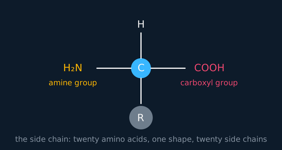
The side chains determine the chemical behaviour of the folded protein. Some are charged at cellular pH, some are polar but uncharged, and some are non-polar hydrocarbon and are excluded from water. Two of the twenty, cysteine and methionine, contain sulfur, and two cysteine side chains anywhere in a chain can form a covalent bond with each other. These properties have no effect in a single amino acid; they act once the chain folds.
- Amino acid
- A molecule containing an amine group, a carboxyl group and a variable side chain attached to the same carbon.
- Polypeptide
- A chain of amino acids joined by peptide bonds.
- Protein
- One or more polypeptides folded into a specific three-dimensional shape.
Joining them: one bond, one water
The amine group of one amino acid reacts with the carboxyl group of the next. A hydroxyl comes off the carboxyl and a hydrogen comes off the amine, the two leave together as water, and what remains is a covalent bond between carbon and nitrogen. That is a peptide bond, and the reaction is condensation, exactly as it is when glucose molecules join into starch.
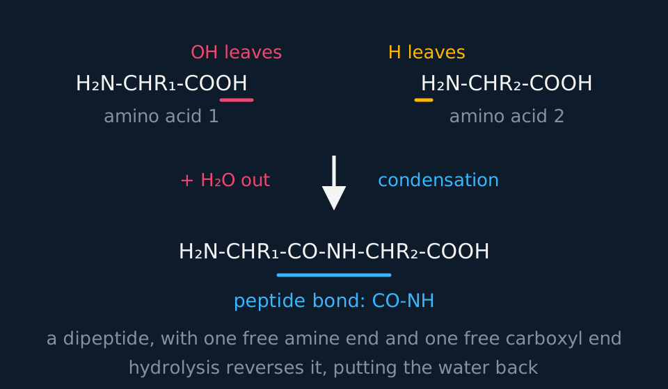
Describe the drawing
Two amino acids written as formulae side by side, each with an amine group at one end, a carboxyl group at the other and a side chain in the middle labelled R one and R two. The hydroxyl of the first carboxyl and a hydrogen of the second amine are marked as leaving. An arrow down leads to the product: a single dipeptide chain in which the two are joined by a CO to NH peptide bond, with one water molecule released.
The reverse reaction, adding water across the bond, is hydrolysis, which is how dietary protein is digested by proteases. A chain of n amino acids has n − 1 peptide bonds, and questions supplying a mass of water released depend on that relationship.
The chain retains one free amine group at one end and one free carboxyl group at the other, so a polypeptide is written and read from the amine end to the carboxyl end. Ribosomes synthesise polypeptides in the same direction.
Counting bonds and water
A polypeptide is 124 amino acids long. How many peptide bonds does it contain, and what mass of water was released as it was assembled? Take the relative molecular mass of water as 18.
Show the working
The bonds sit between amino acids, not on them, so 124 amino acids give 124 − 1 = 123 peptide bonds. Nothing about the sequence changes that.
Each bond released one water, so 123 waters were released, and 123 × 18 = 2214 in relative mass terms.
A chain of n monomers contains n − 1 bonds, not n. Draw three circles in a row and count the gaps between them to confirm the relationship.
The four levels of protein structure
A protein's structure is described at four levels, and different interactions dominate each one, although they overlap: hydrogen bonds appear at more than one level, and the quaternary level involves the same interactions as the tertiary level acting between chains rather than a bond type of its own. Name the bond as well as the level.
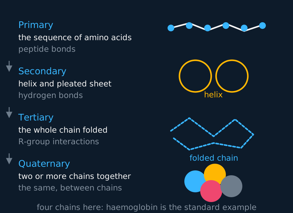
Describe the drawing
The four levels of protein structure listed down the left with a sketch of each on the right. Primary is a chain of amino acids held by peptide bonds. Secondary is a coiled helix or pleated sheet held by hydrogen bonds. Tertiary is the whole chain folded into a shape held by interactions between side chains. Quaternary is several folded chains grouped together, drawn as four subunits.
- Primary structure
- The sequence of amino acids in a polypeptide, held by peptide bonds.
- Secondary structure
- The regular coiling or pleating of the backbone into an α-helix or β-pleated sheet, held by hydrogen bonds between the C=O of one peptide group and the N–H of another.
- Tertiary structure
- The overall three-dimensional fold of a single polypeptide, held by hydrogen bonds, ionic bonds, disulfide bridges and hydrophobic interactions between side chains.
- Quaternary structure
- The arrangement of two or more polypeptides, and any non-protein groups, in a functioning protein.
Be careful with the hydrogen bonds, because they appear at two levels and they are not the same hydrogen bonds. In secondary structure they form between groups in the backbone, which is why the helix and the sheet look the same in every protein that has them. In tertiary structure they form between side chains, which is why every protein's tertiary structure is different.
Only one of the tertiary interactions is covalent: the disulfide bridge, between two cysteine side chains. It is much stronger than the others, and proteins subject to mechanical or chemical stress contain many of them, such as keratin in hair and circulating antibodies.
| Level | What is arranged | Held by | Broken by |
|---|---|---|---|
| Primary | Order of amino acids | Peptide bonds (covalent) | Hydrolysis, not heat |
| Secondary | The backbone, into helix or sheet | Hydrogen bonds along the backbone | Heat, extreme pH |
| Tertiary | The whole chain, into a 3-D shape | Hydrogen, ionic, disulfide, hydrophobic | Heat, extreme pH, reducing agents |
| Quaternary | Several chains, plus prosthetic groups | The same interactions, between chains | Heat, extreme pH |
Globular and fibrous proteins
Proteins fall into two broad structural families, and a named protein can be assigned to one of them from its shape, solubility and function.
Globular proteins fold into compact, roughly spherical shapes with their hydrophobic side chains tucked into the middle and their hydrophilic ones on the outside. That arrangement makes them soluble, and solubility is what a molecule needs if it is going to be carried in blood or dissolved in cytoplasm. Enzymes, antibodies, haemoglobin and the hormone insulin, carried in plasma to its target cells, are all globular.
Haemoglobin is the standard example: four polypeptides, two α and two β, each folded around one haem group, and each haem group holding one iron ion that binds one oxygen molecule. Four subunits, four haem groups, four oxygen molecules carried. The haem group is not made of amino acids at all, which makes it a prosthetic group, and haemoglobin a conjugated protein.
Fibrous proteins have repetitive sequences, form long strands rather than compact shapes, and are insoluble, which suits them to structural rather than transport roles. Collagen is three polypeptides wound round each other like a rope, and about every third amino acid along each chain is glycine, whose side chain is a single hydrogen atom, small enough to sit at the crowded centre of the twist. Neighbouring collagen molecules lie side by side with their ends staggered, so there is no line of weakness, and covalent cross-links between them build fibrils; bundles of fibrils form the fibres that make tendon strong in tension.
Keratin is rich in cysteine, so it carries many disulfide bridges; it is the tough, insoluble protein of hair, nails and the outer layer of skin. Elastin forms cross-linked fibres that stretch and recoil, which is why it is found in artery walls, alveoli and skin.
| Globular | Fibrous | |
|---|---|---|
| Shape | Compact, roughly spherical | Long strands |
| Solubility | Soluble: hydrophilic groups face out | Insoluble |
| Sequence | Irregular | Repetitive |
| Role | Metabolic: enzymes, transport, defence | Structural: support, strength |
| Example | Haemoglobin, catalase, insulin | Collagen, keratin, elastin |
Denaturation and the biuret test
Heat a protein, or move it far from its usual pH, and it denatures. Written carefully, that means the hydrogen bonds, ionic bonds and hydrophobic interactions holding the folded shape are disrupted. The chain unfolds, taking the tertiary structure and any secondary and quaternary structure with it, and the specific shape is lost. Peptide bonds are covalent and survive, so the primary structure does not change: a denatured protein is the same sequence in the wrong shape.
That distinction accounts for several familiar observations. Frying an egg turns the albumen opaque and solid without breaking it down into amino acids. A fever much above 40 °C is dangerous long before anything could be called cooked: human proteins differ widely in how much heat they tolerate, and the least stable of them start to lose their shape only a few degrees above body temperature, so the margin is narrow. Extreme pH acts by adding or removing charge on the side chains, so ionic bonds between them are disrupted and the fold is lost.
- Denaturation
- The loss of a protein's specific three-dimensional shape through disruption of the hydrogen bonds, ionic bonds and hydrophobic interactions holding it, so the function is lost, while the primary structure is unchanged.
The test for protein is the biuret test: add sodium hydroxide and then a few drops of copper(II) sulfate solution. Blue to purple means peptide bonds are present. No heating, no water bath. The reagent detects the peptide bond itself, so a solution of free amino acids stays blue.
TRY IT: Explaining a biuret result after boiling
A student boils a solution of egg albumen for five minutes, cools it, and then carries out the biuret test on it. The solution turns purple. Explain this result.
Check your answer
Purple means peptide bonds are still present, and they are: boiling denatures the protein but does not hydrolyse it.
Boiling disrupts hydrogen bonds, ionic bonds and hydrophobic interactions, so the secondary, tertiary and any quaternary structure is lost and the albumen coagulates. The peptide bonds are covalent and unaffected, so the primary structure survives intact and the biuret reagent still detects the peptide bonds.
State that boiling causes loss of the folded shape, not hydrolysis of the polypeptide chain.
In the exam
- Name the bond as well as the level: 'hydrogen bonds', not 'bonds between the amino acids'. At tertiary level, four interaction types are available: hydrogen, ionic, disulfide and hydrophobic. Include the disulfide bridge.
- State that amino acids differ in their R group, or side chain, when asked how they differ.
- n amino acids give n − 1 peptide bonds and n − 1 water molecules. Use that relationship for questions supplying masses of water released.
- For denaturation, state 'tertiary structure lost, primary structure unchanged'. Describe the change in bonding rather than saying the protein is destroyed, and do not describe a protein as killed: it was never alive.
- For haemoglobin, quote the numbers: four polypeptides, four haem groups, four oxygen molecules. For collagen, quote the glycine and the three chains.
Check yourself
Two proteins are made from exactly the same set of amino acids, in different orders. Explain why one might be an enzyme that works in blood plasma while the other is a structural fibre.
Answer
The order of amino acids is the primary structure, and it fixes which side chains end up next to each other once the chain folds.
In the enzyme, the folding brings hydrophilic side chains to the outside and hydrophobic ones to the core, giving a compact globular shape that dissolves in plasma, with an active site of a specific shape.
In the fibre, a repetitive sequence lets the chain form long regular strands that pack together rather than folding into a ball, so it is insoluble and strong.
The same amino acids in a different order give a different fold and therefore a different function.
Questions
Question 14 marks
Describe the four levels of protein structure, naming in each case the type of bond that holds that level together.
Mark scheme
- B1 primary structure is the sequence of amino acids in the polypeptide, held by peptide bonds
- B1 secondary structure is the regular coiling into an α-helix or pleating into a β-pleated sheet, held by hydrogen bonds between groups in the backbone
- B1 tertiary structure is the overall three-dimensional fold of one polypeptide, held by hydrogen bonds, ionic bonds, disulfide bridges and hydrophobic interactions between side chains
- B1 quaternary structure is the arrangement of two or more polypeptides, together with any prosthetic groups, in the finished protein, held by the same interactions as the tertiary level acting between the chains
Question 24 marks
Compare the structure of haemoglobin with the structure of collagen, and relate the difference in each case to what the protein does.
Mark scheme
- B1 haemoglobin is globular and compact, whereas collagen is fibrous and forms long strands
- B1 haemoglobin has an irregular amino acid sequence, whereas collagen has a repetitive one in which about every third amino acid is glycine
- B1 haemoglobin carries hydrophilic side chains on the outside so it is soluble and can be transported in solution, whereas collagen is insoluble, which suits it to a structural role
- B1 haemoglobin is four polypeptides each holding one haem prosthetic group that binds one oxygen molecule, whereas collagen is three chains wound round each other and cross-linked, giving strength in tension
Question 34 marks
Describe how extreme pH denatures a protein, referring to the charge on side chains and to the bonds affected.
Mark scheme
- B1 extreme pH adds or removes hydrogen ions from the side chains of a protein's amino acids, changing the charge some of them carry
- B1 that change in charge disrupts the ionic bonds that normally form between oppositely charged side chains in the tertiary structure
- B1 hydrogen bonds within the folded protein can also be disrupted as the pattern of charge and polarity around them changes
- B1 the tertiary structure unfolds and the protein loses its specific shape and function, while the covalent peptide bonds of the primary structure remain intact
Question 44 marks
A protein folds into a compact shape with all of its charged and polar side chains on the outside and all of its non-polar side chains buried inside. Suggest what this arrangement indicates about the protein's solubility, and suggest whether it is more likely to be a structural protein or one that is transported in blood.
Mark scheme
- B1 hydrophilic, charged or polar side chains on the outside interact with water molecules, so the protein is likely to be soluble
- B1 hydrophobic, non-polar side chains buried in the interior are shielded from water, which is what compact globular folding achieves
- B1 this compact, soluble arrangement is characteristic of a globular protein rather than a fibrous one, so it is more likely to be a protein that must dissolve and be carried in blood, such as an enzyme or a transport protein
- B1 a structural fibrous protein such as collagen or keratin is by contrast insoluble and does not need this arrangement, since it is not required to dissolve in a body fluid
Question 54 marks
Explain why a prosthetic group such as the haem group of haemoglobin is essential to the protein's function even though it is not made of amino acids.
Mark scheme
- B1 a prosthetic group is a non-protein component that is permanently associated with a protein and is required for it to function
- B1 the haem group of haemoglobin is not built from amino acids, so it is not part of the polypeptide's own primary, secondary or tertiary structure, though it is held within the folded protein by non-covalent interactions
- B1 each haem group contains one iron(II) ion, and it is that ion which actually binds one molecule of oxygen, a job none of haemoglobin's amino acid side chains can do alone
- B1 a haemoglobin molecule that had folded correctly but lacked its four haem groups would be unable to transport oxygen
Question 64 marks
A polypeptide is made from 150 amino acids, each of average relative molecular mass 110 as a free amino acid. Taking the relative molecular mass of water as 18, calculate the number of peptide bonds formed, and calculate the relative molecular mass of the finished polypeptide.
Mark scheme
- M1 number of peptide bonds = 150 − 1 = 149, since bonds form between amino acids rather than on each one
- M1 total mass of the 150 free amino acids before joining = 150 × 110 = 16 500
- M1 total mass of water lost = 149 × 18 = 2682, one water molecule released per peptide bond formed
- A1 mass of the polypeptide = 16 500 − 2682 = 13 818
Question 73 marks
While one molecule of a polypeptide was being assembled, water of total relative mass 3582 was released. Taking the relative molecular mass of water as 18, calculate the number of amino acids in that polypeptide.
Mark scheme
- M1 number of water molecules released = 3582 divided by 18
- M1 = 199 molecules of water, which is also the number of peptide bonds formed
- A1 200 amino acids, because n amino acids give n − 1 peptide bonds
Question 83 marks
A patient's body temperature rises to 43 °C and several of her proteins stop working. Explain what has happened to those proteins, referring to the bonds involved.
Mark scheme
- B1 the heat breaks the hydrogen bonds and ionic bonds holding the tertiary structure together
- B1 the chain unfolds, so the specific three-dimensional shape is lost and with it the function, which is denaturation
- B1 peptide bonds are covalent and survive, so the primary structure is unchanged
Question 93 marks
A protein taken from a bacterium living in a hot spring contains far more cysteine than the equivalent human protein does. Suggest why this helps the bacterial protein keep its shape at high temperature.
Mark scheme
- B1 two cysteine side chains anywhere in the chain can bond to each other, forming a disulfide bridge
- B1 a disulfide bridge is covalent and therefore much stronger than the hydrogen and ionic bonds that also hold the tertiary structure
- B1 heat that breaks those weaker bonds does not break the disulfide bridges, so the fold is held at a temperature that would denature the human protein
Question 103 marks
State the biuret test for protein: state the two reagents added, in order, and state the colour change that shows a positive result.
Mark scheme
- B1 sodium hydroxide solution is added first
- B1 a few drops of copper(II) sulfate solution are then added
- B1 a colour change from blue to purple shows a positive result, meaning peptide bonds are present
Question 113 marks
Explain why a solution of free amino acids gives a negative biuret test, whereas a solution of the polypeptide those same amino acids came from gives a positive one.
Mark scheme
- B1 the biuret reagent detects the peptide bond itself, turning purple in its presence, rather than reacting with amino acids as separate molecules
- B1 free amino acids in solution are not joined by peptide bonds, so none are present for the reagent to detect, and the solution stays blue
- B1 the polypeptide contains many peptide bonds joining its amino acids together, so the reagent gives a positive, purple result
Question 123 marks
Compare primary structure with secondary structure, referring to what is arranged at each level and to the bond holding it.
Mark scheme
- B1 primary structure is the sequence of amino acids in the polypeptide chain, held together by covalent peptide bonds, whereas secondary structure is the coiling or pleating of that chain into an α-helix or a β-pleated sheet
- B1 secondary structure is held by hydrogen bonds between groups in the backbone, rather than by the peptide bonds that hold primary structure
- B1 secondary structure could not exist without primary structure to fold, since it is the same backbone, already joined by peptide bonds, arranged into a specific pattern
Question 133 marks
An enzyme is a single polypeptide chain folded into an active shape. Explain why this enzyme has no quaternary structure, using the definition of quaternary structure in your answer.
Mark scheme
- B1 quaternary structure is the arrangement of two or more polypeptide chains, together with any prosthetic groups, in a functioning protein
- B1 this enzyme is described as a single polypeptide chain, so only one chain is present, and quaternary structure by definition requires more than one
- B1 the enzyme's function therefore depends on its primary, secondary and tertiary structure alone, without any interaction between separate chains
Question 142 marks
State the three groups that every amino acid carries on its central carbon, and state what varies between the twenty amino acids.
Mark scheme
- B1 an amine group, a carboxyl group and a hydrogen atom, all attached to the same central carbon
- B1 they differ only in the R group, also accepted as the side chain
Question 152 marks
Name the bond formed when two amino acids are joined together, and name the molecule released as it forms.
Mark scheme
- A1 a peptide bond, formed between the amine group of one amino acid and the carboxyl group of the next
- A1 water is the molecule released, the reaction being a condensation
Worth remembering
- Amino acids share one skeleton and differ only in the side chain.
- One peptide bond made, one water released; n amino acids give n − 1 bonds.
- Primary is peptide bonds, secondary is hydrogen bonds along the backbone, tertiary is four bond types between side chains, quaternary is several chains together.
- Denaturation loses the tertiary structure and keeps the primary one.
CHECK YOUR PROGRESS
Rate how confident you are with each objective for this lesson. Ratings are saved in this browser, on this device, unless you sign in.
- Draw the general structure of an amino acid and say what varies between the twenty.
- Explain how a peptide bond forms, and what is released when it does.
- Name the four levels of protein structure and the bond holding each one together.
- Account for the difference between a globular and a fibrous protein using haemoglobin and collagen.
- Explain denaturation in terms of bonds rather than in terms of the protein being 'killed'.
Open the full revision checklist to see every objective in the curriculum in one place.
Practise this lesson
Practise protein structure and function with 15 original questions and point-by-point mark schemes
WORKBOOK
The questions from this topic, on paper with room to work, and a separate book of mark schemes. Free to use; please do not redistribute or sell.
Used later in Membrane structure and the fluid mosaic model · Translation and protein synthesis · Haemoglobin and oxygen dissociation curves · Antibodies, vaccination and the four kinds of immunity