Physics › Medical physics › MR scanning and the endoscope
MR scanning and the endoscope
Precessing protons tipped by a radio-frequency pulse give the MR scanner its signal, and the endoscope works by total internal reflection. Relaxation time depends on the tissue the water sits in, and that difference is the contrast. A coherent bundle of fibres is what keeps the image in order.
Choose your exam board to show the notes and questions relevant to your specification. You can change this setting at any time, and content for other boards remains available.
Non-ionising imaging, part 2 of 2. Part 1 is Ultrasound imaging.
Builds on Ultrasound imaging and Refraction and Snell's law.
IN THIS TOPIC
- Outline the endoscope as total internal reflection put to work.
- Describe the principle of the MR scanner: protons precessing in a strong field, a radio-frequency pulse tipping them, and a relaxation time that depends on the tissue.
- Weigh MR against CT and ultrasound for a given diagnostic task.
COMMON MISCONCEPTION
The nuclear in nuclear magnetic resonance means an MR scan is radioactive.
MR uses the nucleus as a magnet, not as a source of radiation. Protons precess in a strong field, absorb a radio-frequency pulse and re-emit it as they relax; no nucleus decays, the photons are far too low in energy to ionise, and the patient takes no dose.
Light down a fibre: the endoscope
The option puts two more non-ionising instruments alongside ultrasound, and the simpler one is the endoscope, which is total internal reflection put to work. Light entering the core of a thin glass fibre strikes the wall of that core at a steep glancing angle. Because the core has the higher refractive index and the surrounding cladding the lower, any ray meeting the wall beyond the critical angle is reflected back inside with nothing escaping, and it goes on reflecting all the way along however the fibre bends.
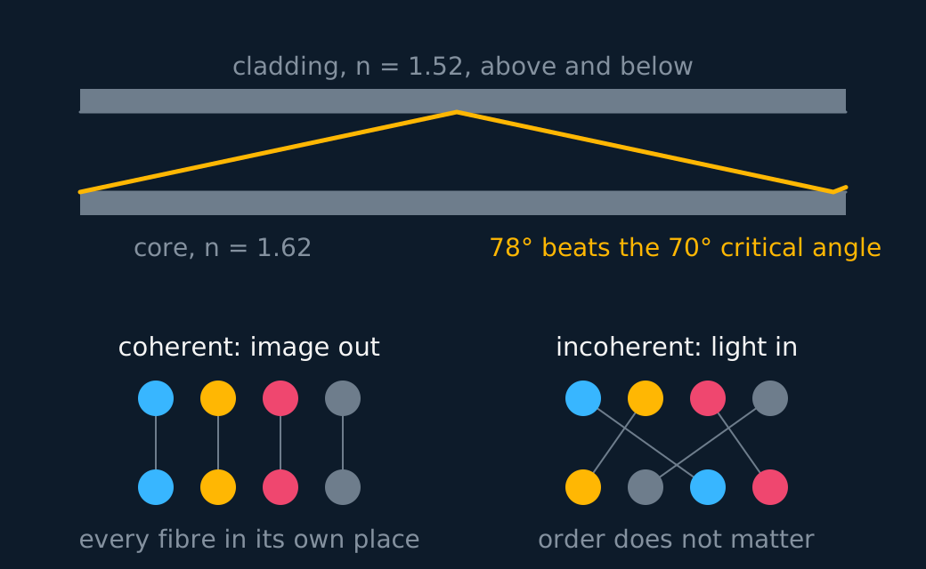
One endoscope carries two bundles of those fibres, and the difference between them is the point. The coherent bundle carries the image out: every fibre holds the same place in the cross-section at both ends, so each fibre delivers its own piece of the picture to the right place and the picture arrives intact. The second bundle only carries light in to illuminate what is being looked at, so it does not matter which fibre ends up where, and it is left incoherent, which is cheaper to make.
Being a physical instrument, the endoscope reaches only where a tube can be threaded, along the gut or the airway or through a small incision, which is exactly what it gains in exchange: the surgeon sees the tissue in real colour and can pass instruments down the same tube.
The MR scanner
The patient lies inside a strong magnetic field, a few teslas from a superconducting magnet. The body is mostly water, every water molecule carries two hydrogen nuclei, and each of those protons behaves as a tiny magnet. In the field they do not simply snap into line. They precess about the field direction, the way a leaning spinning top wheels around the vertical, at a frequency set by the strength of the field, and for the fields an MR magnet supplies that frequency sits in the radio range.
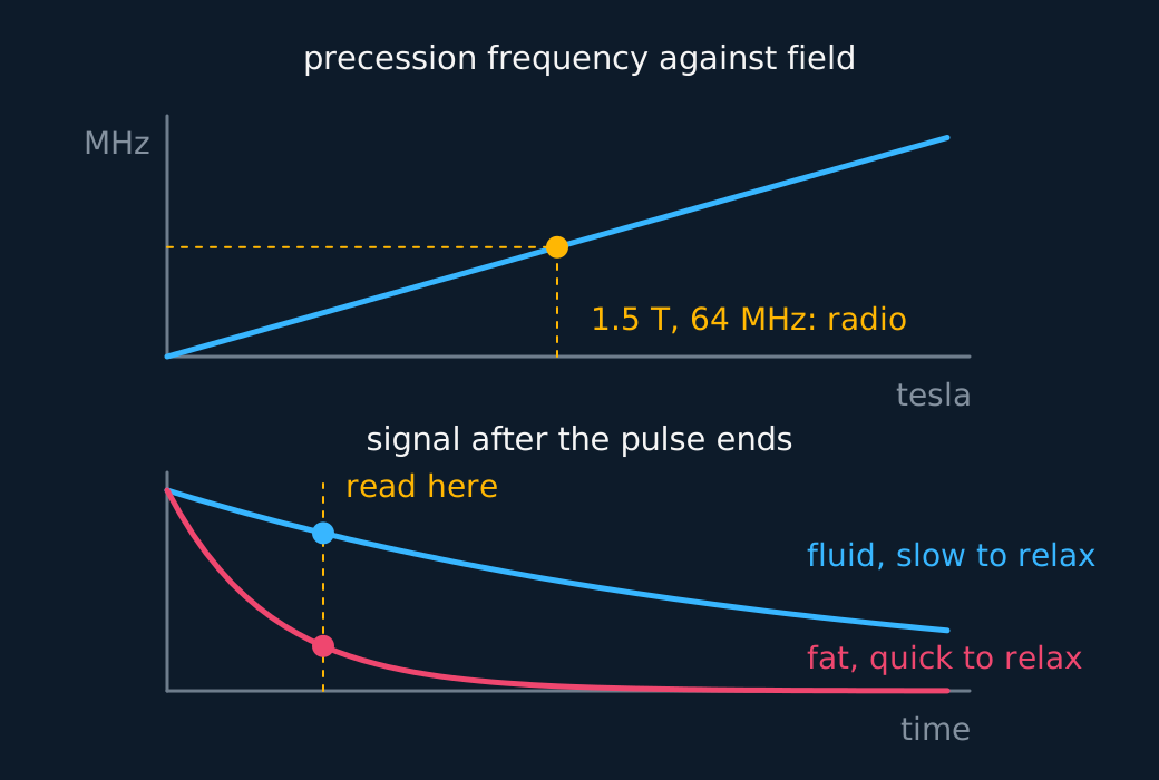
The scanner then supplies energy at exactly that frequency. A radio-frequency pulse at the precession frequency is absorbed by the precessing protons and tips them away from the field direction. When the pulse ends they relax back into line, giving the energy up again as a radio signal that coils around the patient pick up. Nothing here is nuclear decay; the nucleus is being used as a magnet, not broken up, and nobody receives a dose.
The relaxation time, the time the protons take to settle back, depends on the tissue their water sits in. Protons in fat relax at a different rate from protons in fluid or in a tumour, so the timing of the returning signal labels the tissue that sent it. That is the contrast mechanism, and it is why MR tells apart soft tissues that X-rays cross almost identically. One board note: AQA states that de-excitation relaxation times will not be examined, so for AQA this paragraph is the why behind the scan, not a required point.
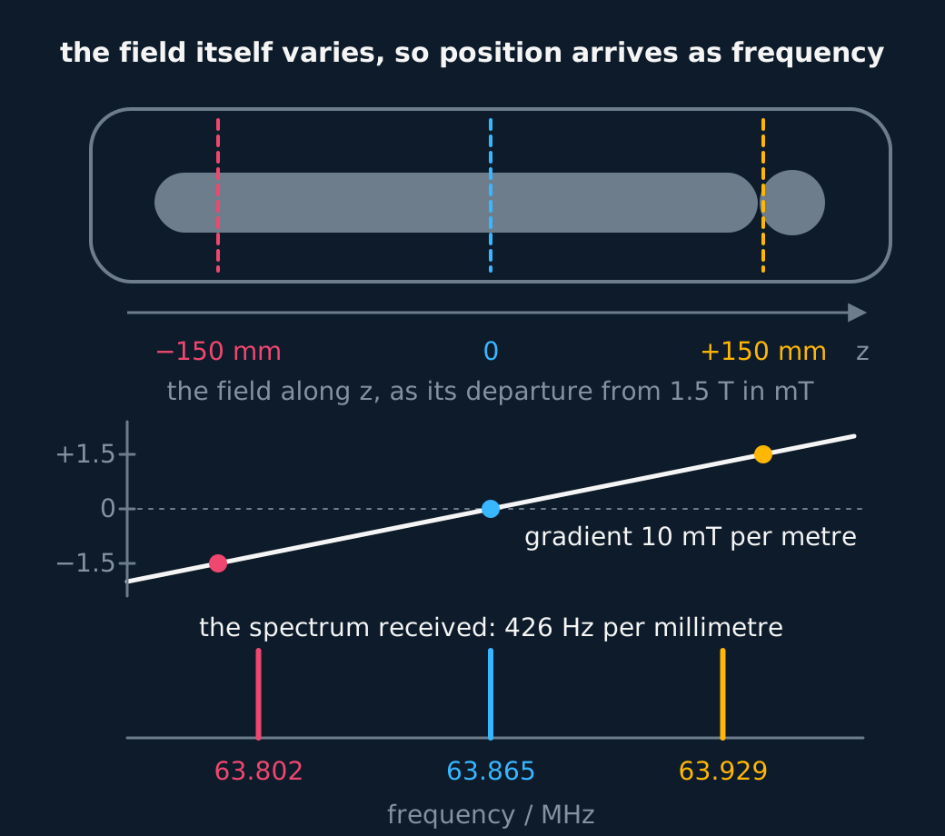
Locating the signal is done with the field itself. The precession frequency depends on the field strength, so gradient coils make the field vary slightly from place to place across the patient, and position along a gradient then maps to frequency. One gradient alone cannot pin down a point, so real scanners switch gradients to encode the remaining directions in the signal's frequency and phase, and a computer reconstructs cross-sectional images in any plane from that encoded signal, with nothing rotating around the patient.
Weigh it as the exam does, both ways at once. In its favour: excellent soft-tissue contrast, the finest of the modalities compared here, sections in any plane, and no ionising radiation, so repeat scans add no radiation dose, though every scan still passes the safety checks on implants, heating and any contrast agent first.
Against it: the superconducting magnet makes it expensive to buy and to run; a scan is slow and loud, with the patient holding still for tens of minutes inside a narrow bore; and the strong field rules out patients with MR-unsafe pacemakers or ferromagnetic implants, which the field would disturb or drag. Many modern implants are MR Conditional and scannable under controlled protocols; the safety label, rather than the presence of a pacemaker, determines whether a scan can proceed.
INDEPENDENT PRACTICE
Choosing the instrument
A patient needs the ligaments of a knee examined after an injury, and a second patient needs a suspected fracture of the same knee confirmed. Say which of MR, CT and ultrasound suits each, with a reason on a named axis.
Show the working
Ligaments are soft tissue, so MR: it gives the finest soft-tissue contrast of the three and adds no radiation dose, and the slowness and the cost are worth paying for detail nothing else supplies.
A fracture is bone, so CT: it resolves fine detail anywhere, bone included, quickly and much more cheaply than MR. The cost is the largest radiation dose of the three, which is accepted for a single diagnostic scan.
Ultrasound suits neither. It cannot see through bone at all, which rules out both the fracture and a joint interior lying behind it.
ASSESSMENT FOCUS
- The MR chain scores in order, like the PET chain does. Strong magnetic field, protons precess about it, a radio-frequency pulse at the precession frequency tips them, and they relax and re-emit a radio signal that is detected and processed into the image. The tissue-dependent relaxation time is the contrast mechanism, but AQA states that de-excitation relaxation times will not be examined, so on that board treat it as understanding behind the chain rather than a mark. If asked how the signal is located, the field is made to vary across the body so each position emits at its own frequency.
- Choosing between MR, CT and ultrasound turns on three axes: dose, soft-tissue detail, and cost. MR gives the finest soft-tissue contrast with no ionising dose but is the most expensive and slowest, and it is barred to patients with MR-unsafe pacemakers or ferromagnetic implants. CT resolves fine detail anywhere, but delivers the largest dose. Ultrasound is cheap, portable, real-time and dose-free, and cannot see through bone or air. Name the axis your reason sits on and the mark follows.
- Say why MR carries no dose when you claim it. The radio-frequency photons are far too low in energy to ionise, and no nucleus decays, so the answer is about photon energy rather than about the machine being gentle.
- The endoscope answer needs the word coherent and the reason it matters. Every fibre in the same place at both ends, so the image arrives in the right order; the illuminating bundle need not be, and saying which is which is where the second mark is.
- Total internal reflection needs its condition stated, not assumed. The core has the higher refractive index and the light must meet the wall beyond the critical angle, and an answer that says light bounces along the fibre without either of those has not explained anything.
CHECK YOURSELF
An MR scan and a CT scan are both offered for imaging the brain. Describe the chain of events inside the MR scanner that produces its signal, and give one reason to prefer MR here and one reason a hospital might use CT instead.
Show a hint
Four links in the chain, in order. Then one axis each way: what MR is best at, and what CT is best at.
Show the answer
The protons in the body's water precess about the scanner's strong magnetic field, at a frequency set by the strength of that field, which for a clinical magnet is in the radio range.
A radio-frequency pulse at exactly that frequency is absorbed and tips the protons away from the field direction.
When the pulse ends the protons relax back into line and re-emit the energy as a radio signal, which receiving coils detect and a computer builds into an image.
Prefer MR for the brain because it gives the finest soft-tissue contrast of the three modalities and adds no ionising dose, so it can be repeated.
A hospital may use CT instead because it is much faster and cheaper, which matters for an unwell or restless patient, and it is the only option for someone with an MR-unsafe pacemaker or a ferromagnetic implant.
An endoscope is total internal reflection put to work, and the bundle carrying the image must be coherent, every fibre in the same place at both ends.
MR times how quickly tipped protons relax, and the relaxation time depends on the tissue: soft-tissue detail with no ionising dose.
Or read them with their mark schemes on the ultrasound imaging questions page.
CHECK YOUR PROGRESS
Rate how confident you feel with each objective for this lesson. Ratings are saved in this browser, on this device, unless you sign in.
- Outline the endoscope as total internal reflection put to work.
- Describe the principle of the MR scanner: protons precessing in a strong field, a radio-frequency pulse tipping them, and a relaxation time that depends on the tissue.
- Weigh MR against CT and ultrasound for a given diagnostic task.
Open the full revision checklist to track your progress across the whole unit.