Physics › Medical physics › Ultrasound imaging
Ultrasound imaging
A piezoelectric transducer sends a pulse of megahertz sound into tissue and times the echo from each boundary; d = ct/2 then gives the boundary's depth. The acoustic impedances Z = ρc at a boundary fix what fraction of the intensity reflects, which is why coupling gel is used. An A-scan gives one line of depths, a B-scan an image.
Choose your exam board to show the notes and questions relevant to your specification. You can change this setting at any time, and content for other boards remains available.
Non-ionising imaging, part 1 of 2. Part 2 is MR scanning and the endoscope.
Builds on Progressive waves and Refraction and Snell's law.
IN THIS TOPIC
- Explain how a piezoelectric transducer generates and detects ultrasound pulses.
- Use the pulse-echo technique and d = ct/2 to locate a boundary in tissue.
- Say how the duration of the pulse and the wavelength limit the detail a pulse-echo scan can recover.
- Distinguish an A-scan from a B-scan.
- Use acoustic impedance Z = ρc and the reflection coefficient to explain what reflects where, and why the gel is needed.
- For CIE, use I = I₀e−μx for the attenuation of ultrasound in tissue.
- For OCR, find the speed of blood from a Doppler shift with Δf/f = 2v cos θ/c.
COMMON MISCONCEPTION
Ultrasound scans photograph the inside of the body with sound.
An ultrasound image is built from echo timings, not captured like a photograph: each boundary in the body reflects a pulse, and d = ct/2 converts each echo's return time into a depth.
A crystal that emits and detects
Ultrasound is sound above about 20 kHz, and medical scanners run in megahertz. One component both makes it and detects it, the piezoelectric transducer. That is a crystal which deforms when a pd is applied across it, and which generates a pd when something deforms it. Drive it with an alternating pd at its resonant frequency and it vibrates, launching a sound pulse. Let a returning echo squeeze it and it produces a measurable pd.
So the transducer alternates between two roles. It transmits for a microsecond, then detects the echoes that return. That switching is what lets the whole technique work with one probe held against the skin.
Pulse and echo
Everything else is timing. A pulse leaves the transducer, part of it reflects off each boundary between tissues, and the echoes arrive back after a delay. Sound of speed c that took time t to go there and back has found a boundary at depth
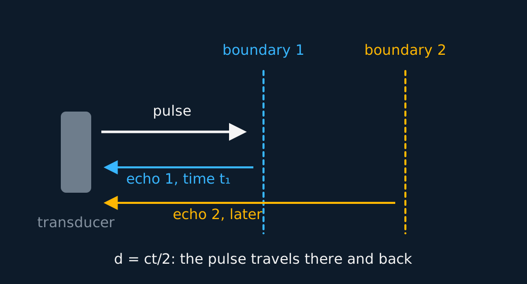
The factor of two is there because the pulse covers the distance twice, out to the boundary and back. An A-scan plots echo strength against time along one line, which suits a single depth measurement such as the length of an eye. A B-scan sweeps the beam and turns each echo into a bright dot on a two-dimensional map, and that is the moving picture of an antenatal scan.
WORKED EXAMPLE
How deep is the organ boundary?
An echo returns 65 μs after the pulse enters soft tissue, where ultrasound travels at 1540 m s−1. Find the depth of the reflecting boundary.
d = ct/2 = (1540 × 65 × 10−6)/2 = 5.0 cm.
Without the factor of two the answer comes out at 10 cm, twice the true depth. Halving the round-trip distance is a required step in the mark scheme.
How much a pulse can tell you
Timing an echo gives a position. It does not give unlimited detail, and the two things that cap the detail are worth knowing because they are the same two on every pulse-echo instrument ever built, from a bat to a ship's sonar to a radar set.
Start with the duration of the pulse. A pulse is not an instant; it lasts some time Δt and therefore occupies a length cΔt of tissue while it travels, and its echo is just as long. Two boundaries a distance Δd apart return echoes separated in time by 2Δd/c, that factor of two arriving for the same reason it arrived in d = ct/2. If that separation is shorter than the pulse itself, the second echo begins before the first has finished, the two run into one blur, and the scanner reports one boundary where there are two. The boundaries are just separable when 2Δd/c equals Δt, so the finest detail the pulse can resolve is about cΔt/2.
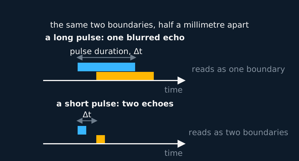
Describe the drawing
Two time traces of the echoes from one pair of boundaries, each echo drawn as a bar spanning the stretch of time it occupies, one bar above the other. In the upper trace the two bars are long and overlap along the time axis, so the receiver hears one unbroken return and the pair reads as a single boundary. In the lower trace the same two echoes are short bars with clear space between them, and the pair is resolved.
So shorter pulses see finer detail, and that pushes you straight into the second limit. A pulse cannot be shorter than about one cycle of the wave carrying it, so the shortest pulse a beam of wavelength can make is roughly long. Detail finer than about a wavelength is out of reach whatever the electronics do, and medical scanners are built around that fact: at 1540 m s−1 a 3 MHz beam has a wavelength near half a millimetre, and half a millimetre is about the resolution such a scan achieves.
Improving either limit costs penetration depth. Raising the frequency shortens both the wavelength and the pulse, which is the gain, but attenuation climbs steeply with frequency, so the finer beam is attenuated sooner. Shallow structures are scanned at high megahertz and deep organs at low, and the sonographer's choice of probe is that trade-off in practice.
WORKED EXAMPLE
How close is too close?
A scanner sends 3.0 MHz pulses, each three cycles long, into tissue where ultrasound travels at 1540 m s−1. Find the wavelength, the duration of one pulse, and how close two boundaries can lie and still be seen separately.
λ = c/f = 1540/(3.0 × 106) = 5.1 × 10⁻⁴ m, so about 0.51 mm.
One cycle lasts 1/f = 3.3 × 10⁻⁷ s, so three of them last Δt = 1.0 × 10⁻⁶ s.
The echoes must arrive at least Δt apart, and they arrive 2Δd/c apart, so Δd = cΔt/2 = (1540 × 1.0 × 10−6)/2 = 7.7 × 10⁻⁴ m, about 0.77 mm.
That is roughly one and a half wavelengths, which is the general result. A pulse-echo instrument resolves down to something near the wavelength of the radiation it uses, and no further, however good the timing.
Acoustic impedance, and the gel
How much of a pulse reflects at a boundary is set by the acoustic impedance of the media either side,
the product of density and sound speed. At every interface between two media the arriving wave splits in two: part of it is reflected straight back and the remainder is transmitted onwards into the second medium. For the head-on, normal-incidence arrival every exam question assumes, with no absorption at the boundary itself, the two impedances alone set the share. Similar impedances let most of the pulse through. Badly mismatched ones reflect most of it. The fraction of the intensity reflected is
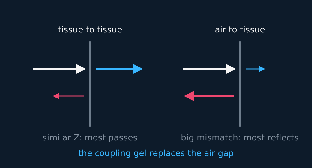
That equation sends the sonographer reaching for the coupling gel. Air's impedance is thousands of times smaller than tissue's, so an air gap between probe and skin reflects nearly all the ultrasound before it ever gets in. The gel displaces the air with a material whose impedance sits close to tissue, letting the pulse through. This is called impedance matching, and the term is worth using. Inside the body, the useful echoes come from mild mismatches between organs, and the strong ones from tissue meeting bone.
Ultrasound also fades as it travels. Absorption and scattering attenuate the pulse exponentially with depth, just as matter attenuates X-rays, and the attenuation rises steeply with frequency, which is the cost of the high frequencies that resolve fine detail. One board asks you to put a number on that fade, and the closing sections take it up.
Attenuation with depth, a CIE equation
CIE alone sets numerical attenuation questions on ultrasound, so read this section only if CIE is your specification. Absorption and scattering strip a fixed fraction of the surviving intensity out of every further millimetre of tissue, which is the recipe for an exponential and gives the law the X-rays lesson already uses:
with μ the attenuation coefficient of the tissue, in per metre, and x the distance travelled through it. None of the mathematics is new. What is new is a feature of the board's own formula sheet: it prints the radioactive-decay exponential, x = x0e−λt, where x stands for an activity or a number of nuclei, and it does not print this one. An exponential on the page is not the exponential you need, so this equation has to be recalled, for ultrasound and for X-rays alike.
The coefficient is where the sonographer's trade-off appears numerically. μ climbs steeply with frequency, so the higher frequency that resolves finer detail is also attenuated faster. μ depends on the tissue too, and bone attenuates so much more strongly than soft tissue that ultrasound cannot usefully see through the skull or into air-filled lungs.
WORKED EXAMPLE
How much of the pulse comes back?
A pulse enters soft tissue whose attenuation coefficient at this frequency is μ = 23 m−1. A boundary lies 4.0 cm below the skin. Find the fraction of the incident intensity arriving at the boundary, and the fraction returning to the transducer.
Going in, x = 0.040 m, so μx = 23 × 0.040 = 0.92 and I/I0 = e−0.92 = 0.40.
The echo crosses the same tissue again, so the round trip is x = 0.080 m, μx = 1.84, and I/I0 = e−1.84 = 0.16, before anything at all is lost at the boundary itself.
The factor of two is back, and for the same reason it appeared in d = ct/2. Decide which distance the question means before you exponentiate, because an attenuation question about an echo almost always means the round trip.
Doppler ultrasound and the speed of blood, an OCR equation
OCR adds one measurement the other boards leave out, so read this section only if OCR A is your specification. Bounce ultrasound off something that is moving and the echo returns at a shifted frequency, the same Doppler effect that drops the pitch of a siren as it passes. Red blood cells are the moving reflectors, and the shift in the returning echo measures how fast they are travelling.
Two features of the equation are worth understanding rather than memorising. First, the shift is doubled. The cells are moving relative to the transmitter, so they meet an already shifted frequency; they then re-radiate that wave while still moving relative to the receiver, so it shifts a second time. Two shifts, one factor of two. Second, only the part of the velocity along the beam shifts anything, and with θ the angle between the beam and the flow that part is v cos θ. Put the two together:
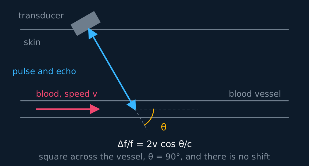
Describe the drawing
A blood vessel drawn as a horizontal channel under the skin, with blood flowing to the right. An angled transducer on the skin sends a beam down into the vessel, meeting the flow direction at an angle theta of sixty degrees, marked with an arc where the beam crosses the vessel axis. Only the component of the blood's velocity along the beam, v cos theta, shifts the frequency, so a beam square across the vessel would report nothing.
Here c is the speed of ultrasound in tissue, about 1540 m s−1, and not the speed of light. The equation is printed on the OCR data sheet, so the work is in choosing the right angle and the right c rather than in recalling it.
The cosine explains how the probe is held. Aim it square across the vessel, θ = 90°, and cos θ is zero: the blood is moving neither towards the probe nor away from it, and the machine reports no flow whatever, however fast the blood is going. Tilt the probe along the flow and the shift grows, which is why scanning angles are chosen deliberately, typically somewhere around 45° to 60°.
WORKED EXAMPLE
How fast is the blood?
A 4.0 MHz beam meets a vessel at 60° to the flow, and the echo returns 780 Hz higher than it left. Taking the speed of ultrasound in tissue as 1540 m s−1, find the speed of the blood.
Rearrange for the unknown first: v = cΔf/(2f cos θ), with cos 60° = 0.50.
v = (1540 × 780)/(2 × 4.0 × 106 × 0.50) = (1.20 × 10⁶)/(4.0 × 106) = 0.30 m s−1.
Notice where that 780 Hz sits: in the audible range. The shift itself is a frequency difference, not a sound, but the machine converts it into a tone through a loudspeaker as well as plotting it, which is why a narrowed artery, where the blood speeds up, can be heard as well as seen.
Light down a fibre: the endoscope has moved to the next part of this lesson.
The MR scanner has moved to the next part of this lesson.
ASSESSMENT FOCUS
- State both halves of the piezoelectric effect. A pd deforms the crystal, which transmits. Deforming the crystal produces a pd, which detects. One sentence in each direction.
- Every pulse-echo calculation carries the factor of two. Write d = ct/2 before substituting, and check the answer against sense, because organs live a few centimetres down and not tens of centimetres.
- Asked what limits the detail in a pulse-echo scan, give both limits and tie each to a length. Two boundaries closer than about half the length of the pulse return echoes that overlap, and no pulse is shorter than about one wavelength, so the resolution stops near . Edexcel writes both limits into the statement itself; on the other boards the same argument is what justifies choosing a high frequency, and the evaluation is complete once the attenuation cost of that choice is stated.
- Z = ρc takes the density and sound speed of the same medium. Taking ρ from one medium and c from the other gives an impedance that belongs to neither, and a question that lists four numbers in a table makes that substitution easy to slip into.
- Explain the gel in impedance language. It excludes the air gap, whose impedance mismatch with tissue would reflect nearly all the intensity straight back at the surface.
- A-scan against B-scan is a two-mark comparison. One line and a depth trace, against a swept beam and a two-dimensional brightness image.
- Ultrasound is the non-ionising option, so there is no photon energy argument and no dose to discuss. State that explicitly in any comparison with X-ray imaging.
- CIE only: I = I₀e−μx covers ultrasound as well as X-rays, and CIE prints neither. The decay exponential on the formula sheet is a different equation about a different quantity, so recall the attenuation one, and check whether the question means the depth or the round trip.
- OCR only: the Doppler shift is doubled because the blood receives a shifted wave and then re-emits one, and cos θ takes the component along the beam. The c in Δf/f = 2v cos θ/c is the speed of ultrasound in tissue, about 1540 m s−1. It is on the data sheet, so the angle is what has to be handled: at θ = 90° the measured shift is zero.
CHECK YOURSELF
Ultrasound crosses a boundary where Z₁ = 1.6 × 10⁶ kg m−2 s−1 and Z₂ = 1.7 × 10⁶ kg m−2 s−1. Roughly what fraction of the intensity reflects, and why is that useful?
Show a hint
Put the numbers into the reflection coefficient. The answer is small.
Show the answer
Ir/I0 = (0.1/3.3)2 ≈ 0.001, so about a tenth of a per cent reflects.
That is useful because imaging needs most of the pulse to carry on to deeper boundaries. Tiny reflections at each interface give faint but detectable echoes from every layer, instead of one blinding echo from the first.
Depth is ct over two, because the echo travelled there and back.
Impedance mismatch sets how much reflects, and the gel exists to remove the worst mismatch of all.
A-scan gives one line of depths. B-scan sweeps that line into a picture.
WORKBOOK
Printable practice for this topic: original exam-style questions with room to work, and a fully worked answer book. Free to use; please do not redistribute or sell.
Or read them with their mark schemes on the ultrasound imaging questions page.
WHERE TO GO NEXT
- Exponentials and logarithms is the maths this lesson leans on, worked through step by step.
CHECK YOUR PROGRESS
Rate how confident you feel with each objective for this lesson. Ratings are saved in this browser, on this device, unless you sign in.
- Explain how a piezoelectric transducer generates and detects ultrasound pulses.
- Use the pulse-echo technique and d = ct/2 to locate a boundary in tissue.
- Say how the duration of the pulse and the wavelength limit the detail a pulse-echo scan can recover.
- Distinguish an A-scan from a B-scan.
- Use acoustic impedance Z = ρc and the reflection coefficient to explain what reflects where, and why the gel is needed.
- For CIE, use I = I₀e−μx for the attenuation of ultrasound in tissue.
- For OCR, find the speed of blood from a Doppler shift with Δf/f = 2v cos θ/c.
Open the full revision checklist to track your progress across the whole unit.