Physics › Medical physics › PET scanning and radiotherapy
PET scanning and radiotherapy
Positrons from a beta-plus tracer annihilate with electrons, and the two 511 keV photons leaving back to back are detected in coincidence. Every coincidence puts the annihilation on the line between one pair of detectors. Radiotherapy aims the same ionising radiation on purpose, from rotated megavoltage beams or a beta-emitting implant.
Choose your exam board to show the notes and questions relevant to your specification. You can change this setting at any time, and content for other boards remains available.
Radionuclide imaging and PET, part 2 of 2. Part 1 is Radionuclide imaging and the gamma camera.
Builds on Radionuclide imaging and the gamma camera and Antimatter and photons.
IN THIS TOPIC
- Explain PET: annihilation into two 511 keV photons, and coincidence detection locating the source.
- Say what PET scanning is used to diagnose, and why it is paired with a CT scanner.
- Explain how high-energy X-ray beams and beta-emitting implants deliver a dose to a tumour and spare the tissue around it.
COMMON MISCONCEPTION
A PET scanner works by sending radiation into the body.
PET sends no radiation in: a positron-emitting tracer inside the body is the source, and the scanner detects the back-to-back 511 keV photons produced when positrons annihilate with electrons.
PET: annihilation as a beacon
Positron emission tomography uses the same tracer principle with antimatter. The tracer is still a radionuclide attached to a molecule the body uses, and the image is still a map of where the biology took it; what changes is the radiation that is detected. The tracer, commonly fluorine-18 in a glucose analogue, is a beta-plus emitter, so what it sends out is positrons.
That looks like a breach of the rule that only photons get out of the patient, and it is not. Each positron travels only a millimetre or so before it meets an electron, so the positron itself never leaves the patient. The pair annihilates, both particles vanish, and their rest energy leaves as two gamma photons of 511 keV each, flying in opposite directions to conserve momentum. Those photons are what the scanner detects, and they escape as readily as any other gamma.
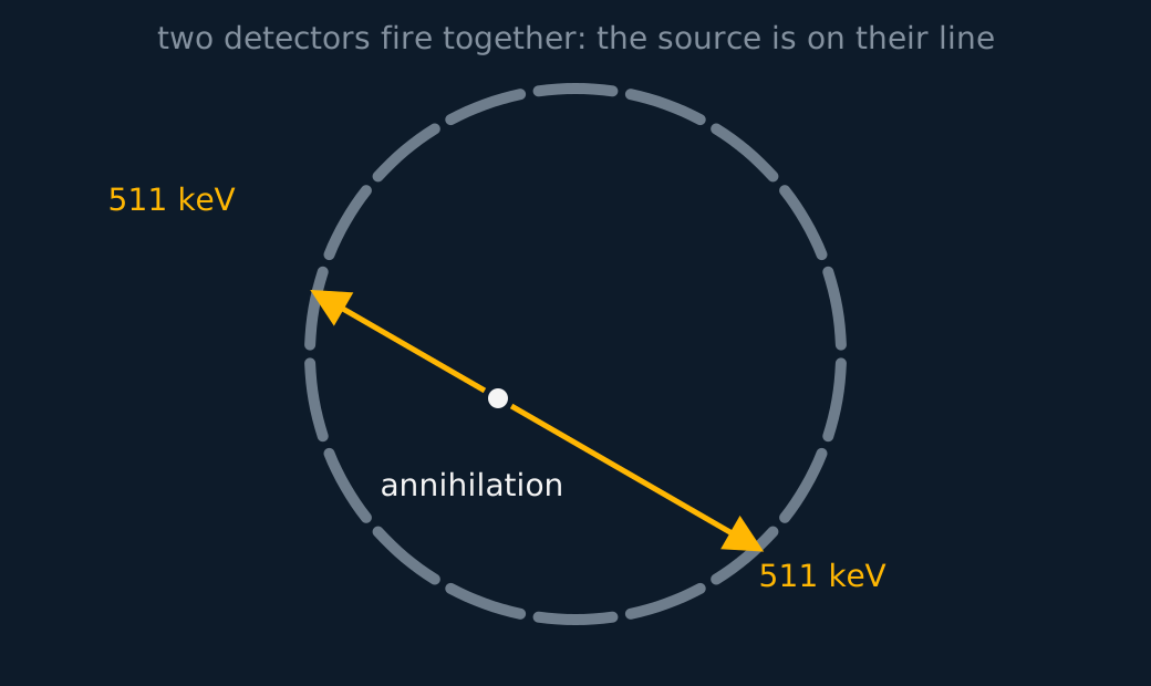
The scanner is a ring of detectors watching for coincidences, meaning two 511 keV photons arriving at opposite sides at effectively the same instant. Each coincidence pins the annihilation to the straight line between the two detectors, and hundreds of thousands of such lines intersect at the places the tracer gathered. The 511 keV is the electron's rest energy m0c2.
What the scan diagnoses depends on the molecule to which the fluorine-18 is attached. In a glucose analogue it goes wherever the body is burning sugar, so the image is a map of metabolic rate. Many tumours consume glucose faster than the tissue around them and show as bright spots, which is why PET, paired with CT, helps to detect certain cancers, to stage them by picking up secondary tumours anywhere in the body, and to check afterwards whether treatment has quietened them.
The uptake is not specific to cancer, though: infection and inflammation burn sugar too and also light up, and not every tumour takes up the tracer strongly, so a PET result is read alongside other evidence rather than as a verdict on its own.
The brain and the heart read the same map differently. Reduced uptake across particular regions of cortex is a marker of dementia, an abnormally active patch can locate where epileptic seizures begin, and heart muscle that still takes up the tracer after a heart attack is muscle worth restoring the blood supply to, while muscle taking up none has died.
Every one of those diagnoses rests on the same property. PET measures function, and it is poor at anatomy, so a bright spot on its own does not say which organ it is in. Scanners are therefore built as PET rings wrapped around a CT scanner, and the two images are laid on top of each other: one says what is happening and the other says where.
When the dose is the point
Diagnostic imaging treats the dose as a cost to be minimised. In treatment the same physics is used the other way, because ionising radiation damages cells: a tumour can be destroyed if it receives a much larger absorbed dose than the tissue around it. Radiotherapy rests on that comparison, and there are two ways of achieving it.
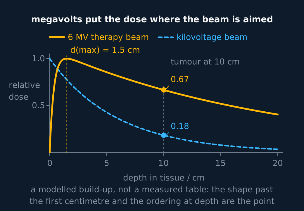
The first is to aim from outside. A beam of high-energy X-rays, megavolts rather than the kilovolts of diagnosis, is rotated around the patient so that it enters through a different path each time while always crossing the tumour. Each path through healthy tissue is used briefly and receives a small share of the dose, while the tumour lies on every path and receives the total. The energy is chosen high deliberately, because a hard beam is attenuated less on the way in and so deposits proportionately more where it is aimed; a megavoltage beam's dose also builds up over the first centimetre or two rather than peaking at the skin, and collimators shape each beam to the tumour's outline.
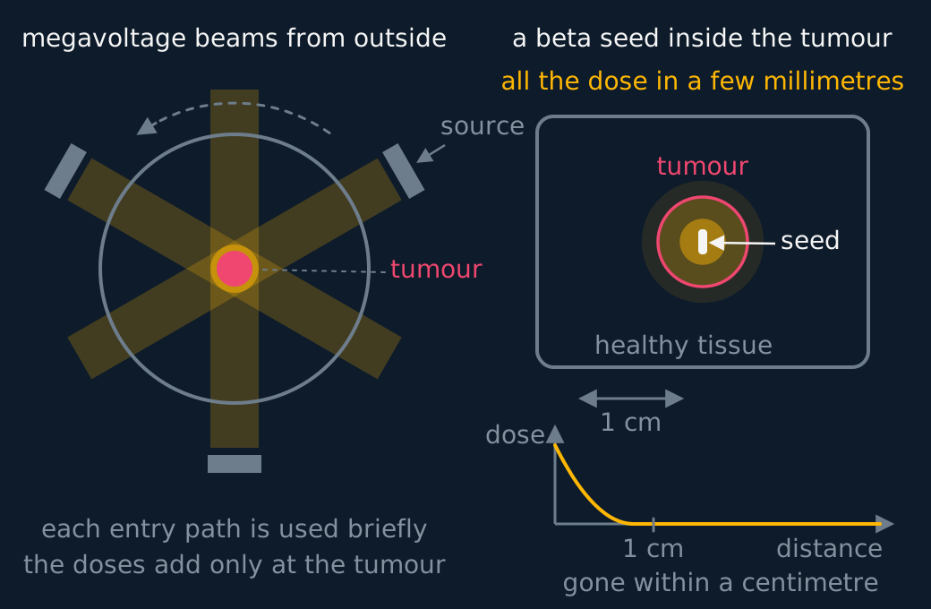
The second is to put the source inside. A radioactive implant is a sealed source, a seed or a small rod, placed in the tumour or against it, and the emitters this specification's model names are beta emitters, yttrium-90 or strontium-90 among them; clinical practice also uses other sources under the same geometry. Beta particles are absorbed within a few millimetres of tissue, so the dose is enormous where the seed sits and negligible a centimetre away, and the healthy tissue beyond that range is untouched however long the source is left.
That is the tracer rule applied in reverse, and it is worth saying so in an answer. A beta-minus emitter is unsuited to gamma-camera imaging precisely because its radiation cannot escape the body, and it is well suited to an implant for precisely the same reason. Unsealed sources exploit chemistry instead of geometry, iodine-131 being swallowed and concentrated by the thyroid itself, and there the tissue to be destroyed does the aiming.
Two numbers then shape the treatment. The activity of the source sets the scale of the dose rate, though the dose delivered also depends on the radiation's energy and on how the surrounding tissue absorbs it, and the half-life fixes how quickly that rate falls.
For a sealed implant the half-life that matters is the physical one, because the body cannot excrete a sealed source; the effective half-life belongs to unsealed tracers that can be cleared. So a permanent seed is chosen with a physical half-life short enough that it has spent itself once the planned dose has been delivered, while a stronger, longer-lived source is put in for a measured number of hours and taken out again.
ASSESSMENT FOCUS
- Keep the two instruments apart when the emission type is asked for. A gamma camera needs a gamma emitter. PET needs a positron emitter, and the radiation that gets out is the pair of annihilation photons, not the positron. Writing "never use a beta emitter" contradicts the whole of PET.
- Give the PET chain in order, in five steps. Positron emitted, annihilates with an electron, two 511 keV photons in opposite directions, detected in coincidence, source located on the line between the two detectors.
- Opposite directions is a momentum statement, so say so. The pair is nearly at rest when it annihilates, so the two photons must carry equal and opposite momentum. The 511 keV itself comes from the electron rest mass through E = mc2, and you can quote it without deriving it unless asked.
- Therapy answers turn on range. A beta-emitting implant delivers its energy within a few millimetres of the seed, so the tumour is destroyed and tissue a centimetre away is spared, while an external beam achieves the same comparison by crossing the tumour from many directions. Saying that a beta emitter is unsuited to gamma-camera imaging for the very reason it suits an implant is the observation that completes the answer.
- Diagnosis questions want the biology of the tracer, not the physics again. Fluorine-18 in a glucose analogue maps how fast tissue burns sugar, so many tumours light up, which helps find and stage a cancer; reduced uptake in the cortex marks dementia, and heart muscle that still takes up the tracer after a heart attack is still alive. Finish by saying PET is paired with CT because function alone does not say where.
CHECK YOURSELF
Explain why the two photons produced in PET annihilation travel in opposite directions, and why both must be detected for the event to be useful.
Show a hint
Start from the momentum of the electron-positron pair just before annihilation.
Show the answer
The pair is very nearly at rest, with total momentum close to zero. Momentum is conserved, so the two photons must leave with equal and opposite momenta, which means back to back.
One photon alone gives only a direction from one detector. Two in coincidence define the whole line through the annihilation point, and it is the intersection of many such lines that locates the tracer.
Annihilation gives two 511 keV photons back to back, and coincidence detection turns each pair into a line. The lines cross where the tracer is.
PET maps function and CT maps structure, which is why the scanner is built as both and the two images are overlaid.
A beta-minus emitter cannot reach a gamma camera because its range is millimetres, and an implant treats for exactly that reason.
Or read them with their mark schemes on the radionuclide imaging and the gamma camera questions page.
CHECK YOUR PROGRESS
Rate how confident you feel with each objective for this lesson. Ratings are saved in this browser, on this device, unless you sign in.
- Explain PET: annihilation into two 511 keV photons, and coincidence detection locating the source.
- Say what PET scanning is used to diagnose, and why it is paired with a CT scanner.
- Explain how high-energy X-ray beams and beta-emitting implants deliver a dose to a tumour and spare the tissue around it.
Open the full revision checklist to track your progress across the whole unit.