Biology › Inheritance and population genetics › Meiosis and the sources of genetic variation
Meiosis and the sources of genetic variation
Meiosis halves the chromosome number and produces four genetically different cells from one. Two processes create that variation: crossing over between homologous chromosomes in prophase I, and the independent assortment of those chromosomes in metaphase I. Random fertilisation then adds a third.
Before this The cell cycle and mitosis · DNA replication · Eukaryotic cell ultrastructure
COMMON MISCONCEPTION
Meiosis is mitosis done twice, so learning it means learning the same four stages again with roman numerals attached.
Meiosis I separates homologous chromosomes; meiosis II separates sister chromatids. Crossing over and independent assortment during meiosis I generate new combinations of alleles, while the two divisions reduce one diploid cell to haploid products.
What you should be able to do
- Say which chromosomes separate in the first division and which in the second, and why that order matters.
- Describe how a bivalent forms and what crossing over does to the chromatids in it.
- Work out how many chromosome combinations a gamete could carry, given the haploid number.
- Track chromosome number and chromatid number through both divisions without losing count.
- Set out the differences between mitosis and meiosis, division by division.
- Name all eight stages in order and say what the spindle, the nuclear envelope and the cell surface membrane are doing in each.
- Name the stage of meiosis that a photomicrograph shows, and justify the naming.
- Explain how non-disjunction leads to polysomy, as in Down's syndrome, and to monosomy, as in Turner's syndrome.
Reduction division and the homologous pair
Meiosis produces haploid cells from a diploid cell through one round of DNA replication followed by two divisions. In meiosis I, homologous chromosomes pair and then separate. In meiosis II, sister chromatids separate. Crossing over and independent assortment generate different combinations of alleles in the products.
A human body cell carries 46 chromosomes as 23 homologous pairs: two copies of chromosome 1, two of chromosome 2, and so on, one member of each pair inherited from each parent. The two members of a pair are the same length and carry the same genes at the same loci, though they can differ in which alleles sit at those loci and in non-coding and structural sequence between them. Mitosis does not pair them; meiosis does, and the pairing is what makes the first division a reduction division.
The halving is necessary. If a sperm carrying 46 chromosomes fertilised an egg carrying 46, the zygote would have 92 and the next generation 184. Gametes are therefore haploid: one chromosome from each pair, 23 in total. The chromosomes are not discarded at random. A gamete missing chromosome 7 and carrying two copies of chromosome 12 lacks the genes of one chromosome and has an extra dose of another, and such gametes usually give inviable zygotes. Pairing the homologues and separating them gives each product exactly one representative of each pair.

Describe the drawing
Meiosis animated through six stages. Prophase one: two homologous pairs lie inside a cell, each chromosome already two chromatids, and one pair is crossed over at a chiasma marked in coral, and from then on one chromatid of each long chromosome carries a tip in the other colour. Metaphase one: both bivalents lie on the equator with spindle fibres running to poles on either side, and the maternal chromosome of one pair faces the opposite way to the maternal chromosome of the other. Anaphase one: whole chromosomes, still two chromatids each, are dragged to opposite poles. Metaphase two: two smaller cells, each with its chromosomes lined up singly on its own equator. Anaphase two: the centromeres divide and the chromatids separate. Finally four haploid cells, each carrying one long and one short chromosome in a different combination, two of the long ones with a swapped tip from the crossover.
DNA replication happens once, in the interphase before meiosis begins, and not again between the two divisions. So a cell entering meiosis has 46 chromosomes and 92 chromatids; the cells leaving meiosis I have 23 chromosomes and 46 chromatids each; the four cells leaving meiosis II have 23 chromosomes and 23 chromatids each. The chromosome number halves at the first division and the DNA content halves at both.
- Homologous pair
- Two chromosomes of the same length carrying the same genes at the same loci, one from each parent, though not necessarily the same alleles.
- Diploid
- Having chromosomes in homologous pairs, written 2n.
- Haploid
- Having one chromosome from each homologous pair, written n.
- Bivalent
- The structure formed when a homologous pair comes together in prophase I: two chromosomes, four chromatids.
- Reduction division
- A division that halves the chromosome number. Meiosis I is one; meiosis II is not.
Prophase I: pairing, and the exchange that follows
Prophase I is the longest and the most eventful stage in the whole process. The chromosomes condense as they do in mitosis, and each chromosome then aligns with its homologue along their whole length. That pairing is called synapsis, and the paired structure is a bivalent. Because both chromosomes were replicated in interphase, a bivalent contains four chromatids, which is why some books call it a tetrad.
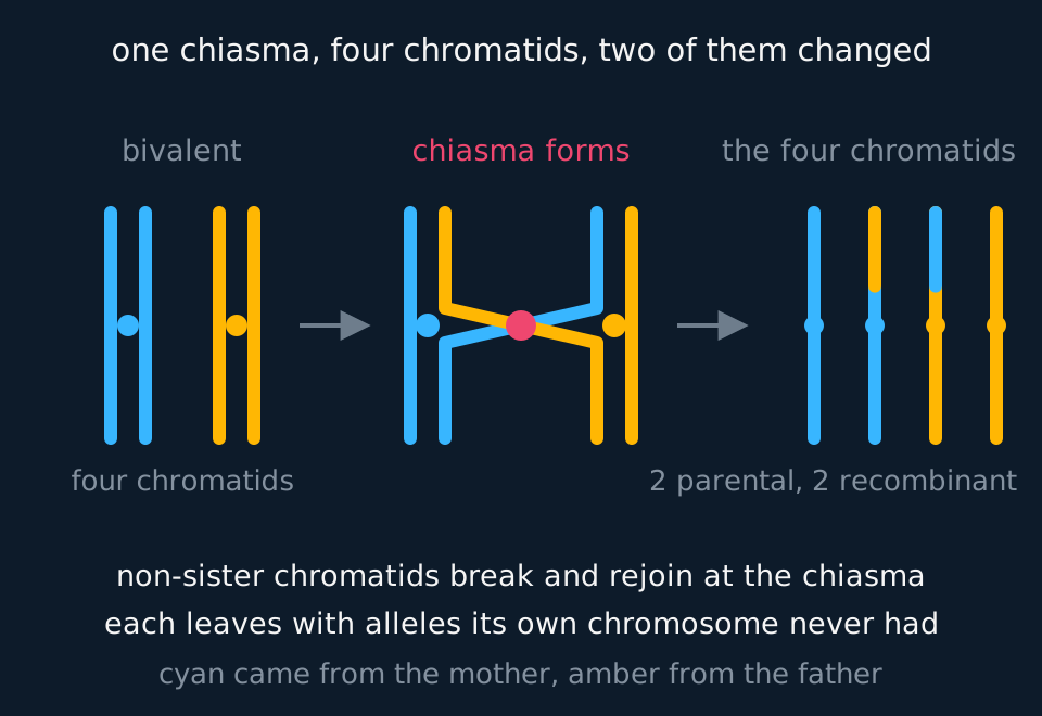
While the homologues are aligned, chromatids break and rejoin. A chromatid of the maternal chromosome and a chromatid of the paternal chromosome, which are non-sister chromatids, break at the same point and each joins to the other's cut end. The point where they cross is a chiasma, plural chiasmata, and it is visible down a good microscope. When the bivalent later comes apart, the two chromatids that took part leave carrying a stretch of DNA from the other parent's chromosome.
Describe the drawing
Three panels reading left to right. On the left a bivalent: one cyan maternal chromosome and one amber paternal chromosome lying side by side, each drawn as two chromatids joined at a centromere, four chromatids in all. In the middle the two inner chromatids cross at a coral dot and continue up the other side, so each runs cyan below the crossing and amber above it or the reverse. On the right the four separated chromatids: one wholly cyan, one wholly amber, and two carrying a tip of the other colour.
After crossing over, sister chromatids are no longer genetically identical. A chromatid can carry the maternal allele of one gene and the paternal allele of a gene further along the same chromosome, a combination that existed on neither chromosome the cell started with. Crossing over is the only mechanism in meiosis that makes new combinations of alleles within a chromosome; without it, alleles of genes on the same chromosome would be inherited together.
- Crossing over
- The exchange of equivalent sections of DNA between non-sister chromatids of a homologous pair during prophase I.
- Chiasma
- The visible point at which two non-sister chromatids cross during a bivalent's crossover.
- Recombinant
- A chromatid, or an offspring, carrying a combination of alleles that neither parental chromosome carried.
Metaphase I and independent assortment
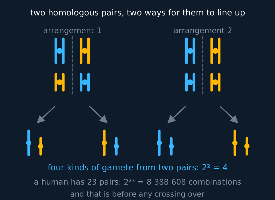
At metaphase I the bivalents move to the equator, and the second source of variation arises there. Each bivalent settles with one member facing one pole and the other facing the opposite pole, and the orientation of each bivalent is independent of the orientation of every other. That is independent assortment. Maternal and paternal chromosomes are therefore not kept together: a gamete can receive any mixture of the two.
Describe the drawing
Two metaphase plates side by side, each with a dashed equator and two homologous pairs lying across it. In the left plate the maternal cyan chromosomes of the two pairs face opposite ways; in the right plate the shorter pair has flipped, so both cyan chromosomes lie on the same side. Arrows lead from each plate down to the poles, and below sit four gametes, each holding one long and one short chromosome in a different colour combination.
number of chromosome combinations in a gamete = 2nn is the haploid number: the number of homologous pairs. Crossing over is not included in this count, and makes the true figure much larger.
With n = 23, that is 223 = 8 388 608 different chromosome combinations a single human gamete could carry. Fertilisation is random, so any one of those sperm could meet any one of those eggs: 223 × 223 = 246, or about 7 × 10¹³ different zygotes from one couple, before crossing over is taken into account.
The number of metaphase I arrangements is not the same quantity as the number of gametes. An arrangement and its mirror image, with every bivalent flipped, produce the same pair of daughter cells, so the number of genuinely distinct metaphase I arrangements is 2n−1. Each arrangement sends a different combination to each of the two poles, so 2n−1 arrangements still give 2n kinds of gamete. Answer whichever the question asked for, and say which you have counted.
Counting combinations in a fruit fly
A fruit fly has a diploid number of 8. Calculate the number of different chromosome combinations possible in its gametes, and the number possible in a zygote formed by two such flies. Ignore crossing over.
Show the working
Diploid number 8 means four homologous pairs, so n = 4 and the number of gamete combinations is 24 = 16.
A zygote takes one gamete from each parent, and the two choices are independent, so 16 × 16 = 256.
The exponent counts pairs, not chromosomes, because the orientation is decided once per pair. Where a question supplies the diploid number, halve it before substituting.
The second division, and the chromosome and chromatid counts
Meiosis II starts with two haploid cells whose chromosomes are still two chromatids each, and it does to them what mitosis would: chromosomes line up singly on a new equator, centromeres divide, chromatids are pulled apart. The outcome is four haploid cells. Because crossing over happened back in prophase I, the two cells produced from each meiosis I product need not be identical to each other either. Where the two chromatids of a chromosome differ, which one goes to which pole depends on how that chromosome orients at metaphase II, and each chromosome orients independently of the others: this is the random orientation of sister chromatids.
The counts are set out below for a cell with a diploid number of 8.
| Stage | Chromosomes per cell | Chromatids per cell | Cells |
|---|---|---|---|
| Before meiosis, after replication | 8 | 16 | 1 |
| End of meiosis I | 4 | 8 | 2 |
| End of meiosis II | 4 | 4 | 4 |
The chromosome number halves once, at the first division. The chromatid number halves twice, once at each division, because the cell divides in two each time; what the centromeres do at anaphase II is not to reduce that count but to turn each chromatid into a chromosome in its own right. A cell at the end of meiosis I is already haploid, even though it contains as much DNA as an unreplicated diploid cell. On a graph of DNA mass per cell against time, the mass doubles during interphase, falls back to the unreplicated diploid level when the cell divides at the end of meiosis I, and halves again when each cell divides at the end of meiosis II.
The differences between mitosis and meiosis are set out below.
| Mitosis | Meiosis | |
|---|---|---|
| Divisions | One | Two |
| Daughter cells | Two | Four |
| Chromosome number | Unchanged: diploid to diploid | Halved: diploid to haploid |
| Homologues pair up? | No | Yes, as bivalents in prophase I |
| Crossing over | No | Yes, in prophase I |
| Genetic result | Daughters identical to the parent cell | Four daughters, usually all different through crossing over and independent assortment |
| Where it happens | Growth, repair, asexual reproduction | Formation of gametes, or of spores in plants and fungi |
In an unfamiliar life cycle, find meiosis by the chromosome number: it sits wherever a diploid stage gives rise to a haploid one, just as fertilisation sits wherever two haploid cells fuse. In many fungi and algae the zygote is the only diploid cell and undergoes meiosis at once, so the organism itself is haploid. In a fern or a moss, meiosis in the diploid generation makes haploid spores, which grow into a haploid generation that later makes gametes by mitosis.
The eight stages, and the structures involved in each
Meiosis is divided into eight named stages, four in each division. Three structures change alongside the chromosomes: the spindle, the nuclear envelope and the cell surface membrane. Interphase, when the DNA is replicated and the organelles are doubled, precedes meiosis and is not one of the eight stages.
Prophase I is the long one, and most of the machinery moves during it. The chromosomes condense and become visible, each already two chromatids joined at a centromere. Homologues pair as bivalents and cross over at chiasmata. The nucleolus disappears, the nuclear envelope breaks up into small vesicles, and a spindle of microtubules is built between two poles of the cell. In an animal cell a pair of centrioles moves to each pole and the spindle grows out from them; most plant cells have no centrioles at all, and the spindle is organised from a region at each pole without them.
Metaphase I: the bivalents move to the equator and settle there, each held by spindle fibres attached to its centromeres, with one member of every pair facing each pole. The nuclear envelope is gone. The orientation of each bivalent is independent of the others, which is independent assortment.
Anaphase I: the spindle fibres shorten and pull whole chromosomes, still two chromatids each, to opposite poles. The centromeres do not divide, and the chiasmata slip apart at the ends of the chromatids as the homologues separate.
Telophase I: the chromosomes arrive at the poles. In many species a nuclear envelope re-forms around each group and the chromosomes partly uncoil; in others neither happens and the cell goes straight on into the second division. The spindle breaks down either way. Cytokinesis usually follows, and it is the stage at which the cell surface membrane changes: in an animal cell a ring of protein filaments just inside the membrane contracts and draws it inwards as a cleavage furrow, deepening until the cell is pinched in two. A plant cell cannot be pinched, because its wall is rigid, so vesicles from the Golgi apparatus line up along the old equator and fuse into a cell plate that becomes the new cell surface membranes and, between them, the new wall.
A short gap called interkinesis may follow. No DNA replication occurs in it, and some species have no gap at all.
Prophase II: the chromosomes condense again; any nuclear envelope that re-formed breaks down once more; and a new spindle is built in each of the two cells, usually with its axis at right angles to the axis of the first one.
Metaphase II: the chromosomes line up singly on the equator of each cell, not in pairs, with spindle fibres from both poles attached to each centromere.
Anaphase II: now the centromeres divide. The spindle fibres shorten, the sister chromatids are pulled to opposite poles, and each one, once separated, is a chromosome in its own right.
Telophase II: the chromosomes reach the poles and uncoil, nuclear envelopes form around four haploid nuclei, the nucleoli reappear and the spindles break down. Cytokinesis, by furrow or by cell plate, completes four haploid cells. In a flowering plant those four are the products of a pollen mother cell in the anther, and they often stay together as a group of four for a while before separating.
| Stage | The spindle | The nuclear envelope | The cell surface membrane |
|---|---|---|---|
| Prophase I | Forms between the poles | Breaks down | Unchanged |
| Metaphase I | Attached to the centromeres of each bivalent | Absent | Unchanged |
| Anaphase I | Fibres shorten, pulling homologues apart | Absent | Unchanged |
| Telophase I | Breaks down | May re-form around each group | Furrows in, or a cell plate forms in a plant |
| Prophase II | A new spindle forms in each cell | Breaks down again if it re-formed | Unchanged |
| Metaphase II | Attached to centromeres on the equator | Absent | Unchanged |
| Anaphase II | Fibres shorten, pulling chromatids apart | Absent | Unchanged |
| Telophase II | Breaks down | Re-forms around four nuclei | Furrows in, or a cell plate forms, giving four cells |
- Spindle
- A framework of microtubules running between the poles of a dividing cell, which attaches to centromeres and moves the chromosomes.
- Cytokinesis
- The division of the cytoplasm that follows nuclear division: by a cleavage furrow in an animal cell and by a cell plate in a plant cell.
- Cell plate
- The line of vesicles that fuse across the equator of a dividing plant cell, forming the new cell surface membranes and the new cell wall between the daughter cells.
A stage can be identified from a photomicrograph in three steps. First, count the cells: two cells lying side by side, each with its own set of chromosomes, indicate the second division. Second, locate the chromosomes. Thick and tangled with the envelope breaking up indicates prophase; in a line across the middle, metaphase; strung out between the middle and the poles, anaphase; gathered in clumps at the poles, telophase. Third, for metaphase and anaphase only, separate the first division from the second. At metaphase I the chromosomes lie on the equator in homologous pairs, so the line is two chromosomes deep, while at metaphase II they lie in a single line. At anaphase I each moving object is a whole chromosome of two chromatids, often a V or a rod with a visible split along it; at anaphase II each is a single chromatid, and there are twice as many of them per cell as there are chromosomes in a gamete.
Naming a stage from what the picture shows
A photomicrograph of a lily anther shows a single cell containing chromosomes arranged across the middle of the cell, with no nuclear envelope visible. The chromosomes are arranged in pairs, and spindle fibres run from the two members of each pair to opposite ends of the cell. Name the stage, and give two features of the photograph that rule out the alternatives.
Show the working
The stage is metaphase I.
The first feature is the pairing. The chromosomes are on the equator as bivalents, two deep, which happens only in the first division; at metaphase II they would lie in a single line, because the homologues have already been separated.
The second is that there is one cell rather than two. By the second division of an animal or plant cell that has completed cytokinesis there are two cells, each with its own spindle, so a single cell with one equator rules out metaphase II on its own.
Two further checks confirm the identification. There is no nuclear envelope, which rules out prophase in a species where it is still breaking down, and the chromosomes are on the equator rather than strung out towards the poles, which rules out anaphase.
Three sources of variation, and non-disjunction
Sexual reproduction generates genetic variation at three points. Where a question asks how meiosis produces variation, name the first two; include the third where the question refers to sexual reproduction rather than to meiosis alone.
Crossing over in prophase I makes new combinations of alleles along a single chromosome. Independent assortment at metaphase I makes new combinations of whole chromosomes. Random fertilisation then combines any one gamete with any other. Mutation supplies the alleles that these three processes recombine; it is a separate process and is not a product of meiosis.
Separation sometimes fails. If a homologous pair does not separate at anaphase I, or a pair of sister chromatids does not separate at anaphase II, the result is non-disjunction: gametes with one chromosome too many and gametes with one too few. What those gametes give at fertilisation is the subject of the next section. The same failure at anaphase II affects only two of the four products rather than all four, which is a distinction some specifications ask you to draw.
TRY IT: Explaining an unexpected count
A plant cell with a diploid number of 12 undergoes meiosis. One of the four resulting cells is found to contain 7 chromosomes. Explain how this could have happened, and state the chromosome numbers you would expect in the other three cells.
Check your answer
Non-disjunction has occurred: a pair of chromosomes has failed to separate, so one cell received both members of that pair and another received neither.
The normal haploid number is 6. A cell with 7 has one extra, so one other cell has 5. Whether the remaining two cells are affected shows when it happened: if the failure was at anaphase I, all four cells are abnormal and the counts are 7, 7, 5, 5. If it was at anaphase II, only the two products of the affected cell are wrong, so the counts are 7, 5, 6, 6.
A complete answer names non-disjunction, states which anaphase it occurred at, and gives the four chromosome numbers that follow from that choice.
Polysomy, monosomy and Turner's syndrome
A gamete produced by non-disjunction has n + 1 or n − 1 chromosomes. When it fuses with a normal gamete, the zygote has 2n + 1 or 2n − 1, and every cell that divides from it by mitosis carries the same count.
Polysomy is the presence of one or more extra copies of a chromosome; three copies is a trisomy. Monosomy is the presence of a single copy of a chromosome where there should be a pair. Both are changes in chromosome number by less than a whole set, called aneuploidy, and both are chromosome mutations rather than changes to a base sequence.
A karyotype is written as the total chromosome count followed by the sex chromosomes, with any other change after them. A typical female is 46,XX and a typical male 46,XY. A boy with Down's syndrome is 47,XY,+21, meaning 47 chromosomes with an extra chromosome 21, and a girl with Turner's syndrome is 45,X, meaning 45 chromosomes with a single X and no second sex chromosome.
Down's syndrome is the standard example of polysomy. In most cases the person has three copies of chromosome 21 in every cell, and in most of those the extra copy came from the egg. The risk rises with the mother's age, which is consistent with a non-disjunction that happens in the egg rather than the sperm.
The third copy of the genes on chromosome 21 raises the amount of many of their products, and that imbalance gives the features of the syndrome, which include learning difficulties that vary widely between people and a higher risk of heart defects present from birth.
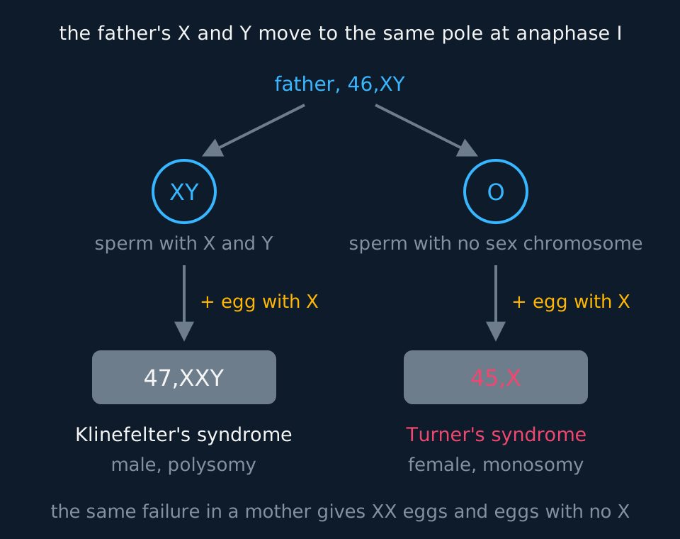
Monosomy is far less often seen at birth, because losing one copy of a chromosome halves the dose of every gene on it. An embryo with a single copy of any autosome almost never survives to be born. The one complete monosomy regularly found in live births is a single X: Turner's syndrome, 45,X. Even so, most 45,X embryos are lost during pregnancy, and about one girl in every 2500 born has the condition.
Describe the drawing
A father with the chromosomes 46,XY at the top. Arrows lead to two kinds of sperm produced when his X and Y fail to separate at anaphase I: one carrying both X and Y, and one, marked O, carrying no sex chromosome. Each is fertilised by a normal egg carrying an X. The first gives a zygote 47,XXY, a male with Klinefelter's syndrome, an example of polysomy. The second gives 45,X, a female with Turner's syndrome, an example of monosomy, shown in coral. A line at the bottom notes that the same failure in a mother gives eggs with two X chromosomes and eggs with none.
A 45,X zygote forms when a gamete with no sex chromosome at all fuses with a normal gamete carrying an X. The gamete with no sex chromosome can be a sperm, when the father's X and Y failed to separate, or an egg, when the mother's two X chromosomes failed to separate. Either division of meiosis can be the one at fault.
The partner gamete from the same non-disjunction has the extra sex chromosome, and if it is fertilised it gives a polysomy instead, such as 47,XXY, which is Klinefelter's syndrome in a male.
A single X is survivable where a single autosome is not because a female cell already works with one active X. In each cell of a 46,XX female one of the two X chromosomes is largely switched off early in development, so a 45,X cell has close to the usual dose of most X-linked gene products.
A small number of genes on the X escape the switching off and are normally active in two copies; having one copy of these is thought to produce the features of the syndrome. SHOX, a gene that affects the growth of the long bones, is the one linked to short stature.
The usual features of Turner's syndrome are short stature and ovaries that do not develop normally, so that most girls with it do not go through puberty without hormone treatment and most are infertile. Some have a narrowing of the aorta or differences in the kidneys. Intelligence is usually within the normal range.
Which parent's meiosis went wrong
A girl with Turner's syndrome, 45,X, has red-green colour blindness, which is caused by a recessive allele on the X chromosome. Her father is colour-blind and her mother is homozygous for normal colour vision. Explain in which parent the non-disjunction happened.
Show the working
The girl has one X and shows the recessive phenotype, so that X carries the colour-blindness allele, Xb.
Her mother is XBXB and has no Xb to give. Her father is XbY, so the girl's X came from the sperm.
The egg therefore carried no sex chromosome at all. The non-disjunction happened in the mother: her two X chromosomes went to the same pole at anaphase I, or two X chromatids did at anaphase II, leaving an egg with neither.
The method generalises. A sex-linked allele that can only have come from one parent identifies which parent supplied the single X, and so which parent's gamete lacked one.
- Non-disjunction
- The failure of a homologous pair to separate at anaphase I, or of sister chromatids to separate at anaphase II, giving gametes with one chromosome too many or too few.
- Polysomy
- The presence of one or more extra copies of a chromosome in a cell, as in trisomy 21.
- Monosomy
- The presence of only one copy of a chromosome where a diploid cell should have a pair, as in 45,X.
- Aneuploidy
- A chromosome number that differs from the normal by less than a whole set.
- Turner's syndrome
- The condition of a female with a single X chromosome and no second sex chromosome, 45,X: a monosomy.
- Down's syndrome
- The condition of a person with three copies of chromosome 21: a trisomy, and so a polysomy.
In the exam
- State which structures separate in each division: homologous chromosomes in meiosis I, sister chromatids in meiosis II.
- Specify that crossing over is between non-sister chromatids of a homologous pair. Exchange between sister chromatids, which carry identical sequence before crossing over, produces no new combination of alleles.
- For the number of gamete combinations, the exponent is the number of homologous pairs. Halve a diploid number before substituting it.
- State that a cell at the end of meiosis I is haploid while its chromosomes are still two chromatids each. Graphs of DNA content against time are read against that statement.
- In a photomicrograph, count the cells, locate the chromosomes, then check whether they are paired. Bivalents two deep on the equator indicate metaphase I; a single line indicates metaphase II.
- Give cytokinesis in a plant cell as a cell plate built from Golgi vesicles rather than a furrow, and give the rigid cell wall as the reason.
- Where a question asks how meiosis produces genetic variation, give crossing over and independent assortment and name the stage of each. Include random fertilisation where the question refers to sexual reproduction.
- Give the chromosome number with the named condition: Down's syndrome is trisomy 21, 47 chromosomes, a polysomy; Turner's syndrome is 45,X, a monosomy.
Check yourself
A mouse has a diploid number of 40. State the number of chromosomes and the number of chromatids in one cell at the end of meiosis I, and in one cell at the end of meiosis II. Then state how many different chromosome combinations its gametes could carry, ignoring crossing over, and explain where that number comes from.
Answer
At the end of meiosis I each cell has 20 chromosomes and 40 chromatids. The homologous pairs have separated, so the chromosome number has halved, but the centromeres have not divided and each chromosome is still two chromatids.
At the end of meiosis II each cell has 20 chromosomes and 20 chromatids. The centromeres have now divided and the sister chromatids have gone to opposite poles, so each is a chromosome in its own right.
A diploid number of 40 means 20 homologous pairs, so n = 20 and the number of combinations is 220 = 1 048 576.
The exponent is 20 rather than 40 because each homologous pair is decided once, at metaphase I, and the pairs are decided independently of one another. Twenty independent two-way choices give 220 outcomes.
Questions
Question 14 marks
Describe how crossing over happens during prophase I of meiosis, and describe its effect on the chromatids involved.
Mark scheme
- B1 each chromosome finds its homologue and the two lie alongside each other along their whole length, forming a bivalent of four chromatids
- B1 non-sister chromatids, one maternal and one paternal, break at the same point and each joins to the other's cut end
- B1 the visible point at which they cross is a chiasma, and equivalent sections of DNA have been exchanged
- A1 the two chromatids that took part leave carrying a combination of alleles that neither parental chromosome had, so sister chromatids are no longer genetically identical; the other two leave unchanged
Question 24 marks
Explain how meiosis produces gametes that are genetically different from one another, naming the stage at which each process happens.
Mark scheme
- B1 crossing over in prophase I exchanges sections of DNA between non-sister chromatids of a homologous pair
- B1 this makes new combinations of alleles along a single chromosome, combinations that were on neither chromosome the cell started with
- B1 independent assortment at metaphase I decides which member of each bivalent faces which pole, and the decision is made separately for every bivalent
- A1 this makes new combinations of whole chromosomes, giving 2 to the power n kinds of gamete where n is the number of homologous pairs
Question 34 marks
A species of plant has a diploid number of 14. Calculate the number of different chromosome combinations its gametes could carry from independent assortment alone, and calculate the number of different chromosome combinations possible in a zygote formed by two such plants.
Mark scheme
- M1 halve the diploid number to get the number of homologous pairs, so n = 7, because one decision is made per pair
- A1 2 to the power 7 is 128 chromosome combinations in a gamete
- M1 a zygote takes one gamete from each parent and the two are independent, so multiply 128 by 128
- A1 16 384 combinations, and crossing over would make the true figure larger still
Question 44 marks
Compare mitosis with meiosis, referring to the number of divisions, the chromosome number of the daughter cells, the behaviour of homologous chromosomes and the genetic result.
Mark scheme
- B1 mitosis is one division producing two daughter cells, whereas meiosis is two divisions producing four
- B1 mitosis leaves the chromosome number unchanged, diploid to diploid, whereas meiosis halves it, diploid to haploid
- B1 homologous chromosomes do not pair in mitosis, whereas in meiosis they pair as bivalents in prophase I and cross over
- B1 the daughters of mitosis are genetically identical to the parent cell, whereas the four products of meiosis all differ from one another
Question 54 marks
Describe what happens to the spindle and to the nuclear envelope during prophase I and during telophase II of meiosis.
Mark scheme
- B1 in prophase I the nuclear envelope breaks up into small vesicles and the nucleolus disappears
- B1 a spindle of microtubules is built between the two poles of the cell, growing from a pair of centrioles at each pole in an animal cell and from a region without centrioles in most plant cells
- B1 in telophase II the spindles break down
- A1 a nuclear envelope re-forms around each of the four groups of chromosomes, the chromosomes uncoil and the nucleoli reappear
Question 64 marks
A cell of a species with a diploid number of 20 is about to begin meiosis, immediately after DNA replication. Calculate the number of chromosomes and the number of chromatids present at the start of meiosis, at the end of meiosis I, and at the end of meiosis II.
Mark scheme
- M1 a diploid number of 20 means the cell, after replication, has 20 chromosomes and 40 chromatids at the start of meiosis (each of the 20 chromosomes now two chromatids)
- A1 at the end of meiosis I: 10 chromosomes, 20 chromatids, since homologues have separated but the centromeres have not divided
- M1 chromosome number does not change again at the second division: the centromeres divide so each chromatid becomes a chromosome, and the cell then divides, so the chromatid count per cell halves
- A1 at the end of meiosis II: 10 chromosomes, 10 chromatids in each of the four cells
Question 74 marks
In a species with a diploid number of 16, non-disjunction of one homologous pair occurs at anaphase I of meiosis in one cell, and the rest of the division is normal. Describe the chromosome number you would expect to find in each of the four cells that result from this meiosis.
Mark scheme
- B1 the normal haploid number for this species is 8
- B1 because non-disjunction happened at anaphase I, both homologues of the affected pair went to the same pole, so one of the two cells produced by meiosis I has one extra chromosome, 9, and the other has one too few, 7
- B1 meiosis II proceeds normally on each of these two cells, simply separating the sister chromatids of each without changing the chromosome number further
- B1 the four final cells therefore have chromosome numbers 9, 9, 7 and 7, because non-disjunction at anaphase I affects all four products, unlike non-disjunction at anaphase II, which would affect only two of the four
Question 84 marks
A person with Turner's syndrome has 45 chromosomes, with a single X chromosome and no other sex chromosome. Explain how non-disjunction during meiosis in the father can produce a child with Turner's syndrome, and explain why the child is female.
Mark scheme
- B1 the father's X and Y chromosomes fail to separate at anaphase I, or the sister chromatids of a sex chromosome fail to separate at anaphase II
- B1 so a sperm is produced with no sex chromosome, carrying 22 chromosomes instead of 23
- B1 it fertilises a normal egg carrying 22 autosomes and an X, giving a zygote with 45 chromosomes and one X: a monosomy
- B1 the zygote has no Y chromosome, so it lacks the gene on the Y that switches development onto the male route, and develops as a female
Question 94 marks
State what is meant by polysomy and by monosomy, and state the chromosome change found in Down's syndrome and in Turner's syndrome.
Mark scheme
- B1 polysomy is the presence of one or more extra copies of a chromosome in a cell
- B1 Down's syndrome is a polysomy: three copies of chromosome 21 (trisomy 21), 47 chromosomes in all
- B1 monosomy is the presence of only one copy of a chromosome where there should be a pair
- B1 Turner's syndrome is a monosomy: a single X chromosome and no second sex chromosome, 45,X
Question 103 marks
A chemical prevents chiasmata from forming in prophase I but allows the rest of meiosis to run normally. Suggest what this does to the genetic variation of the gametes produced, and suggest why they would still not all be the same.
Mark scheme
- B1 no crossing over would occur, so no new combinations of alleles would be made along a chromosome and each chromatid would carry the alleles of the parental chromosome it came from
- B1 sister chromatids would therefore stay genetically identical, so the two cells produced by each meiosis II would be identical to each other
- A1 independent assortment at metaphase I would still combine whole chromosomes in new ways, giving 2 to the power n kinds of gamete, so variation would be reduced rather than abolished
Question 113 marks
Compare the way the cytoplasm divides at the end of meiosis in an animal cell with the way it divides in a plant cell.
Mark scheme
- B1 in an animal cell a ring of protein filaments just inside the cell surface membrane contracts and draws the membrane inwards as a cleavage furrow, which deepens until the cell is pinched in two
- B1 in a plant cell vesicles from the Golgi apparatus line up along the old equator and fuse into a cell plate, which becomes the new cell surface membranes and the new cell wall between the daughter cells
- A1 the difference exists because the rigid cell wall of a plant cell cannot be pinched inwards
Question 123 marks
The drawing shows a single cell in which the chromosomes lie across the middle of the cell in homologous pairs, with no nuclear envelope and with spindle fibres running to each end. Identify the stage shown, and give two features of the drawing that rule out the other stages.
Mark scheme
- B1 the stage is metaphase I
- B1 the chromosomes are on the equator in pairs, two chromosomes deep, which happens only in the first division; at metaphase II they would lie in a single line
- A1 there is a single cell rather than two, and the chromosomes are on the equator rather than strung out towards the poles, which rules out the second division and rules out anaphase
Question 133 marks
Before crossing over takes place, the two sister chromatids of one chromosome are genetically identical. Suggest why, once crossing over has taken place in a bivalent, this is no longer necessarily true, and suggest what would be different about the cells produced by meiosis II from a bivalent in which no crossing over occurred at all.
Mark scheme
- B1 crossing over exchanges a section of DNA between one chromatid of the maternal chromosome and one non-sister chromatid of the paternal chromosome, so those two participating chromatids each carry a new combination of maternal and paternal alleles and are no longer identical to their own sister chromatid
- B1 where no crossing over takes place in a particular bivalent, each chromatid remains identical to its sister, carrying only the alleles of the parental chromosome it came from
- B1 the two cells produced by meiosis II from that bivalent would therefore be genetically identical to each other at those loci, whereas if crossing over had occurred the two meiosis II products from that cell could differ from each other at those loci
Question 143 marks
Haemophilia A is caused by a recessive allele on the X chromosome. A girl has a single X chromosome and no other sex chromosome, and she does not have haemophilia. Her father has haemophilia and her mother is a carrier. Explain in which parent non-disjunction occurred.
Mark scheme
- B1 the girl is unaffected with only one X, so her X carries the dominant allele, XH
- B1 her father is XhY and has no XH to give, so her X came from her mother's egg
- B1 the sperm therefore carried no sex chromosome, so non-disjunction occurred in the father, his X and Y failing to separate at anaphase I or a pair of sister chromatids failing to separate at anaphase II
Question 152 marks
State the stage of meiosis at which homologous chromosomes are separated, and state the stage at which sister chromatids are separated.
Mark scheme
- B1 homologous chromosomes are separated at anaphase I
- B1 sister chromatids are separated at anaphase II, when the centromeres divide
Question 162 marks
State what is meant by a bivalent, and state how many chromatids it contains.
Mark scheme
- B1 a bivalent is the structure formed when a homologous pair of chromosomes pairs up (synapsis) in prophase I of meiosis
- B1 a bivalent contains four chromatids, two from each of the two homologous chromosomes, because both chromosomes were already replicated before pairing
Question 172 marks
In a dividing cell, two separate daughter cells each contain single chromatids being pulled to opposite poles, with the number moving to each pole equal to the haploid number of the species. Identify the stage described in each cell, and identify one feature of that description which shows this is the second division rather than the first.
Mark scheme
- B1 the stage is anaphase II
- B1 there are two separate cells, each already a product of meiosis I, rather than one, and what is moving to each pole is a single chromatid becoming its own chromosome rather than a whole chromosome of two chromatids, both of which occur only in the second division
Question 182 marks
State when DNA replication occurs relative to the two divisions of meiosis, and state the chromosome number and chromatid number of a cell immediately after that replication in a species with a diploid number of 24.
Mark scheme
- B1 DNA replication occurs once, during the interphase before meiosis begins, and does not happen again between meiosis I and meiosis II
- B1 immediately after that replication such a cell has 24 chromosomes and 48 chromatids, since each of the 24 chromosomes now consists of two sister chromatids
Worth remembering
- One replication, two divisions, four haploid cells.
- Meiosis I separates homologues and halves the chromosome number; meiosis II separates chromatids and does not.
- Crossing over happens in prophase I, between non-sister chromatids, and makes sister chromatids no longer identical.
- Independent assortment happens at metaphase I and gives 2 to the power n combinations, where n is the number of pairs.
- Non-disjunction at anaphase I affects all four products; at anaphase II it affects two.
- Eight stages: know what the spindle, the nuclear envelope and the cell surface membrane are doing in each.
- An n + 1 gamete gives polysomy (trisomy 21, Down's syndrome); an n − 1 gamete gives monosomy (45,X, Turner's syndrome).
CHECK YOUR PROGRESS
Rate how confident you are with each objective for this lesson. Ratings are saved in this browser, on this device, unless you sign in.
- Say which chromosomes separate in the first division and which in the second, and why that order matters.
- Describe how a bivalent forms and what crossing over does to the chromatids in it.
- Work out how many chromosome combinations a gamete could carry, given the haploid number.
- Track chromosome number and chromatid number through both divisions without losing count.
- Set out the differences between mitosis and meiosis, division by division.
- Name all eight stages in order and say what the spindle, the nuclear envelope and the cell surface membrane are doing in each.
- Name the stage of meiosis that a photomicrograph shows, and justify the naming.
- Explain how non-disjunction leads to polysomy, as in Down's syndrome, and to monosomy, as in Turner's syndrome.
Open the full revision checklist to see every objective in the curriculum in one place.
Practise this lesson
WORKBOOK
The written questions from this topic, on paper with room to work, and a separate book of mark schemes. The question that is set on a diagram stays on this page, where the diagram can be drawn. Free to use; please do not redistribute or sell.
Used later in Gametes, fertilisation and early development · Sexual reproduction in flowering plants · Natural selection: variation, selection and allele frequency · Speciation and reproductive isolation