Chemistry › Organic analysis › Proton and carbon-13 NMR spectroscopy
Proton and carbon-13 NMR spectroscopy
In a strong magnetic field, hydrogen-1 and carbon-13 nuclei absorb radio waves at a frequency set by the electrons around them, so each chemical environment in a molecule gives its own peak. Carbon-13 NMR counts the carbon environments; proton NMR adds how many hydrogens are in each environment and how many sit next door.
Key terms Every definition in organic analysis, lesson by lesson
Before this Infrared spectroscopy – functional groups from absorptions · Structural isomerism and E/Z isomerism – drawing the structural isomers of a formula · Electronegativity, polarity and intermolecular forces – electronegative atoms drawing electron density
COMMON MISCONCEPTION
The number of peaks in a proton NMR spectrum is the number of hydrogen atoms in the molecule.
Each peak is one environment, not one atom. Ethanol has six hydrogens in three environments, CH₃, CH₂ and OH, so three peaks, and the area under each is in proportion to the number of hydrogens in it, 3 : 2 : 1. Splitting then adds detail: the CH₃ peak is a triplet because of the two neighbouring CH₂ hydrogens.
What you should be able to do
- Explain why TMS is used as the standard and why the solvent contains no hydrogen-1 atoms.
- Deduce the number of carbon environments in a molecule and assign carbon-13 shifts with the data sheet.
- Use the chemical shift, the integration ratio and the splitting of each peak in a proton NMR spectrum to describe the hydrogen environments.
- Apply the n + 1 rule to predict the splitting of a peak from the number of hydrogens on the neighbouring carbons.
- Deduce the structure of a compound from its molecular formula and its NMR spectra.
Nuclei in a magnet
A hospital MRI scanner places a patient inside a magnet tens of thousands of times stronger than the Earth's field and pulses radio waves through them. The hydrogen nuclei in water and fat absorb those radio waves, and the scanner maps where. An NMR spectrometer in a chemistry laboratory does the same to a few milligrams of a compound in a narrow glass tube, and asks a different question: not where the hydrogen nuclei are, but what each one is bonded to.
Nuclei with an odd number of nucleons, such as ¹H and ¹³C, have a property called spin and behave like tiny magnets. In a strong external field each one lines up either with the field or against it, and the two arrangements differ slightly in energy. Radio waves of exactly the right frequency flip a nucleus from the lower energy state to the higher one, and that absorption is what is recorded.
The gap between the two states is tiny, so the radiation that matches it is in the radio-frequency range, hundreds of megahertz, and a stronger magnet gives a larger gap and a higher frequency: a '400 MHz' spectrometer is one whose magnet makes hydrogen nuclei absorb near 400 MHz. Carbon-12, with six protons and six neutrons, has an even number of nucleons and no spin, so it gives no signal at all.
The frequency is not the same for every hydrogen in a molecule. Electrons around a nucleus set up a small field of their own that opposes the applied one, shielding the nucleus. An electronegative atom nearby, such as oxygen, draws electron density away, so a hydrogen on a carbon bonded to oxygen is less shielded and absorbs at a higher shift. Each different chemical environment gives its own peak.
- Chemical environment
- A set of atoms in a molecule that are bonded to identical surroundings, so they absorb at the same chemical shift.
- Chemical shift
- The position of an NMR absorption relative to the TMS peak, δ, measured in parts per million (ppm).
- TMS
- Tetramethylsilane, Si(CH₃)₄, the reference compound whose single peak is defined as δ = 0.
- Nuclear spin
- The property of some nuclei, including every nucleus with an odd number of nucleons such as ¹H or ¹³C, that lets them align with or against an applied magnetic field.
- Shielding
- The reduction in the magnetic field felt by a nucleus caused by the electrons around it; a less shielded nucleus absorbs at a higher chemical shift.
Shifts are measured from tetramethylsilane, TMS. Its twelve hydrogens are all equivalent, as are its four carbons, so it gives one sharp peak in each kind of spectrum. Silicon is less electronegative than carbon, so those nuclei are more shielded than in almost any organic compound and the TMS peak sits to the right of everything else. TMS is also inert, non-toxic and volatile, boiling at 27 °C, so it is easily removed from the sample afterwards.
The sample is dissolved in a solvent with no ¹H atoms, so that the solvent gives no peaks of its own in the proton spectrum, which would swamp the sample's: the solvent molecules outnumber the sample's by hundreds to one. Deuterium, ²H, has a nucleus with spin, but it absorbs at a frequency far from that of ¹H, so a deuterated solvent is invisible in a proton spectrum. Each choice has its reason.
| Substance | Role | Why it is used |
|---|---|---|
| TMS, Si(CH₃)₄ | Reference, added in a trace | One sharp peak, set at δ = 0, to the right of almost everything; inert and volatile |
| CDCl₃ | The usual solvent | No ¹H atoms; dissolves most organic compounds; its carbon gives a known peak at 77 ppm in carbon-13 spectra |
| CCl₄ | Solvent | No hydrogen at all; dissolves non-polar compounds, but few polar ones, and it is toxic |
| D₂O | Solvent for polar compounds | No ¹H atoms; dissolves sugars, amino acids and salts |
Say 'no ¹H atoms' or 'no hydrogen-1' for the solvent, not 'no hydrogen': CDCl₃ contains deuterium, which is hydrogen.
Carbon-13 NMR: counting carbon environments
About 1.1% of carbon atoms are carbon-13, and that is enough for a spectrum. Each carbon environment gives one peak, and the spectra met at A level show no splitting. Peak heights are not proportional to the number of carbons, so a carbon-13 spectrum is read for two things only: how many peaks, and where.
The lack of splitting follows from that 1.1%. Two carbon-13 atoms are almost never bonded to each other in the same molecule, so one carbon peak is not split by another carbon, and the splitting that attached hydrogens would cause is switched off by the instrument as the spectrum is recorded.
Count environments by looking for symmetry. In propan-2-ol, CH₃CH(OH)CH₃, the two methyl carbons are bonded to identical surroundings and give one peak; the carbon bearing the OH gives a second. Three carbons, two peaks.
The test for equivalence is whether swapping two carbons, by rotating or reflecting the molecule, gives the same molecule. Propanone has two methyl carbons that swap in this way, so it has two peaks, one at about 30 ppm and the C=O at about 205 ppm. Propan-1-ol, CH₃CH₂CH₂OH, has three carbons with three different sets of neighbours and gives three peaks. Benzene, with six identical carbons in a ring, gives just one.
Counting peaks separates isomers that every other quick test confuses. Pentan-2-one, CH₃COCH₂CH₂CH₃, and pentan-3-one, CH₃CH₂COCH₂CH₃, are both C₅H₁₀O ketones with a C=O near 210 ppm. Pentan-2-one has no symmetry and gives five peaks. Pentan-3-one is symmetrical about its C=O: its two CH₃ carbons are equivalent, and so are its two CH₂ carbons, and it gives three peaks.
| Type of carbon | δ / ppm |
|---|---|
| C–C (alkyl) | 5–40 |
| C next to C=O | 20–50 |
| C–O (alcohols, ethers, esters) | 50–90 |
| C=C (alkenes) | 90–150 |
| Aromatic carbons | 110–160 |
| C=O (esters, acids) | 160–185 |
| C=O (aldehydes, ketones) | 190–220 |
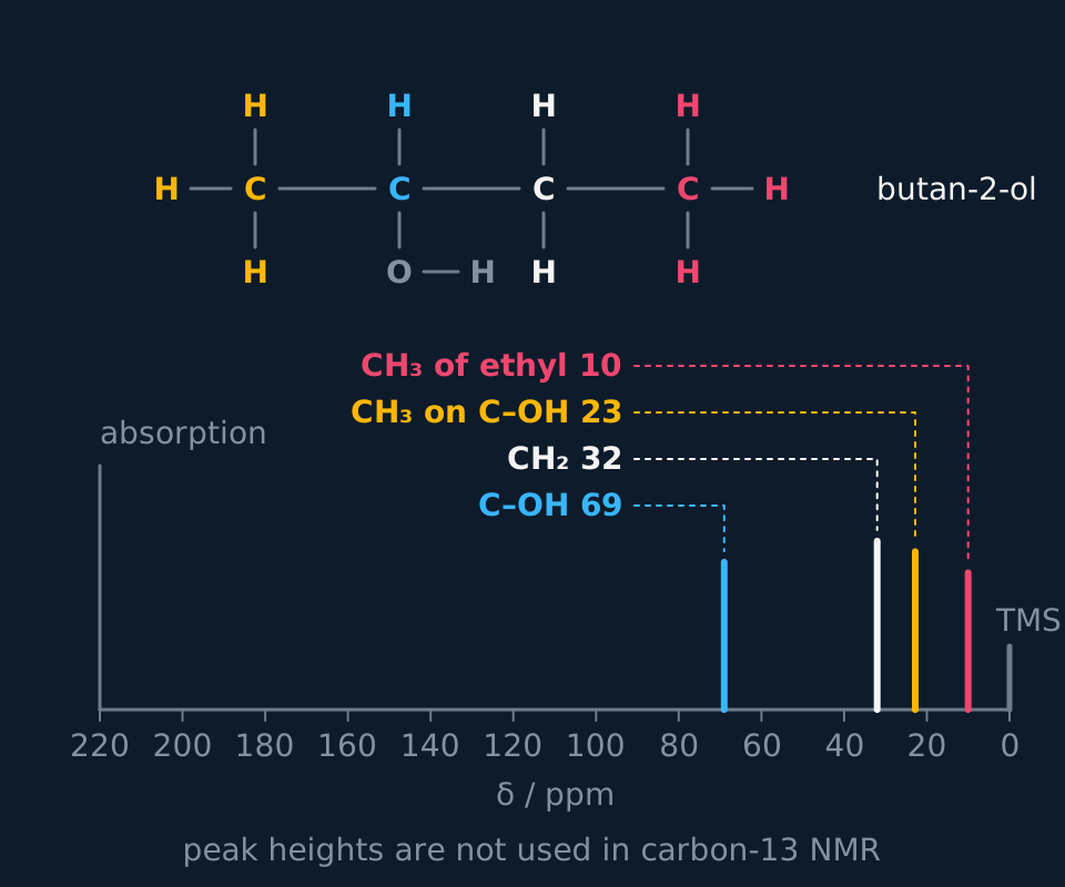
Describe the drawing
A carbon-13 NMR spectrum of butan-2-ol with δ from 220 on the left to 0 on the right: four single lines and a small TMS line at 0. The carbon bonded to oxygen is at 69, the CH₂ at 32, the CH₃ on the C–OH carbon at 23 and the CH₃ of the ethyl group at 10. Above, the displayed formula colours each carbon to match its line, and a note says peak heights are not used in carbon-13 NMR.
Carbon environments in three isomers of C₄H₁₀O
Give the number of peaks in the carbon-13 spectrum of butan-1-ol, 2-methylpropan-1-ol and 2-methylpropan-2-ol, and say which peak in each lies between 50 and 90 ppm.
Show the working
Butan-1-ol, CH₃CH₂CH₂CH₂OH, has no symmetry: every carbon has different neighbours, so there are 4 peaks. The CH₂ bonded to OH absorbs near 62 ppm.
2-Methylpropan-1-ol, (CH₃)₂CHCH₂OH, has two methyl groups on the same CH. They are equivalent and share a peak, so there are 3 peaks. The CH₂OH carbon absorbs near 70 ppm.
2-Methylpropan-2-ol, (CH₃)₃COH, has three equivalent methyls on one carbon, so there are 2 peaks: the methyls near 31 ppm and the carbon bonded to OH near 69 ppm.
So the three isomers give 4, 3 and 2 peaks, and a carbon-13 spectrum alone tells them apart.
Proton NMR: shift and integration
A proton NMR spectrum gives more than a carbon-13 spectrum. Each hydrogen environment gives a peak, and the peaks carry three pieces of information: the shift, the area and the splitting.
| Carbon-13 NMR | Proton NMR | |
|---|---|---|
| Nucleus observed | ¹³C, 1.1% of carbon atoms | ¹H, almost all hydrogen atoms |
| Shift range | 0 to about 220 ppm | 0 to about 12 ppm |
| Number of peaks | Number of carbon environments | Number of hydrogen environments |
| Peak area | Not used | Ratio of hydrogens in each environment |
| Splitting | None in A-level spectra | n + 1 lines from n hydrogens on adjacent carbons |
Hydrogens are counted by environment, not one by one. Propan-2-ol, (CH₃)₂CHOH, has eight hydrogens but only three environments: the six hydrogens of the two equivalent methyl groups, the one on the central carbon, and the one on the oxygen. Its spectrum has three peaks. Propan-1-ol, CH₃CH₂CH₂OH, with the same formula, has four environments and four peaks, which is enough to tell the two isomers apart before any shift is read.
The shift says what the hydrogens are near. The data sheet gives ranges: R–CH₃ at 0.7–1.2 ppm, H–C–C=O at 2.1–2.6, H–C–O at 3.1–3.9, RCOO–C–H at 3.7–4.1, an aldehyde CHO at 9.0–10.0 and a carboxylic acid COOH at 10.0–12.0. An O–H in an alcohol can appear anywhere from 0.5 to 5.0 ppm, depending on concentration and solvent.
The area under each peak is proportional to the number of hydrogens in that environment. The spectrometer draws an integration trace that steps up at each peak; the heights of the steps give the ratio. In ethanol the steps are in the ratio 3:2:1, for CH₃, CH₂ and OH.
- Integration ratio
- The ratio of the areas under the peaks in a proton NMR spectrum, equal to the ratio of the numbers of hydrogens in each environment.
Splitting and the n + 1 rule
Under high resolution most peaks are split into a cluster of lines. The splitting comes from the hydrogens on the neighbouring carbon atoms, whose small magnetic fields add to or subtract from the field the observed hydrogens feel. A hydrogen environment with n hydrogens on the adjacent carbons gives a peak split into n + 1 lines. This is the n + 1 rule, and n counts the hydrogens on every adjacent carbon together, on both sides.
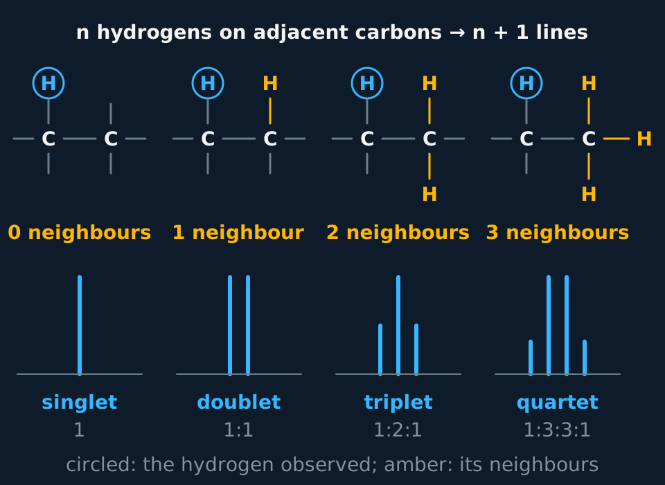
Describe the drawing
Four columns, each showing two bonded carbons with one hydrogen circled on the left carbon and zero, one, two or three amber hydrogens on the carbon next door. Beneath each, the peak for the circled hydrogen: a singlet of one line, a doublet of two equal lines, a triplet in the ratio 1:2:1, and a quartet in the ratio 1:3:3:1. A banner reads n hydrogens on adjacent carbons give n + 1 lines.
| Neighbouring H | Lines | Name |
|---|---|---|
| 0 | 1 | Singlet |
| 1 | 2 | Doublet |
| 2 | 3 | Triplet |
| 3 | 4 | Quartet |
The lines in a cluster are not of equal height. Each neighbouring hydrogen is aligned either with or against the field, and the lines record how many ways each total can arise. One neighbour gives a doublet of 1:1; two give a triplet of 1:2:1, since the middle line can arise in two ways; three give a quartet of 1:3:3:1. A symmetrical cluster with the tallest lines in the middle is the sign of a genuine multiplet.
Two rules stop the n + 1 rule giving wrong answers. Equivalent hydrogens do not split each other, so the three hydrogens of a CH₃ group count only as neighbours to something else. And the hydrogen of an O–H group usually exchanges rapidly between molecules, so it neither splits nor is split: an alcohol O–H is normally a singlet.
Splitting reaches only to the next carbon. A CH₃ bonded to a C=O carbon, which carries no hydrogens, is a singlet whatever lies beyond it, and so is a CH₃ bonded to an ester oxygen. In propan-2-ol the six methyl hydrogens have one neighbour, the CH, and give a doublet of area 6. The CH has six neighbours and is split into seven lines. A doublet of area 6 beside a small multiplet of area 1 is the sign of a (CH₃)₂CH group.
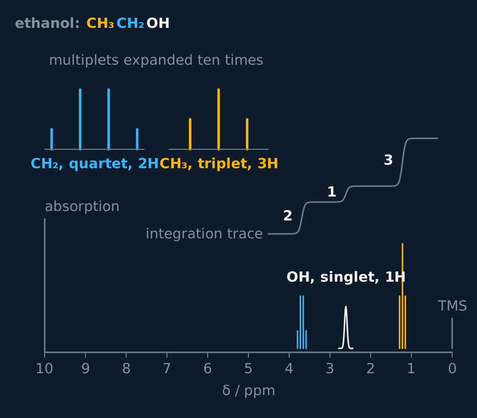
Describe the drawing
A proton NMR spectrum of ethanol with δ from 10 on the left to 0 on the right. A cyan quartet of four close lines at 3.69 is the CH₂, a single slightly broadened line at 2.61 is the OH, and an amber triplet of three lines at 1.22 is the CH₃, with TMS at 0. A stepped integration trace rises by 2, then 1, then 3 across the three peaks, and expanded insets show the quartet in the ratio 1:3:3:1 and the triplet 1:2:1.
The proton NMR spectrum of ethanol
Describe and explain the proton NMR spectrum of ethanol, CH₃CH₂OH.
Show the working
There are three hydrogen environments, CH₃, CH₂ and OH, so there are three peaks, with areas in the ratio 3:2:1.
The CH₃ is at about 1.2 ppm, in the R–CH₃ range. Its neighbour is the CH₂ with 2 hydrogens, so it is split into 2 + 1 = 3: a triplet.
The CH₂ is at about 3.7 ppm, in the H–C–O range, because it is bonded to the oxygen. Its neighbour is the CH₃ with 3 hydrogens, so it is split into 3 + 1 = 4: a quartet. The OH hydrogen is not counted, because it exchanges too fast to split anything.
The OH is a singlet near 2.6 ppm, its exact position depending on concentration.
Assigning the spectrum of propan-1-ol
The proton NMR spectrum of propan-1-ol, CH₃CH₂CH₂OH, has four peaks: δ 3.6, triplet, 2H; δ 2.3, singlet, 1H; δ 1.6, sextet, 2H; δ 0.9, triplet, 3H. Assign each peak to a group of hydrogens.
Show the working
There are four environments, CH₃, the middle CH₂, the CH₂ bonded to O and the OH, and the integration, 2 : 1 : 2 : 3, adds to the eight hydrogens of C₃H₈O.
The triplet of 2H at 3.6 ppm is in the H–C–O range: the CH₂ bonded to oxygen. Its only neighbouring hydrogens are the two on the middle carbon, because the OH does not split it: 2 + 1 = 3 lines.
The sextet of 2H at 1.6 ppm is the middle CH₂. It has three hydrogens on one side and two on the other, n = 5, so 6 lines.
The triplet of 3H at 0.9 ppm is the CH₃, split by the two hydrogens of the middle CH₂. The singlet of 1H at 2.3 ppm is the O–H, which neither splits nor is split.
- Spin-spin splitting
- The splitting of an NMR peak into lines by the hydrogens on adjacent carbon atoms, n neighbouring hydrogens giving n + 1 lines.
- Deuterated solvent
- A solvent such as CDCl₃ in which ¹H atoms are replaced by deuterium, so that it gives no peaks in a proton NMR spectrum.
From formula and spectrum to structure
A structure question gives a molecular formula and one or two spectra. Work in a fixed order. Count the peaks, which gives the environments. Turn the integration ratio into actual numbers of hydrogens, using the formula. Read each splitting as a statement about the neighbouring carbon. Use the shifts to place each group next to O, next to C=O or in a chain. Then draw a structure and check it against every piece of data.
Ethyl ethanoate from its spectra
Compound Q, C₄H₈O₂, has these proton NMR peaks: δ 4.1, quartet, 2H; δ 2.1, singlet, 3H; δ 1.2, triplet, 3H. Its carbon-13 spectrum has peaks at 171, 60, 21 and 14 ppm. Deduce its structure.
Show the working
The integration totals 2 + 3 + 3 = 8, matching the 8 hydrogens of C₄H₈O₂. The carbon-13 peak at 171 ppm is a C=O of an ester or acid; with no peak between 10 and 12 ppm in the proton spectrum there is no COOH, so Q is an ester.
The quartet (2H) and the triplet (3H) together are an ethyl group, CH₃CH₂: the CH₂ is split by 3 neighbours and the CH₃ by 2.
The CH₂ is at 4.1 ppm, in the RCOO–C–H range, so it is bonded to the ester oxygen; its carbon is the 60 ppm peak. The singlet (3H) at 2.1 ppm is a CH₃ next to the C=O with no hydrogens on its neighbour.
Q is CH₃COOCH₂CH₃, ethyl ethanoate. Its isomer methyl propanoate, CH₃CH₂COOCH₃, would put the quartet near 2.3 ppm, next to the C=O, and the singlet near 3.7 ppm, next to O.
A ketone with a branched chain
Compound U, C₅H₁₀O, has these proton NMR peaks: δ 2.6, septet; δ 2.1, singlet; δ 1.1, doublet. The integration trace steps are 0.6 cm, 1.8 cm and 3.6 cm in the same order. Its carbon-13 spectrum has four peaks, one of them at 212 ppm. Deduce its structure.
Show the working
The steps are in the ratio 0.6 : 1.8 : 3.6 = 1 : 3 : 6, a total of 10 parts, matching the ten hydrogens of C₅H₁₀O. The environments hold 1, 3 and 6 hydrogens.
The carbon-13 peak at 212 ppm is in the 190–220 range of an aldehyde or ketone C=O. There is no proton peak at 9–10 ppm, so there is no CHO and U is a ketone.
The doublet of 6H and the septet of 1H are a (CH₃)₂CH group: six equivalent hydrogens split by one neighbour, and one hydrogen split by six. The CH at 2.6 ppm is in the H–C–C=O range, so it is bonded to the carbonyl carbon.
The singlet of 3H at 2.1 ppm is a CH₃ on the other side of the C=O, with no hydrogens on its neighbour.
U is (CH₃)₂CHCOCH₃, 3-methylbutan-2-one. Its two methyls on the CH are equivalent, so it has four carbon environments, matching the four carbon-13 peaks.
TRY IT: An acid or an ester
A compound C₃H₆O₂ gives three proton NMR peaks: δ 11.7, singlet, 1H; δ 2.4, quartet, 2H; δ 1.2, triplet, 3H. Deduce its structure.
Check your answer
The peak at 11.7 ppm is in the 10.0–12.0 range of a carboxylic acid COOH, and is a singlet with 1H.
A quartet of 2H and a triplet of 3H are an ethyl group, and the CH₂ at 2.4 ppm is in the H–C–C=O range, so it is bonded to the carbonyl carbon.
The compound is CH₃CH₂COOH, propanoic acid. Its isomers methyl ethanoate and ethyl methanoate have no peak above 10 ppm.
Other boards: D₂O exchange
OCR and Cambridge add a way of proving which peak belongs to an O–H or N–H hydrogen. The sample is shaken with a few drops of D₂O and the spectrum run again. The O–H hydrogen exchanges with deuterium to give O–D, which does not absorb in a proton spectrum, so the O–H peak disappears; an N–H peak behaves the same way, and the C–H peaks are unchanged. In propan-1-ol the singlet at 2.3 ppm goes and the other three peaks stay, which fixes it as the O–H.
CH₃CH₂CH₂OH(l) + D₂O(l) ⇌ CH₃CH₂CH₂OD(l) + HDO(l)The O–H hydrogen exchanges; the C–H hydrogens do not.
In the exam
- TMS questions award a mark each for a single peak, a position to the right of almost every other compound, and being inert and volatile. 'TMS is a standard' repeats the question and earns no mark.
- For each proton NMR peak, state the shift, the integration and the splitting, and say what the splitting means: 'quartet, so three H on the adjacent carbon'. A splitting named without that inference is only half the point.
- The n in n + 1 is the number of hydrogens on the adjacent carbons, not on the carbon itself.
- In a structure question, the scheme gives credit for showing that the integration adds up to the number of hydrogens in the formula, and for a structure checked against every peak.
- For a deuterated solvent, schemes accept 'no ¹H' or 'no hydrogen-1 atoms' and reject 'no hydrogen'.
Check yourself
An isomer of C₄H₁₀O gives a proton NMR spectrum with only two peaks: δ 1.2, singlet, 9H, and δ 2.0, singlet, 1H. Its infrared spectrum has a broad absorption at 3230–3550 cm⁻¹ and none at 1680–1750 cm⁻¹. Explain what the compound is.
Answer
The broad infrared absorption is an alcohol O–H, and the 1H singlet at 2.0 ppm, inside the 0.5–5.0 range, is that O–H hydrogen. It accounts for 1 of the 10 hydrogens.
The other 9 hydrogens are in one environment and give a singlet, so they are three equivalent CH₃ groups with no hydrogens on the carbon they share. Three CH₃ groups on one carbon is (CH₃)₃C.
(CH₃)₃C– with OH is (CH₃)₃COH, 2-methylpropan-2-ol. It accounts for all four carbons, ten hydrogens and one oxygen, and its carbon-13 spectrum would show 2 peaks.
Questions
Question 16 marks
Explain how the NMR data in the table show that compound W, C₄H₈O₂, is methyl propanoate and not its isomer ethyl ethanoate.
| Spectrum | δ / ppm | Splitting | Integration |
|---|---|---|---|
| Proton | 3.7 | Singlet | 3 |
| Proton | 2.3 | Quartet | 2 |
| Proton | 1.1 | Triplet | 3 |
| Carbon-13 | 175, 52, 27, 9 | not split | not used |
Mark scheme
Indicative content
- the carbon-13 peak at 175 ppm is the C=O of an ester or acid, and with no proton peak at 10–12 ppm there is no COOH, so W is an ester
- the integration 3 + 2 + 3 = 8 accounts for all eight hydrogens of C₄H₈O₂, and four carbon-13 peaks for four carbon environments
- the quartet of 2H and the triplet of 3H show an ethyl group, the CH₂ split by three neighbours and the CH₃ by two
- the quartet at 2.3 ppm is in the H–C–C=O range, so the CH₂ of the ethyl group is bonded to the carbonyl carbon
- the singlet of 3H at 3.7 ppm is in the RCOO–C–H range, a CH₃ bonded to the ester oxygen with no neighbouring hydrogens
- in ethyl ethanoate the quartet would be near 4.1 ppm, next to O, and the singlet near 2.1 ppm, next to C=O, so the data fit only methyl propanoate, CH₃CH₂COOCH₃
Levels
- Level 3 (5–6 marks) the ester is identified from the carbon-13 and the absence of an acid proton, every proton signal is assigned with its splitting and its shift range, the integration is checked against the formula, and the answer shows where the signals of ethyl ethanoate would differ; the structure follows from the data rather than being asserted
- Level 2 (3–4 marks) the quartet and triplet are read as an ethyl group and the singlet as OCH₃; the shift ranges are not used to place the groups either side of the ester link, or ethyl ethanoate is never compared
- Level 1 (1–2 marks) a quartet and a triplet are said to show an ethyl group and the compound is then named ethyl ethanoate, or it is named correctly with no data behind it
Question 24 marks
Deduce the number of peaks in the carbon-13 NMR spectrum of each of the four isomers of C₃H₆Cl₂: 1,1-dichloropropane, 1,2-dichloropropane, 1,3-dichloropropane and 2,2-dichloropropane.
Mark scheme
- B1 1,1-dichloropropane, CHCl₂CH₂CH₃: three different carbons, 3 peaks
- B1 1,2-dichloropropane, CH₂ClCHClCH₃: three different carbons, 3 peaks
- B1 1,3-dichloropropane, ClCH₂CH₂CH₂Cl: the two CH₂Cl carbons are equivalent, 2 peaks
- B1 2,2-dichloropropane, CH₃CCl₂CH₃: the two CH₃ carbons are equivalent, 2 peaks
Question 34 marks
Explain why a sample for proton NMR is dissolved in CDCl₃ rather than CHCl₃, why CCl₄ is sometimes used instead, and why D₂O is chosen for a sample of glucose.
Mark scheme
- B1 CDCl₃ has deuterium, ²H, in place of ¹H, and deuterium does not absorb at the frequency of ¹H
- B1 so the solvent, present in large excess, gives no peak that could swamp or be confused with the sample's peaks
- B1 CCl₄ contains no hydrogen at all, so it gives no proton peak, though it dissolves only non-polar compounds well
- B1 glucose is polar and dissolves in D₂O, which has no ¹H, where it would barely dissolve in CDCl₃ or CCl₄
All 21 questions with mark schemes, the other 18 set out in full on the question page.
Worth remembering
- Counting hydrogens instead of environments gives too many peaks.
- Read only the number of peaks and their shifts from a carbon-13 spectrum; its peak heights are not a ratio.
- An integration ratio not scaled to the formula gives the wrong number of hydrogens.
- Count the hydrogens on the neighbouring carbons for n + 1, not those on the carbon itself.
- An O–H counted as a neighbour gives a splitting the spectrum does not show.
CHECK YOUR PROGRESS
Rate how confident you are with each objective for this lesson. Ratings are saved in this browser, on this device, unless you sign in.
- Explain why TMS is used as the standard and why the solvent contains no hydrogen-1 atoms.
- Deduce the number of carbon environments in a molecule and assign carbon-13 shifts with the data sheet.
- Use the chemical shift, the integration ratio and the splitting of each peak in a proton NMR spectrum to describe the hydrogen environments.
- Apply the n + 1 rule to predict the splitting of a peak from the number of hydrogens on the neighbouring carbons.
- Deduce the structure of a compound from its molecular formula and its NMR spectra.
Open the full revision checklist to see every objective in the curriculum in one place.
Practise this lesson
WORKBOOK
The written questions from this topic, on paper with room to work, and a separate book of mark schemes. The 5 questions that are set on a diagram stay on this page, where the diagram can be drawn. Free to use; please do not redistribute or sell.
Used later in Putting the analysis together: structure from spectra