Chemistry › Transition metals › Colour in transition metal complexes and colorimetry
Colour in transition metal complexes and colorimetry
Copper(II) sulfate solution is blue because it absorbs orange and red light, not because it gives out blue. The energy it absorbs is the gap between d orbitals that the ligands have split apart, and that gap changes with the ligand, the oxidation state and the shape. Measured in a colorimeter, the same absorption gives the concentration of a coloured ion from a calibration curve.
Key terms Every definition in transition metals, lesson by lesson
Before this Transition metal characteristics and complex formation – the electron configurations of the ions · Ligands, the shapes of complexes, isomerism and ligand substitution – the colours of the substitution products · Concentration, volumetric solutions and titration calculations – diluting a stock solution
COMMON MISCONCEPTION
A blue solution is blue because it absorbs blue light.
A solution has the colour of the light it lets through. [Cu(H₂O)₆]²⁺ absorbs orange and red light, promoting a 3d electron to the higher of the split d orbitals, and transmits blue. Change the ligand and the gap between the orbitals changes, so a different colour is absorbed and a different one is seen.
What you should be able to do
- Explain why transition metal complexes are coloured, using d-orbital splitting, the ground and excited states, and complementary colours.
- Use ΔE = hν = hc/λ to calculate the energy, frequency or wavelength of the light absorbed.
- Explain how a change of oxidation state, ligand or coordination number changes ΔE and the colour, with an example of each.
- Explain why the compounds of Sc³⁺, Zn²⁺ and Cu⁺ are colourless.
- Describe how to find the concentration of a coloured ion with a colorimeter and a calibration curve.
Light absorbed and the colour seen
A coloured solution is the colour of the light it does not absorb. White light, a mixture of every visible wavelength, enters copper(II) sulfate solution. What comes out is blue, because the solution has absorbed some wavelengths, mostly at the orange and red end of the spectrum, and let the rest through; the eye sees what is left. Say 'absorbs' and 'transmits' in an answer: a solution that 'gives out blue light' would glow in the dark.
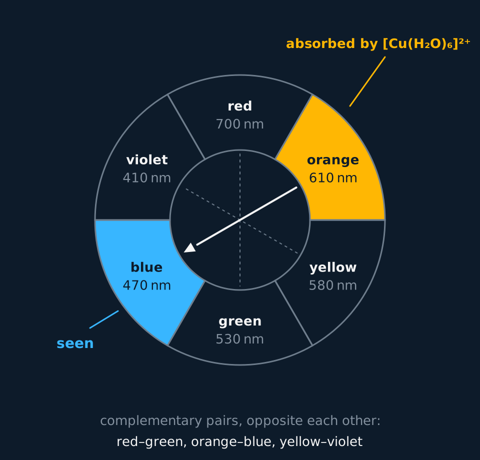
Colours that together make white light are complementary, and on a colour wheel they sit opposite each other. A substance that absorbs red light appears green; one that absorbs orange appears blue; one that absorbs violet appears yellow. The wheel gives the colour seen from the colour absorbed, and the colour absorbed from the colour seen.
Describe the drawing
A colour wheel of six segments in order round the circle: red 700 nm at the top, then orange 610, yellow 580, green 530, blue 470 and violet 410 nanometres. Complementary colours sit opposite each other, red and green, orange and blue, yellow and violet, joined by faint lines through the centre. The orange segment is labelled as absorbed by the hexaaquacopper(II) ion and an arrow runs across the centre to the blue segment opposite, labelled seen.
- Complementary colours
- Two colours of light that together make white light; a substance that absorbs one appears the other.
- Transmitted light
- The light that passes through a sample without being absorbed; its colour is the colour the sample appears.
Splitting the d orbitals
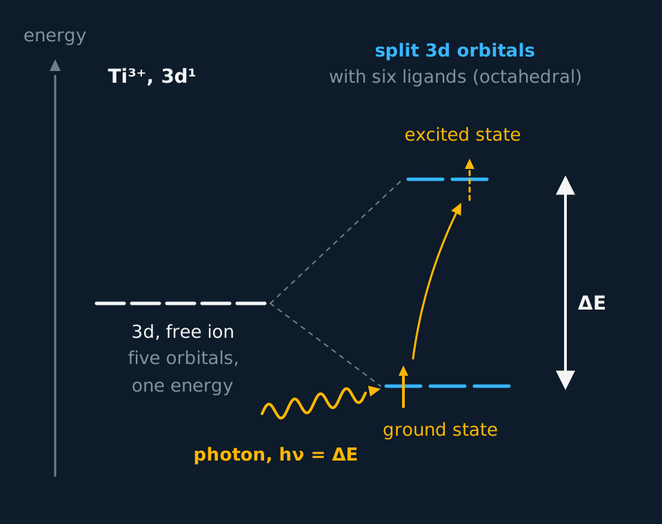
In an isolated metal ion the five 3d orbitals all have the same energy. Surround the ion with six ligands and that changes. The ligands' lone pairs approach along three axes, and the two d orbitals that point along those axes are repelled more strongly than the three that point between them. The 3d orbitals split into two groups, two at a higher energy and three at a lower one. The gap between them is ΔE.
A d electron in the lower group can move to the higher group if it absorbs exactly the energy ΔE. It moves from its ground state to an excited state, taking the energy from a photon. For most complexes of titanium to copper, ΔE matches the energy of a photon of visible light, so a band of visible wavelengths is absorbed and the complex is coloured.
ΔE = hν = hc/λh is the Planck constant, 6.63 × 10⁻³⁴ J s; ν is the frequency in Hz; c is the speed of light, 3.00 × 10⁸ m s⁻¹; λ is the wavelength in m.
The equation sets the direction of every comparison. A larger ΔE means a higher frequency and a shorter wavelength of light absorbed, towards the violet end of the spectrum. A smaller ΔE means a lower frequency and a longer wavelength, towards the red end.
A UV–visible spectrometer shows the absorption directly. It scans through the wavelengths and plots absorbance against wavelength. The spectrum of [Cu(H₂O)₆]²⁺ is a single broad band across the orange and red, peaking near 800 nm at the far red end and trailing down into the yellow; absorbance is low across the blue, which is the light that gets through. The wavelength at the peak is the one that corresponds to ΔE, and it is the wavelength a colorimeter filter is chosen to pass.
Describe the drawing
An energy level diagram, energy increasing upwards. On the left five short lines at one level are the five 3d orbitals of a free ion. On the right, joined to them by dashed guide lines, the same orbitals with six ligands in place: three lower and two higher, the upper pair further above the original level than the lower three are below it. A double arrow between the groups is labelled ΔE. The single electron of Ti³⁺ sits in a lower orbital, a wavy photon of energy ΔE arrives, and a curved arrow carries the electron to an upper orbital, the excited state.
- d-orbital splitting
- The separation of the five d orbitals of a metal ion into groups of different energy by the lone pairs of the ligands; the gap between the groups is ΔE.
- Ground state
- The lowest-energy arrangement of the electrons in an atom or ion.
- Excited state
- An arrangement of the electrons at higher energy than the ground state, reached when an electron absorbs energy and moves to a higher orbital.
The light absorbed for a given ΔE
The energy gap between the split d orbitals in a complex is 3.0 × 10⁻¹⁹ J. Calculate the frequency and the wavelength of the light absorbed, and predict the colour of the complex. h = 6.63 × 10⁻³⁴ J s and c = 3.00 × 10⁸ m s⁻¹.
Show the working
Rearrange ΔE = hν for the frequency: ν = ΔE ÷ h = 3.0 × 10⁻¹⁹ ÷ 6.63 × 10⁻³⁴ = 4.52 × 10¹⁴ Hz.
To two significant figures, matching the data, ν = 4.5 × 10¹⁴ Hz.
λ = c ÷ ν = 3.00 × 10⁸ ÷ 4.52 × 10¹⁴ = 6.63 × 10⁻⁷ m, which is 660 nm to two significant figures.
Light of 660 nm is red. The complex absorbs red light and lets the rest through, so it appears green, the colour opposite red on the wheel.
From a wavelength to kJ mol⁻¹
[Ti(H₂O)₆]³⁺ absorbs most strongly at 495 nm. Ti³⁺ is [Ar] 3d¹, so the absorption is its single d electron crossing ΔE. Calculate ΔE for one ion and for a mole of ions in kJ mol⁻¹, and explain the colour of the solution. h = 6.63 × 10⁻³⁴ J s, c = 3.00 × 10⁸ m s⁻¹ and L = 6.022 × 10²³ mol⁻¹.
Show the working
Convert the wavelength to metres: λ = 495 × 10⁻⁹ m = 4.95 × 10⁻⁷ m.
ΔE = hc/λ = 6.63 × 10⁻³⁴ × 3.00 × 10⁸ ÷ 4.95 × 10⁻⁷ = 4.018 × 10⁻¹⁹ J for one ion, kept unrounded for the next line.
For a mole of ions, multiply by the Avogadro constant: 4.018 × 10⁻¹⁹ × 6.022 × 10²³ = 2.42 × 10⁵ J mol⁻¹, which is 242 kJ mol⁻¹ to three significant figures.
Light of 495 nm is blue-green. The solution absorbs it and lets the red and the violet-blue ends of the spectrum through, which together look violet.
What changes ΔE
ΔE depends on how strongly the ligands interact with the d orbitals, and three things change it. Each one changes the colour, which is how a change in a complex is first noticed in a test tube.
| Change | Example | Colour change |
|---|---|---|
| Oxidation state of the metal | [Fe(H₂O)₆]²⁺ to [Fe(H₂O)₆]³⁺ | Pale green solution to yellow-brown solution |
| Ligand | [Cu(H₂O)₆]²⁺ to [Cu(NH₃)₄(H₂O)₂]²⁺ | Pale blue solution to deep blue solution |
| Coordination number and shape | [Co(H₂O)₆]²⁺ to [CoCl₄]²⁻ | Pink solution to blue solution |
Ammonia interacts more strongly with the d orbitals of Cu²⁺ than water does, so ΔE is larger in the ammine complex. The absorption moves to a shorter wavelength, and the light let through changes from pale blue to a deeper, more violet blue. A higher charge on the metal ion pulls the ligands closer and also increases ΔE, which is one reason the iron(II) and iron(III) aqua ions differ.
Cobalt shows the direction of the change clearly. Pink [Co(H₂O)₆]²⁺ absorbs green light, around 510 nm, and lets red and blue through. Blue [CoCl₄]²⁻ has the smaller tetrahedral splitting, so it absorbs at a longer wavelength, in the orange and red near 690 nm, and lets blue through. A smaller ΔE has moved the absorption towards the red, and the colour seen towards the blue.
Changing the coordination number changes the geometry, and with it the pattern of splitting. In a tetrahedral complex only four ligands approach and none of them points straight at a d orbital, so the splitting is smaller, a little under half the octahedral value for the same metal and ligand. A change of coordination number therefore changes ΔE, and the colour with it.
Mixtures add a further complication. The green seen partway through adding concentrated hydrochloric acid to copper(II) sulfate solution is blue [Cu(H₂O)₆]²⁺ and yellow [CuCl₄]²⁻ in the same tube, not a third complex with its own ΔE.
Colourless ions
ΔE is a gap that a d electron crosses, so an ion with no d electron to move, or with no room in the upper orbitals to move into, cannot absorb visible light this way. Sc³⁺ is [Ar] 3d⁰: there is no d electron to promote. Zn²⁺ and Cu⁺ are [Ar] 3d¹⁰: the upper orbitals are full. Their compounds are white and their solutions colourless. Titanium(IV) oxide, with Ti⁴⁺ as 3d⁰, is the white pigment in paint, toothpaste and sun cream.
The reverse does not always hold. A partly filled d subshell gives colour only when ΔE falls in the visible region. Where the gap is larger, the absorption is in the ultraviolet, every visible wavelength passes through, and the compound looks white or colourless.
Manganate(VII) and dichromate(VI) look like exceptions. Manganese in MnO₄⁻ is +7, [Ar] 3d⁰, yet the ion is an intense purple; chromium in Cr₂O₇²⁻ is +6, also 3d⁰, and the ion is orange. Their colour does not come from a d electron moving between split d orbitals. The light moves an electron from an oxygen atom onto the metal, a charge transfer, which absorbs far more strongly than a d-to-d change, and it makes 0.02 mol dm⁻³ manganate(VII) too dark to see through in a burette. No board examines the mechanism; the 3d⁰ and 3d¹⁰ rule is about d-to-d colour in complexes such as the aqua ions.
TRY IT: Predicting colour from a configuration
Suggest which of these solutions are coloured, giving the 3d configuration of each metal ion: titanium(III) sulfate, titanium(IV) chloride in acid, copper(I) chloride in concentrated hydrochloric acid, and nickel(II) sulfate.
Check your answer
Ti³⁺ is [Ar] 3d¹. Its one d electron can be promoted across ΔE, so the solution is coloured: it is violet.
Ti⁴⁺ is [Ar] 3d⁰, with no d electron to promote, so its solution is colourless. Cu⁺ is [Ar] 3d¹⁰, with no vacancy in the upper d orbitals, so its solution is colourless too.
Ni²⁺ is [Ar] 3d⁸, a partly filled subshell, so the solution is coloured: green.
Colorimetry
The more of a coloured complex there is in the path of a light beam, the more light it absorbs, and a colorimeter measures this. Light passes through a coloured filter, then through a cuvette of the solution, then onto a photocell. The meter gives the absorbance, which for a dilute solution in a cuvette of fixed width is proportional to the concentration of the coloured species.
The filter passes the colour of light the solution absorbs most strongly, which is the complementary colour of the solution. A blue copper solution is measured with an orange or red filter, and a pink cobalt solution with a green one. With the wrong filter the light passes straight through whatever the concentration, and the readings barely change.
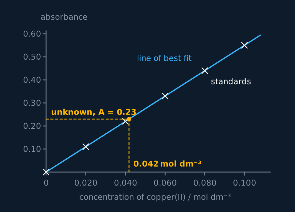
To find an unknown concentration, make a set of standard solutions of known concentration by diluting a stock solution. Zero the colorimeter with a cuvette of the solvent alone, which removes the absorbance of the solvent and the cuvette from every reading. Measure each standard and plot a calibration curve of absorbance against concentration. Measure the unknown with the same filter and cuvette, and read its concentration from the graph. The unknown must lie within the range of the standards, because the line may not stay straight beyond them.
Pale solutions give small absorbances and large percentage uncertainties, so a ligand is often added first to make a more intensely coloured complex. Excess ammonia turns pale blue copper(II) into deep blue [Cu(NH₃)₄(H₂O)₂]²⁺, and thiocyanate ions turn pale iron(III) solutions blood red. The same reagent, in the same excess, goes into every standard and the unknown.
Each standard is measured more than once, and the cuvette is rinsed with the next solution and wiped clean on its clear faces before each reading, with the clear faces in the beam every time. A point well off the line is measured again. If the best-fit line through the standards curves at the top, the unknown is diluted by a known factor until it falls on the straight part, and the concentration read off is multiplied by that factor.
Describe the drawing
A calibration graph of absorbance against concentration of copper(II), with six standards plotted as crosses from zero to 0.100 mol dm⁻³ lying on a straight line through the origin, absorbance equal to 5.5 times the concentration. A dashed line runs across from the unknown's absorbance of 0.23 to the line and then down to the concentration axis at 0.042 mol dm⁻³.
- Colorimeter
- An instrument that measures the absorbance of a solution for light of a chosen colour, selected by a filter.
- Absorbance
- A measure of how much light of a given wavelength a sample absorbs, proportional to the concentration of the absorbing species.
- Calibration curve
- A graph of a measured quantity, here absorbance, against the known concentrations of standard solutions, used to find an unknown concentration.
Copper in a brass screw
A 1.00 g brass screw is dissolved in nitric acid, excess ammonia is added and the solution is made up to 250.0 cm³. Copper(II) standards from 0.020 to 0.100 mol dm⁻³, given the same treatment, give a straight line through the origin with absorbance = 5.5 × concentration. The unknown has an absorbance of 0.23 with an orange filter. Calculate the percentage by mass of copper in the brass. Ar of Cu = 63.5.
Show the working
Concentration from the line: c = 0.23 ÷ 5.5 = 0.0418 mol dm⁻³.
Moles of copper in 250.0 cm³: n = c × V = 0.0418 × 0.2500 = 0.01045 mol.
Mass of copper: m = n × Ar = 0.01045 × 63.5 = 0.664 g.
Percentage by mass = 0.664 ÷ 1.00 × 100 = 66.4%, which is 66% to two significant figures, the precision of the absorbance reading. Brass is copper and zinc, and the zinc, as colourless Zn²⁺, does not absorb.
Other boards: naming the split orbitals
Cambridge asks for the orbitals in the octahedral splitting to be named; AQA and Edexcel ask only that the d orbitals split. The two that rise are 3dz² and 3dx²−y², whose lobes lie along the axes and point straight at the ligands' lone pairs. The three that fall are 3dxy, 3dxz and 3dyz, whose lobes lie between the axes and avoid the ligands.
Cambridge also examines the tetrahedral pattern. With four ligands approaching between the axes rather than along them, the splitting is the other way up: 3dxy, 3dxz and 3dyz are the three higher orbitals and 3dz² and 3dx²−y² the two lower ones. The gap is smaller, a little under half the octahedral value for the same metal and ligand, and for that reason [CoCl₄]²⁻ absorbs at a longer wavelength than [Co(H₂O)₆]²⁺.
| Octahedral, six ligands | Tetrahedral, four ligands | |
|---|---|---|
| Higher-energy orbitals | 3dz², 3dx²−y² (two) | 3dxy, 3dxz, 3dyz (three) |
| Lower-energy orbitals | 3dxy, 3dxz, 3dyz (three) | 3dz², 3dx²−y² (two) |
| Size of ΔE | Larger | Smaller, a little under half |
| Example | [Co(H₂O)₆]²⁺, pink, absorbs near 510 nm | [CoCl₄]²⁻, blue, absorbs near 690 nm |
In the exam
- A full explanation of colour is marked on four points: the ligands split the d orbitals, light of energy ΔE is absorbed, an electron moves from the ground state to an excited state, and the complementary colour is transmitted. An answer saying the ion gives out (emits) blue light gets no credit for the last point.
- In a ΔE = hc/λ calculation the answer mark needs the wavelength in metres, so convert nanometres with × 10⁻⁹ first. Energy per mole is ΔE × L, divided by 1000 for kJ mol⁻¹; a frequency may be given in Hz or s⁻¹.
- Zn²⁺ is colourless because its 3d subshell is full, so no d electron can be promoted. 'It is not a transition metal' restates the question and the scheme does not accept it.
- A colorimetry method needs a filter of the complementary colour, zeroing on the solvent, standards of known concentration plotted as a calibration curve, and the unknown read from that curve within the range of the standards. Each is a separate marking point.
Check yourself
[Cu(H₂O)₆]²⁺ is a pale blue solution and [Cu(NH₃)₄(H₂O)₂]²⁺ a deep blue solution. Explain why the two complexes differ in colour, and explain why copper(I) iodide is white.
Answer
Both complexes are coloured because the ligands split the 3d orbitals of Cu²⁺, which is [Ar] 3d⁹. Light of energy ΔE is absorbed as a d electron moves from the ground state to an excited state, and the complementary colour is seen.
Ammonia is a different ligand from water and interacts more strongly with the d orbitals, so ΔE is larger in the ammine complex. A larger ΔE means light of higher frequency and shorter wavelength is absorbed, so the colour let through changes, from pale blue to deep blue.
In copper(I) iodide the copper is Cu⁺, [Ar] 3d¹⁰. The 3d subshell is full, so no d electron can move to a higher d orbital, no visible light is absorbed, and the solid is white.
Questions
Question 16 marks
Describe how a colorimeter can be used to find the concentration of Cu²⁺ ions in a solution, and explain why excess ammonia is added to every solution before it is measured.
Mark scheme
Indicative content
- make a series of standard solutions of known Cu²⁺ concentration by diluting a stock solution, each with the same excess of ammonia
- choose a filter of the complementary colour, orange or red, the light that the deep blue complex absorbs most strongly
- zero the colorimeter with a cuvette of the solvent alone, so that the cuvette and solvent contribute no absorbance
- measure the absorbance of each standard and plot a calibration curve of absorbance against concentration, with a line of best fit
- measure the unknown with the same filter and cuvette, and read its concentration from the curve, within the range of the standards
- ammonia forms deep blue [Cu(NH₃)₄(H₂O)₂]²⁺, which absorbs far more strongly than the aqua ion, so absorbances are larger and percentage uncertainties smaller
Levels
- Level 3 (5–6 marks) the standards with the same excess of ammonia, the complementary filter, zeroing with the solvent, the calibration curve and reading the unknown within its range are all present, and the ammonia is explained through the much stronger absorbance of the deep blue complex and the smaller percentage uncertainty
- Level 2 (3–4 marks) the calibration curve from standards, the filter and the reading of the unknown are described; zeroing, keeping the unknown inside the range of the standards, or the reason for the ammonia is left out
- Level 1 (1–2 marks) absorbance is compared with known solutions; the ammonia is typically said to be there to precipitate the copper, and calibration and filter are largely missing
Question 24 marksAQA · Edexcel · CAIE
State what happens to a d electron in a complex ion when the ion absorbs visible light, and state the equation that links the energy absorbed with the frequency of the light, giving the meaning of each symbol.
Mark scheme
- B1 the d electron is promoted from its ground state to an excited state, a higher-energy d orbital
- B1 the energy absorbed equals ΔE, the gap between the split d orbitals
- B1 the equation is ΔE = hν; allow ΔE = hc/λ with λ defined
- B1 h is the Planck constant and ν the frequency of the light absorbed, in Hz or s⁻¹
Question 34 marksAQA · Edexcel · CAIE
Explain why a solution containing [Cu(H₂O)₆]²⁺ ions is blue.
Mark scheme
- B1 the water ligands split the 3d orbitals of the Cu²⁺ ion into two groups of different energy
- B1 light of energy ΔE is absorbed as a d electron is promoted to the higher group
- B1 the light absorbed is at the orange and red end of the visible spectrum
- B1 the remaining light, which appears blue, is transmitted
All 18 questions with mark schemes, the other 15 set out in full on the question page.
Worth remembering
- A blue solution absorbs orange and red light; 'it absorbs blue' is the opposite of the truth.
- Convert λ to metres before ΔE = hc/λ; left in nanometres it gives an answer 10⁹ times too small.
- 'Larger ΔE, longer wavelength' reverses hc/λ: a larger gap absorbs a shorter wavelength.
- 'Zn²⁺ is not a transition metal ion' does not explain its lack of colour; the full 3d subshell does.
- Choose the complementary filter, the colour the solution absorbs; a filter of the solution's own colour lets the light through.
CHECK YOUR PROGRESS
Rate how confident you are with each objective for this lesson. Ratings are saved in this browser, on this device, unless you sign in.
- Explain why transition metal complexes are coloured, using d-orbital splitting, the ground and excited states, and complementary colours.
- Use ΔE = hν = hc/λ to calculate the energy, frequency or wavelength of the light absorbed.
- Explain how a change of oxidation state, ligand or coordination number changes ΔE and the colour, with an example of each.
- Explain why the compounds of Sc³⁺, Zn²⁺ and Cu⁺ are colourless.
- Describe how to find the concentration of a coloured ion with a colorimeter and a calibration curve.
Open the full revision checklist to see every objective in the curriculum in one place.
Practise this lesson
WORKBOOK
The written questions from this topic, on paper with room to work, and a separate book of mark schemes. The question that is set on a diagram stays on this page, where the diagram can be drawn. Free to use; please do not redistribute or sell.