Biology › Nucleic acids, genomes and protein synthesis › DNA and RNA structure
DNA and RNA structure
A phosphate, a sugar and a base make one nucleotide, and DNA is two antiparallel strands of millions of nucleotides wound into a double helix. The result is a molecule long enough to hold the instructions for an entire organism and simple enough to copy without losing them.
Before this Carbohydrates: condensation and hydrolysis · Water and inorganic ions: hydrogen bonding · The nucleus and the nuclear envelope
COMMON MISCONCEPTION
The two strands of a DNA molecule are identical, so either one is a spare copy of the other.
Complementary, not identical. Where one strand reads 5′-ATGGC-3′, the strand facing it reads 3′-TACCG-5′, and no base matches the one it faces. That is what makes the spare useful: the fixed pairing rule lets either strand specify the other exactly.
What you should be able to do
- Draw a nucleotide and name its three components.
- Explain how the sugar-phosphate backbone is built and what is released as it forms.
- State which bases pair, how many hydrogen bonds hold each pair, and why the pairing cannot be swapped around.
- Say what 5′ and 3′ mean, and why the two strands are called antiparallel.
- Account for four features of DNA structure in terms of the job the molecule does.
- Compare DNA with mRNA, tRNA and rRNA, and give each kind of RNA its role.
Three parts, one nucleotide
A nucleotide is three things joined together: a phosphate group, a pentose sugar with five carbons, and a nitrogen-containing base. The sugar sits in the middle, with the phosphate attached on one side and the base on the other. That arrangement is worth learning by position, because the phosphate and the sugar are what build the backbone while the base is the part that carries meaning.
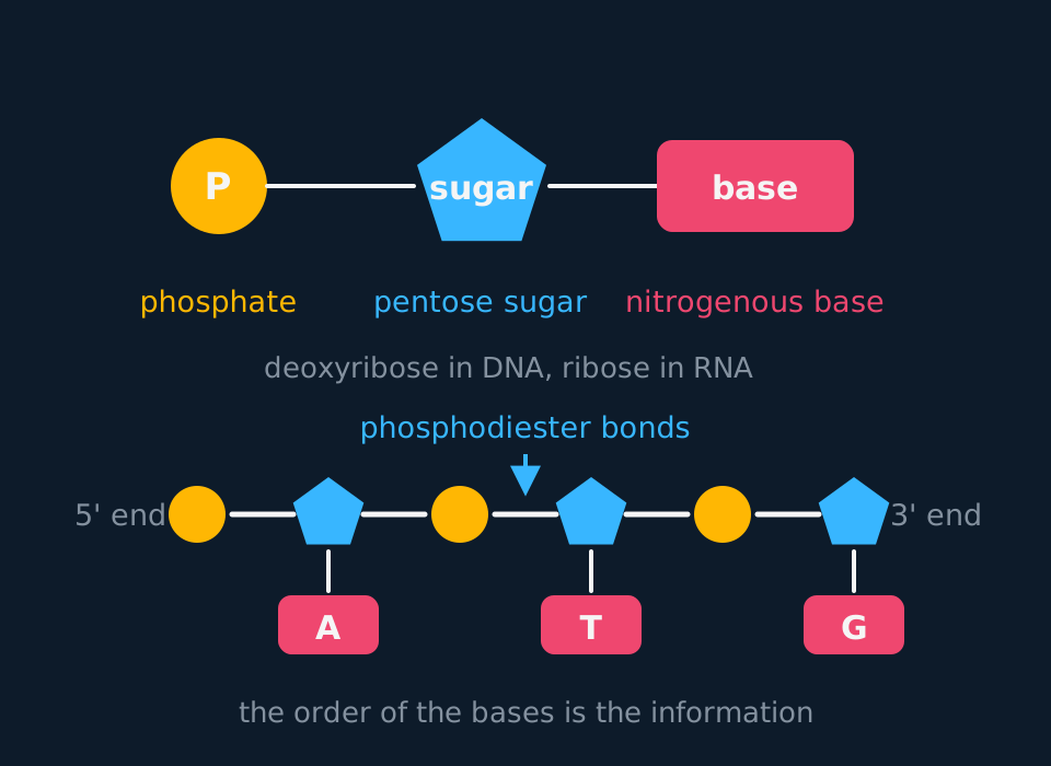
Describe the drawing
A nucleotide drawn as three joined shapes: a circle for the phosphate group, a pentagon for the pentose sugar and a rectangle for the nitrogenous base. Below it, three nucleotides are joined into a strand in which phosphate and sugar alternate along the backbone, with the bases adenine, thymine and guanine hanging off the sugars and the two ends of the strand marked five prime and three prime.
The sugar differs between DNA and RNA. DNA contains deoxyribose, which is ribose with one oxygen fewer at carbon 2. The absence of that hydroxyl group makes DNA less reactive than RNA and therefore more stable over the lifetime of a cell.
- Nucleotide
- A phosphate group, a pentose sugar and a nitrogenous base joined together.
- Polynucleotide
- A chain of nucleotides joined by phosphodiester bonds between the sugar of one and the phosphate of the next.
- Phosphodiester bond
- The covalent bond joining the phosphate group of one nucleotide to the sugar of the next, formed by condensation.
Nucleotides join by condensation, as other biological monomers do. The phosphate of one nucleotide reacts with a hydroxyl group on the sugar of the next, water is released, and a covalent phosphodiester bond forms. Repeat that a few million times and you have a polynucleotide whose backbone is a strict alternation of sugar, phosphate, sugar, phosphate, with the bases projecting sideways. Hydrolysis reverses the reaction, which is how dietary DNA is digested before the nucleotides are absorbed.
Human chromosome 1 is a single DNA molecule roughly 249 million base pairs long. Stretched out it would be about 8 cm. Every phosphodiester bond along its backbone was made by the same condensation reaction.
The rungs: which base goes with which
Four bases appear in DNA: adenine, thymine, cytosine and guanine. They are not interchangeable. Adenine only ever pairs with thymine, and guanine only ever pairs with cytosine, and the reason is a matter of shape and of where the hydrogen-bond donors and acceptors sit on each ring system.
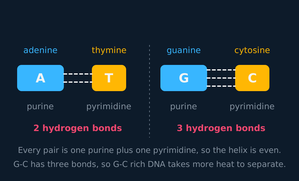
Adenine and guanine are purines, built from two fused rings. Thymine and cytosine are pyrimidines, built from one. So is uracil, the base RNA uses in thymine's place: the split into purines and pyrimidines covers all five bases, not just the four in DNA. Because every pair is a purine opposite a pyrimidine, every rung of the ladder is the same width, and the two backbones stay a constant distance apart all the way along. A purine facing a purine would bulge; two pyrimidines would pinch.
- Purine
- A base built from two fused rings: adenine and guanine.
- Pyrimidine
- A base built from a single ring: cytosine, thymine and, in RNA, uracil.
The hydrogen-bond counts are worth memorising as numbers rather than as an idea. A–T is two hydrogen bonds. G–C is three. Individually these are weak interactions, easily broken by an enzyme or by heat, which is what allows the strands to be separated whenever the sequence needs reading. Collectively, across millions of pairs, they hold the molecule together firmly.
Working out the other three bases
A sample of double-stranded DNA is analysed and found to contain 22% adenine. Calculate the percentage of each of the other three bases, and state what result you would expect if the sample had been single-stranded.
Show the working
Adenine pairs only with thymine, so in a double-stranded molecule every adenine has a thymine opposite it. Thymine is therefore also 22%.
Together A and T account for 44% of the bases, leaving 56% shared between guanine and cytosine. Those two also pair one-for-one, so they split the remainder equally: G = 28% and C = 28%.
For a single-stranded molecule none of this holds. Nothing forces an adenine to have a partner, so the four percentages could be anything at all. A sample in which A does not equal T is a sample that is not double-stranded, and that observation is a real analytical tool.
Two strands, pointing opposite ways
The carbons in the pentose sugar are numbered 1 to 5, and the numbering is why the ends of a strand have names. At one end of a polynucleotide carbon 5 of the final sugar faces outwards, usually carrying a phosphate; that is the 5′ end. At the other, carbon 3 faces outwards, usually with a free hydroxyl group; that is the 3′ end. The names come from the carbon numbering rather than from whatever happens to be attached there, and a strand therefore has a direction, in the same way a sentence does.
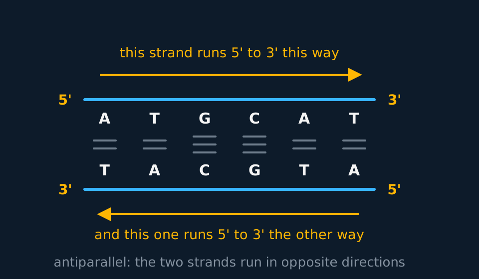
Describe the drawing
A short stretch of double-stranded DNA drawn as a flat ladder. The upper strand reads A T G C A T from left to right with its five prime end on the left; the lower strand reads T A C G T A with its five prime end on the right. Between each pair of bases sit two short rungs for an A to T pair and three for a G to C pair, and an arrow beside each strand shows the two directions pointing opposite ways.
The two strands of a DNA molecule run in opposite directions. Wherever one strand has its 5′ end, the other has its 3′ end. That is what antiparallel means. DNA polymerase adds nucleotides in one direction only, so the two strands are not replicated in the same way, as the next lesson shows.
Wind the whole thing into a right-handed spiral and you have the double helix, with roughly ten base pairs per complete turn. The bases are on the inside, shielded from the water around them; the charged sugar-phosphate backbones are on the outside, where being charged lets them interact with water and, in eukaryotes, bind the positively charged histones.
- Antiparallel
- The two strands of a DNA molecule run in opposite directions: the 5′ end of one lies alongside the 3′ end of the other.
- Complementary base pairing
- Adenine pairs with thymine by two hydrogen bonds and guanine pairs with cytosine by three, so the sequence of one strand fixes the sequence of the other.
- Double helix
- Two antiparallel polynucleotide strands wound into a spiral, bases inside and backbones outside.
Relating DNA structure to its function
Link each structural feature to a specific requirement of the molecule. Four links cover most cases.
It is stable. The backbone is held by covalent phosphodiester bonds, the bases lie inside the helix away from water, and deoxyribose is less reactive than ribose, so the base sequence is preserved over long periods.
It is very long. There is no limit to how many nucleotides can be joined, so an enormous quantity of information fits into one molecule. The human genome runs to about 3.1 billion base pairs.
The base sequence carries the information. The backbone is identical everywhere and carries no information; the order of the four bases is free to vary and carries all of it. A stretch of only ten bases can be arranged in 4¹⁰ ways, a little over a million. Many scientists once doubted this. With only four kinds of nucleotide on a repeating backbone, DNA looked too simple to carry the genetic code, and proteins, built from twenty kinds of amino acid, seemed the likelier candidate until experiments in the 1940s and early 1950s showed that DNA is the genetic material.
The strands are complementary. Because the pairing rule is fixed, each strand is a full set of instructions for rebuilding the other. That is what makes accurate copying possible, and it is also why damage to one strand can often be repaired using the other as the reference.
One further feature applies where a question mentions packing: in eukaryotes the DNA is wound around histone proteins and coiled repeatedly, which is how 2 metres of DNA fits inside a nucleus about 6 μm across. In the bacteria this course describes, the DNA is circular, shorter and not wound around histones, though it is still supercoiled to fit. (The other prokaryote domain, the archaea, breaks the tidy rule: many archaeal species do carry histone proteins.)
RNA structure and the three kinds of RNA
RNA is a polynucleotide too, made the same way, with two changes. The sugar is ribose rather than deoxyribose, and uracil replaces thymine. Uracil pairs with adenine, using the same two hydrogen bonds thymine would have used, and since it is a single-ring pyrimidine like the thymine it stands in for, an A-U pair keeps the purine-against-pyrimidine geometry and the same width as every other pair.
| DNA | RNA | |
|---|---|---|
| Sugar | Deoxyribose | Ribose |
| Bases | A, T, C, G | A, U, C, G |
| Strands | Two, antiparallel, wound into a helix | One, though it often folds back on itself |
| Length | Up to hundreds of millions of nucleotides | A few tens to many thousands of nucleotides |
| Stability | Stable enough to last the life of the cell | Broken down quickly; some mRNA lasts minutes |
| Where | Nucleus, mitochondria, chloroplasts | Made in the nucleus, used in the cytoplasm |
| Job | Long-term store of the base sequence | Carrying, reading and assembling |
DNA is a stable long-term store of sequence information; many RNA molecules are shorter-lived intermediates or functional products. The short lifetime of mRNA limits how long a protein continues to be synthesised after transcription of its gene stops.
- Messenger RNA (mRNA)
- A single-stranded copy of one gene, made in the nucleus and carrying codons to a ribosome in the cytoplasm.
- Transfer RNA (tRNA)
- A small folded RNA of about 80 nucleotides with an anticodon at one end and a binding site for a specific amino acid at the other.
- Ribosomal RNA (rRNA)
- RNA which, with proteins, forms the two subunits of a ribosome and catalyses the formation of peptide bonds.
Each of the three has a shape suited to what it does. mRNA is drawn as a straight run of codons because that is how the ribosome treats it; the real molecule folds back on itself in places and carries untranslated stretches at both ends, but its job needs no overall working shape, only a readable sequence. tRNA folds into a clover leaf held together by hydrogen bonds between complementary stretches of its own sequence, which puts the anticodon at one end and the amino acid at the other. rRNA does not carry a coding sequence; it is a structural and catalytic component of the ribosome.
TRY IT: Deducing base composition from melting temperature
Two samples of nucleic acid are heated slowly and the temperature at which the strands separate is recorded. Sample X separates at 68 °C and sample Y at 83 °C. Both are double-stranded DNA of the same length. What can you conclude about their base composition? Explain your reasoning.
Check your answer
Sample Y has the higher proportion of guanine and cytosine.
Heating separates the strands by breaking the hydrogen bonds between paired bases. Each G–C pair is held by three hydrogen bonds while each A–T pair is held by only two, so a molecule with more G–C pairs needs more energy, and therefore a higher temperature, before its strands come apart.
Since the two samples are the same length, the difference cannot be a matter of one molecule having more base pairs than the other. State the limit of the conclusion: the data give the proportion of G-C pairs, not the base sequence. The reasoning also assumes all other factors are equal, which holds here because length and conditions were matched. In general, base stacking and the ionic conditions of the solution also shift melting temperatures.
In the exam
- Two hydrogen bonds for A-T, three for G-C. Use those numbers for questions about heat stability, strand separation and melting temperature.
- 'Complementary' and 'identical' are not synonyms. A strand and its partner carry the same information in complementary form, not the same sequence.
- For antiparallel, state that the strands run in opposite directions with 5′ against 3′.
- Pair each structural feature with the function it makes possible: covalent backbone with stability, base sequence with information, complementary strands with accurate replication.
- In RNA the base is uracil, not thymine. Write U rather than T in an mRNA sequence.
- Chargaff calculations follow one rule: %A = %T and %G = %C, and the four add to 100. Work out the given pair first, then divide the remainder.
Check yourself
A student writes: 'DNA is a good molecule for storing genetic information because it has a double helix shape.' Rewrite this as a complete explanation, referring to at least three features of the molecule.
Answer
The helical shape alone does not explain information storage. Tie each feature to a specific requirement of the molecule.
The backbone is held together by covalent phosphodiester bonds, so the molecule is chemically stable and the information is not lost over time. The bases are also positioned on the inside of the helix, shielded from the water and from reactive molecules in the nucleus.
The sequence of the four bases can vary without limit, and the molecule can be extremely long, so an enormous amount of information can be encoded in one molecule. Ten bases alone allow over a million arrangements.
The two strands are complementary, held by hydrogen bonds that are weak enough to break when the sequence has to be read or copied. Because the pairing rule is fixed, either strand acts as a template for rebuilding the other, which is what makes replication accurate and repair possible.
Name the bond in each case: covalent phosphodiester bonds along the backbone, hydrogen bonds between the paired bases. The backbone is therefore stable while the strands can still be separated.
Questions
Question 14 marks
Compare the structure of a DNA molecule with the structure of a messenger RNA molecule.
Mark scheme
- B1 DNA contains the pentose sugar deoxyribose, whereas mRNA contains ribose
- B1 DNA contains thymine, whereas mRNA carries uracil in its place
- B1 DNA is two antiparallel strands wound into a helix, whereas mRNA is a single strand
- B1 DNA can be hundreds of millions of nucleotides long, whereas an mRNA molecule is far shorter, usually hundreds to tens of thousands of nucleotides long
Question 24 marks
A nucleic acid extracted from a virus contains adenine 31%, guanine 19%, cytosine 27% and thymine 23%. Suggest what these figures tell you about the structure of the molecule, and suggest one further measurement that would test your conclusion.
Mark scheme
- B1 in a double-stranded molecule adenine would equal thymine and guanine would equal cytosine, because each base has a fixed partner opposite it
- B1 here adenine at 31% does not equal thymine at 23%, and guanine at 19% does not equal cytosine at 27%
- B1 the molecule cannot therefore be double-stranded; it is single-stranded DNA, since thymine rather than uracil is present
- B1 any one workable further measurement: heat the sample and look for a temperature at which strands separate, since a single-stranded molecule has no paired bases to separate
Question 34 marks
Describe the double helix formed by DNA, and describe how DNA is packaged to fit inside a eukaryotic nucleus about 6 μm across.
Mark scheme
- B1 the two antiparallel polynucleotide strands are wound around each other into a right-handed spiral, with roughly ten base pairs per complete turn
- B1 the bases lie on the inside of the helix, shielded from the surrounding water, while the charged sugar-phosphate backbones lie on the outside
- B1 in a eukaryotic cell the DNA is wound around histone proteins
- B1 that DNA-histone complex is then coiled repeatedly, so that about two metres of DNA is compacted to fit inside a nucleus only a few micrometres across
Question 44 marks
Compare the organisation of DNA in a eukaryotic cell with its organisation in a bacterial cell.
Mark scheme
- B1 eukaryotic DNA is linear and organised into several separate molecules, chromosomes, whereas a bacterium's DNA is usually one circular molecule
- B1 eukaryotic DNA is wound around histone proteins, whereas bacterial DNA is not wound around histones, though it is still supercoiled to fit inside the cell
- B1 eukaryotic DNA is held within a membrane-bound nucleus, whereas bacterial DNA lies free in the cytoplasm, in a region called the nucleoid
- B1 eukaryotic DNA is very much longer in total than bacterial DNA, so eukaryotic packaging must condense a far greater length into a comparable or larger volume
Question 54 marks
Explain how the length of a DNA molecule and the sequence of its bases together allow it to store a very large amount of information.
Mark scheme
- B1 there is no limit to how many nucleotides can be joined into a polynucleotide, so an extremely long molecule, such as human chromosome 1 at about 249 million base pairs, can exist
- B1 the backbone is identical at every position along the molecule and carries no information itself; it is the order of the four bases that is free to vary
- B1 a short stretch of only ten bases can already be arranged in 4¹⁰ ways, a little over a million, so the number of possible sequences grows enormously with length
- B1 combining a very long molecule with a base sequence that can vary at every position gives a capacity for information that is, for practical purposes, unlimited
Question 64 marks
A student says that the two strands of a DNA molecule are identical, so either one is simply a spare copy of the other. Discuss whether this is an accurate description of the relationship between the two strands.
Mark scheme
- B1 what is right in the claim: each strand does carry the full information needed to rebuild the other, since the identity of every base on one strand fixes the identity of the base opposite it
- B1 against: the two strands are not identical in sequence; adenine on one strand is paired with thymine on the other, and guanine with cytosine, so the base at a given position on one strand is not the same base as the one at the corresponding position on the other
- B1 against: the two strands also run in opposite, antiparallel directions, so even where the base sequences are complementary they are not read in the same direction, and are not copied in the same way when the molecule is replicated
- B1 judgement: 'complementary' and 'identical' are not synonyms; the strands carry the same information in complementary rather than identical form, which is what makes accurate copying and repair possible
Question 73 marks
Explain why every base pair in a DNA molecule is the same width, and explain what this means for the shape of the molecule as a whole.
Mark scheme
- B1 adenine and guanine are purines, built from two fused rings, while thymine and cytosine are pyrimidines, built from one
- B1 the pairing rule puts a purine opposite a pyrimidine in every pair, so each rung spans the same distance
- B1 the two sugar-phosphate backbones therefore stay a constant distance apart along the whole molecule, whereas two purines would bulge and two pyrimidines would pinch
Question 83 marks
A sample of double-stranded DNA contains 3200 bases in total, of which 960 are guanine. Calculate the number of adenine bases in the sample.
Mark scheme
- M1 cytosine equals guanine, so there are 960 cytosine bases, giving 1920 bases in the two of them together
- M1 subtracts from the total to leave the adenine and thymine: 3200 − 1920 = 1280
- A1 adenine equals thymine, so 1280 ÷ 2 gives 640 adenine bases
Question 93 marks
Two double-stranded DNA samples of the same length are heated slowly. Sample X separates into single strands at 68 °C and sample Y at 83 °C. Explain what this difference tells you about the base composition of the two samples.
Mark scheme
- B1 heating separates the strands by breaking the hydrogen bonds between paired bases
- B1 each G–C pair is held by three hydrogen bonds while each A–T pair is held by only two
- B1 sample Y needs more energy, and so a higher temperature, before its strands come apart, so it has the higher proportion of guanine and cytosine
Question 103 marks
Describe how two nucleotides are joined together during the synthesis of a polynucleotide, naming the reaction, the bond formed and the parts of each nucleotide involved.
Mark scheme
- B1 the phosphate group of one nucleotide reacts with a hydroxyl group on the pentose sugar of the next
- B1 the reaction is a condensation, in which a molecule of water is released
- B1 the bond formed is a covalent phosphodiester bond, giving a backbone that strictly alternates sugar and phosphate
Question 113 marks
Explain what is meant by describing the two strands of a DNA molecule as antiparallel, and explain what this means for the direction in which each strand is copied during replication.
Mark scheme
- B1 antiparallel means the two strands run in opposite directions, so wherever one strand has its 5′ end, the other strand has its 3′ end at that same position
- B1 running in opposite directions is what lines each base up opposite its complementary partner, so hydrogen bonds form between the pairs along the whole length
- B1 DNA polymerase can only build a new strand in one direction, so the antiparallel arrangement of the two template strands is why the two new strands cannot be synthesised in the same way as each other
Question 123 marks
Describe the role of ribosomal RNA (rRNA) in a ribosome, and describe how its role differs from that of messenger RNA.
Mark scheme
- B1 rRNA, together with protein, forms the two subunits of a ribosome, giving the ribosome its structure
- B1 rRNA also catalyses the formation of peptide bonds between adjacent amino acids during translation
- B1 unlike messenger RNA, rRNA does not carry a coding sequence read as codons; its role is structural and catalytic rather than carrying genetic information from gene to ribosome
Question 132 marks
Name the three components of a single DNA nucleotide, and name the bond that joins one nucleotide to the next along a strand.
Mark scheme
- B1 a phosphate group, a pentose sugar (deoxyribose) and a nitrogenous base
- B1 a phosphodiester bond, formed by condensation between the phosphate of one nucleotide and the sugar of the next
Question 142 marks
State two ways in which a molecule of transfer RNA differs from a molecule of messenger RNA.
Mark scheme
- B1 any one of: tRNA is short, at about 80 nucleotides; it folds into a clover leaf held by hydrogen bonds between complementary stretches of its own sequence; it carries an anticodon; it has a binding site for a specific amino acid
- B1 any second, different point from the same list, or the matching contrast that mRNA is a run of codons with no clover-leaf fold and no amino acid binding site
Question 152 marks
State what is meant by the 5′ end of a DNA strand, and state what is meant by the 3′ end.
Mark scheme
- B1 the 5′ end is the end at which carbon 5 of the terminal sugar faces outward, usually carrying a phosphate group
- B1 the 3′ end is the end at which carbon 3 of the terminal sugar faces outward, usually carrying a free hydroxyl group
Worth remembering
- One nucleotide is a phosphate, a pentose sugar and a base.
- The backbone alternates sugar and phosphate, joined by phosphodiester bonds made by condensation.
- A pairs with T through two hydrogen bonds; G pairs with C through three.
- The strands are antiparallel: 5′ on one side lies against 3′ on the other.
- RNA has ribose instead of deoxyribose and uracil instead of thymine, and it is single-stranded.
CHECK YOUR PROGRESS
Rate how confident you are with each objective for this lesson. Ratings are saved in this browser, on this device, unless you sign in.
- Draw a nucleotide and name its three components.
- Explain how the sugar-phosphate backbone is built and what is released as it forms.
- State which bases pair, how many hydrogen bonds hold each pair, and why the pairing cannot be swapped around.
- Say what 5′ and 3′ mean, and why the two strands are called antiparallel.
- Account for four features of DNA structure in terms of the job the molecule does.
- Compare DNA with mRNA, tRNA and rRNA, and give each kind of RNA its role.
Open the full revision checklist to see every objective in the curriculum in one place.
Practise this lesson
Practise DNA and RNA structure with 15 original questions and point-by-point mark schemes
WORKBOOK
The questions from this topic, on paper with room to work, and a separate book of mark schemes. Free to use; please do not redistribute or sell.
Used later in The genetic code and transcription · Recombinant DNA technology · Genome analysis, genetic screening and gene therapy · Nutrient cycles: nitrogen, phosphorus and eutrophication