Chemistry › Aromatic chemistry and amines › Amino acids, proteins, enzymes and DNA
Amino acids, proteins, enzymes and DNA
An amino acid carries an amine group and a carboxylic acid group on the same carbon, so it is an acid and a base in one molecule. Joined by peptide links, amino acids make proteins, whose shapes are held by hydrogen bonds, ionic bonds and sulfur bridges; and enzymes are proteins whose shape makes them catalysts. DNA is a second polymer, two strands held together by hydrogen bonds between bases, and the anticancer drug cisplatin works by binding to it.
Key terms Every definition in aromatic chemistry and amines, lesson by lesson
Before this Amines: preparation, basicity and nucleophilic reactions – the amine group as a base · Optical isomerism – a chiral centre and enantiomers · Condensation polymers and the disposal of polymers – the amide link and hydrolysis · Ligands, the shapes of complexes, isomerism and ligand substitution – cis–trans isomers of square planar complexes
COMMON MISCONCEPTION
An amino acid is an acid, so a solution of glycine must be acidic, and solid glycine is a molecular solid like ethanoic acid.
Glycine carries a base as well, and its COOH donates a proton to its own NH₂. In the solid it exists as the zwitterion ⁺H₃NCH₂COO⁻, held in an ionic lattice, so it is a crystalline solid that decomposes above 230 °C rather than melting. In water the zwitterion dominates near pH 6, and the solution is close to neutral.
What you should be able to do
- Describe the structure of an amino acid, identify its chiral centre, and draw the zwitterion and the ions it forms in acid and in alkali.
- Explain how a peptide link forms by condensation, and give the two dipeptides made from two different amino acids.
- Describe the primary, secondary and tertiary structure of a protein and the bonds that hold each in place.
- Describe how a protein is hydrolysed and its amino acids identified by thin-layer chromatography, calculate Rf values, and predict how amino acids move in electrophoresis.
- Explain how enzymes act as stereospecific catalysts and how a drug can act as an enzyme inhibitor.
- Describe the structure of DNA, and explain how cisplatin prevents DNA replication and why its use carries risks.
Amino acids
Hair, egg white and muscle are made largely of protein, and every protein is a chain of amino acids. Living things build their proteins from twenty amino acids, all with the same skeleton: an amine group, NH₂, and a carboxylic acid group, COOH, bonded to the same carbon atom. The general formula is H₂NCH(R)COOH, and the amino acids differ only in the side chain, R.
Glycine, the simplest, has R = H and is aminoethanoic acid. Alanine has R = CH₃ and is 2-aminopropanoic acid. Serine has CH₂OH, cysteine CH₂SH, lysine a chain ending in a second NH₂ group, and aspartic acid a second COOH group. A question gives the structure of any amino acid it uses, so the skill is recognising the pattern, not learning the side chains.
In every amino acid except glycine, the central carbon has four different groups attached: H, NH₂, COOH and R. It is a chiral centre, and the amino acid exists as two enantiomers that rotate plane-polarised light in opposite directions. Glycine has two hydrogen atoms on that carbon, so it has no chiral centre and is not optically active. Proteins in living things are built from one enantiomer of each amino acid only.
- Amino acid
- A compound with an amine group and a carboxylic acid group; in the α-amino acids that make proteins both are on the same carbon atom.
Zwitterions and the isoelectric point
Glycine is a white crystalline solid that decomposes above about 230 °C without melting. Propanoic acid, with almost the same Mr, is a liquid that freezes at −21 °C. The difference is that solid glycine is ionic. The COOH group donates a proton and the NH₂ group accepts one, within the same molecule, giving ⁺H₃NCH₂COO⁻, an ion with a positive and a negative charge and no overall charge, called a zwitterion. The solid is a lattice of zwitterions held by strong attractions between opposite charges, so a great deal of energy is needed to break it down, and the charged groups make glycine dissolve well in water.
In solution the form an amino acid takes depends on the pH. In acid the COO⁻ group takes a proton back, and the amino acid becomes a positive ion, ⁺H₃NCH(R)COOH. In alkali the NH₃⁺ group loses its proton, and the amino acid becomes a negative ion, H₂NCH(R)COO⁻. Between the two there is a pH at which the amino acid is present almost entirely as the zwitterion, with no overall charge: the isoelectric point.
⁺H₃NCH₂COO⁻(aq) + H⁺(aq) → ⁺H₃NCH₂COOH(aq)In acid: the carboxylate group accepts a proton.
⁺H₃NCH₂COO⁻(aq) + OH⁻(aq) → H₂NCH₂COO⁻(aq) + H₂O(l)In alkali: the ammonium group loses a proton to hydroxide.
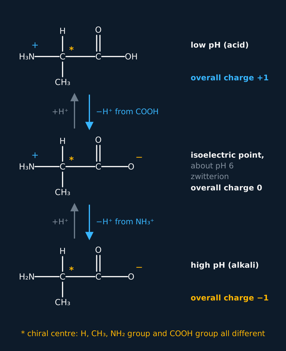
Describe the drawing
Three displayed structures of alanine stacked from low pH at the top to high pH at the bottom. At low pH, in acid, the amine group is NH₃⁺ and the acid group COOH, overall charge plus one. At the isoelectric point, about pH 6, the zwitterion has NH₃⁺ and COO⁻, overall charge zero. At high pH, in alkali, the amine is NH₂ and the acid group COO⁻, overall charge minus one. Downward arrows read minus H⁺ from COOH and then minus H⁺ from NH₃⁺, with plus H⁺ arrows back up, and the central carbon in each is starred as the chiral centre.
For glycine and alanine the isoelectric point is close to pH 6. An amino acid with an extra COOH in its side chain, such as aspartic acid, has a lower isoelectric point, about pH 2.8, because a lower pH is needed to keep its second acid group protonated. Lysine, with an extra NH₂, has an isoelectric point of about pH 9.7.
The three forms of alanine
Give the condensed formula of the species alanine, H₂NCH(CH₃)COOH, forms at pH 1, at its isoelectric point, and at pH 13.
Show the working
At pH 1 the solution is strongly acidic, so both groups are protonated: ⁺H₃NCH(CH₃)COOH, overall charge +1.
At the isoelectric point, about pH 6, the zwitterion: ⁺H₃NCH(CH₃)COO⁻, overall charge 0.
At pH 13 the solution is strongly alkaline, so both groups have lost their protons: H₂NCH(CH₃)COO⁻, overall charge −1.
The charge falls by one at each step as the pH rises, and the chiral centre is unchanged in all three.
A drawn answer must show the charges on the right atoms: the positive charge on the nitrogen of NH₃⁺ and the negative charge on an oxygen of COO⁻. A zwitterion drawn with the charges but with the H still on the COOH group is a different species and does not earn the mark.
- Zwitterion
- An ion with both a positive and a negative charge and no overall charge, as in ⁺H₃NCH(R)COO⁻.
- Isoelectric point
- The pH at which an amino acid exists as the zwitterion and has no overall charge.
Peptides and proteins
The amine group of one amino acid can react with the carboxylic acid group of another, losing a molecule of water. The new –CONH– group is a peptide link, the same amide link as in nylon, and the reaction is a condensation. Two amino acids joined this way make a dipeptide, which still has a free NH₂ at one end and a free COOH at the other, so more amino acids can join on.
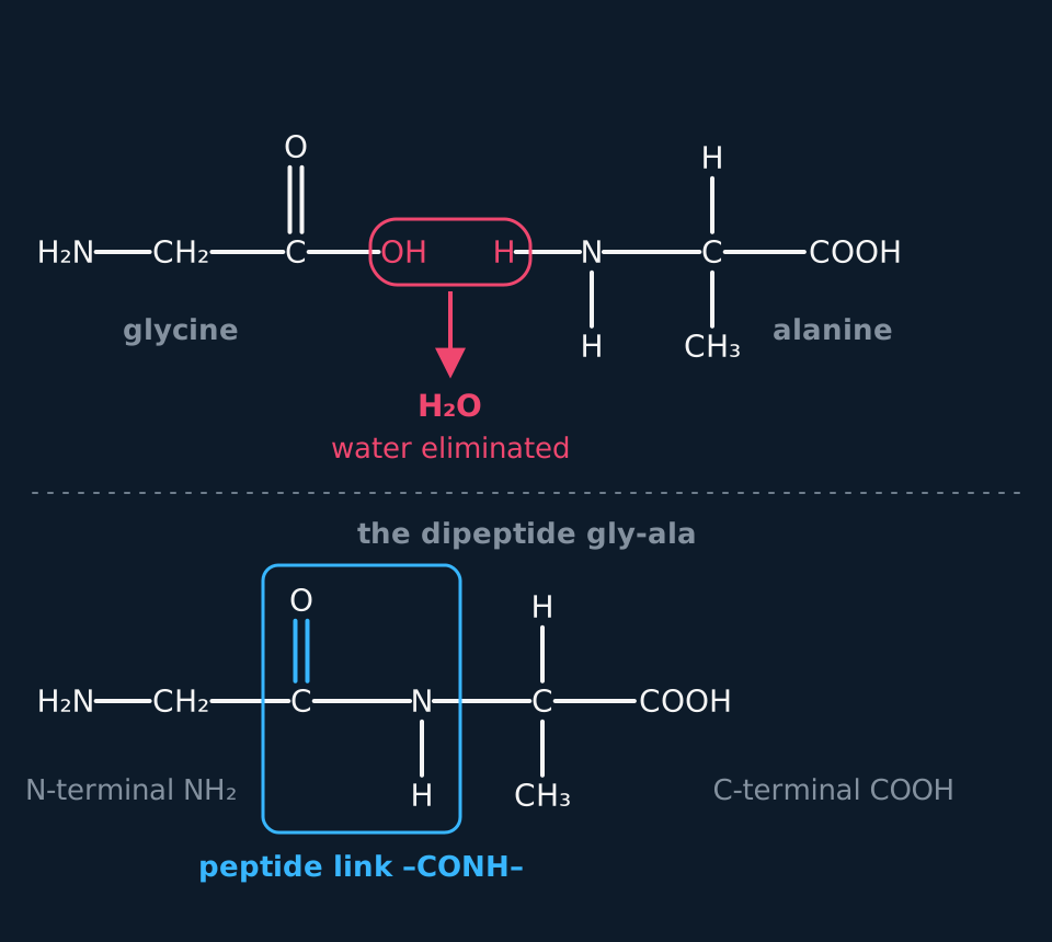
Describe the drawing
Glycine, H₂N–CH₂–C(=O)–OH, and alanine, with its amine group drawn as H–N–H and a CH₃ side chain, side by side. The OH of glycine's acid group and one H of alanine's amine group are ringed together in coral, with an arrow down to H₂O, water eliminated. Below, the dipeptide gly-ala is drawn in full, H₂N–CH₂–C(=O)–N(H)–CH(CH₃)–COOH, with the C=O and N–H group enclosed in a cyan box labelled peptide link, and the free NH₂ and COOH ends labelled N-terminal and C-terminal.
Two different amino acids can join in two ways. Glycine's COOH can react with alanine's NH₂, giving H₂NCH₂CONHCH(CH₃)COOH, or alanine's COOH can react with glycine's NH₂, giving H₂NCH(CH₃)CONHCH₂COOH. These are different compounds, gly-ala and ala-gly. A mixture of the two amino acids can also give gly-gly and ala-ala, so four dipeptides are possible in all.
A protein is a polypeptide chain of fifty or more amino acids, and its structure is described at three levels. The primary structure is the sequence of amino acids in the chain. The secondary structure is the way the chain coils into an α-helix or lies side by side in a β-pleated sheet, held in place by hydrogen bonds between the N–H of one peptide link and the C=O of another. The tertiary structure is the folding of the whole chain into a three-dimensional shape.
The tertiary structure is held by bonds between the side chains. Hydrogen bonds form between groups such as OH and NH₂. Ionic bonds form between an NH₃⁺ on one side chain and a COO⁻ on another. Disulfide bridges, covalent S–S bonds, form between the side chains of two cysteine residues. Weaker van der Waals forces hold hydrocarbon side chains together inside the fold. An answer about tertiary structure must name hydrogen bonding and the sulfur–sulfur bonds; the ionic bonds and van der Waals forces complete the picture.
| Level | What it is | What holds it |
|---|---|---|
| Primary | the sequence of amino acids | covalent peptide links |
| Secondary | α-helix or β-pleated sheet | hydrogen bonds between N–H and C=O of peptide links |
| Tertiary | the folding of the whole chain | hydrogen bonds, ionic bonds, disulfide bridges, van der Waals forces |
Heating a protein under reflux with 6 mol dm⁻³ hydrochloric acid for about a day hydrolyses every peptide link, the reverse of the condensation. In the acid the amino acids come out as their positive ions. A drop of the mixture is spotted on a thin-layer chromatography plate beside drops of known amino acids, and the plate is stood in a solvent. Amino acids are colourless, so the dried plate is sprayed with ninhydrin, which turns them purple, or viewed under ultraviolet light. Each amino acid is identified by matching its Rf value, the distance it moved divided by the distance the solvent moved, with a known one on the same plate.
TRY IT: Identifying an amino acid by TLC
After hydrolysis, one spot moved 3.6 cm while the solvent front moved 8.0 cm. Standards on the same plate gave Rf values of 0.26 for glycine, 0.45 for alanine and 0.70 for leucine. Calculate the Rf value of the spot and identify the amino acid.
Check your answer
Rf = distance moved by the spot ÷ distance moved by the solvent front = 3.6 ÷ 8.0 = 0.45.
The value matches the alanine standard run on the same plate, so the amino acid is alanine.
What hydrolysis can and cannot show
A dipeptide is hydrolysed and the mixture run on a TLC plate beside the same three standards: glycine 0.26, alanine 0.45, leucine 0.70. Two spots appear, at 2.1 cm and 5.6 cm, and the solvent front is at 8.0 cm. Identify the amino acids and give the possible structures of the dipeptide. Leucine's side chain is CH₂CH(CH₃)₂.
Show the working
Rf of the first spot = 2.1 ÷ 8.0 = 0.26, which matches glycine.
Rf of the second spot = 5.6 ÷ 8.0 = 0.70, which matches leucine.
Hydrolysis breaks the peptide link and releases both amino acids but says nothing about which one carried the free NH₂ group. The dipeptide is gly-leu, H₂NCH₂CONHCH(CH₂CH(CH₃)₂)COOH, or leu-gly, H₂NCH(CH₂CH(CH₃)₂)CONHCH₂COOH.
The amino acids are glycine and leucine, and a further method that removes one amino acid at a time from one end is needed to fix the order.
- Peptide link
- The –CONH– group that joins two amino acids, formed by a condensation reaction that eliminates water.
- Primary structure
- The sequence of amino acids in a polypeptide chain.
- Secondary structure
- The coiling of a polypeptide chain into an α-helix or its folding into a β-pleated sheet, held by hydrogen bonds between peptide links.
- Tertiary structure
- The folding of the whole polypeptide chain into a three-dimensional shape, held by hydrogen bonds, ionic bonds and disulfide bridges between side chains.
Enzymes and drugs
Enzymes are proteins that catalyse reactions in living cells. Each has an active site, a pocket in its tertiary structure lined with the side chains of a few amino acids. A substrate binds in the site by the same interactions that hold the protein's fold: hydrogen bonds between OH, NH and C=O groups, ionic attractions between charged groups such as NH₃⁺ and COO⁻, and van der Waals forces between hydrocarbon parts. Held there, the substrate forms an enzyme–substrate complex in which the bonds that must break are strained and the groups that react are brought together, so the reaction has a lower activation energy.
The simplest picture of this is the lock and key model: the active site has a shape that only its substrate fits, as only one key fits a lock, and a molecule of a different shape is not held and does not react.
The active site is chiral, because the protein is built from one enantiomer of each amino acid. A substrate is usually held at three or more points at once, each group on its chiral centre matched to a binding group in the site. The mirror image of the substrate can match at most two of those points at a time, with the third group pointing the wrong way, so it does not bind properly and is not converted. Enzymes are therefore stereospecific: the enzyme that oxidises lactic acid in muscle acts on one of its two enantiomers and leaves the other untouched.
Use 'stereospecific' and 'the active site is chiral' in an answer. 'The wrong enantiomer is the wrong shape' does not say why the site can tell two molecules with identical bonds apart.
A drug can work by blocking an active site. A molecule shaped enough like the substrate to bind in the site, with groups placed to make the same hydrogen bonds and ionic attractions, but which does not react, occupies the site and stops the substrate binding. The enzyme is inhibited. The ACE inhibitors that lower blood pressure work this way: they bind the active site of the enzyme that makes a hormone which narrows blood vessels. Because the site is chiral, a chiral drug usually has to be the right enantiomer, and the other may do nothing or something harmful.
Chemists now design such molecules by computer before any of them is made. The three-dimensional structure of the enzyme, often found by X-ray crystallography, is loaded into modelling software. Candidate molecules are fitted into the active site on screen, and each is scored on how well its shape fills the pocket and how many hydrogen bonds and ionic attractions it can make with the side chains there. Only the best-scoring candidates are synthesised and tested, which saves years of making compounds that would never have fitted. Modelling cannot show whether a molecule is absorbed, reaches the enzyme or is toxic, so laboratory and clinical testing still follow.
- Active site
- The region of an enzyme, formed by its tertiary structure, to which the substrate binds and where the reaction is catalysed.
- Stereospecific active site
- An active site that can bond to only one enantiomeric form of a substrate or drug, because the site itself is chiral.
DNA, and how cisplatin acts on it
DNA is a polymer of nucleotides. Each nucleotide has three parts: a phosphate group, a sugar called 2-deoxyribose, and a base, one of adenine, cytosine, guanine and thymine. The phosphate of one nucleotide is joined to the sugar of the next by a condensation reaction, so a single strand has a backbone of sugar and phosphate groups in turn, with a base hanging from every sugar.
Two strands wind round each other in a double helix, with the bases on the inside. The bases pair by hydrogen bonding: adenine with thymine through two hydrogen bonds, and cytosine with guanine through three. Only these pairs fit, because only they line up hydrogen bond donors opposite acceptors at the right distance. The sequence of one strand therefore fixes the sequence of the other, which is how DNA is copied when a cell divides.
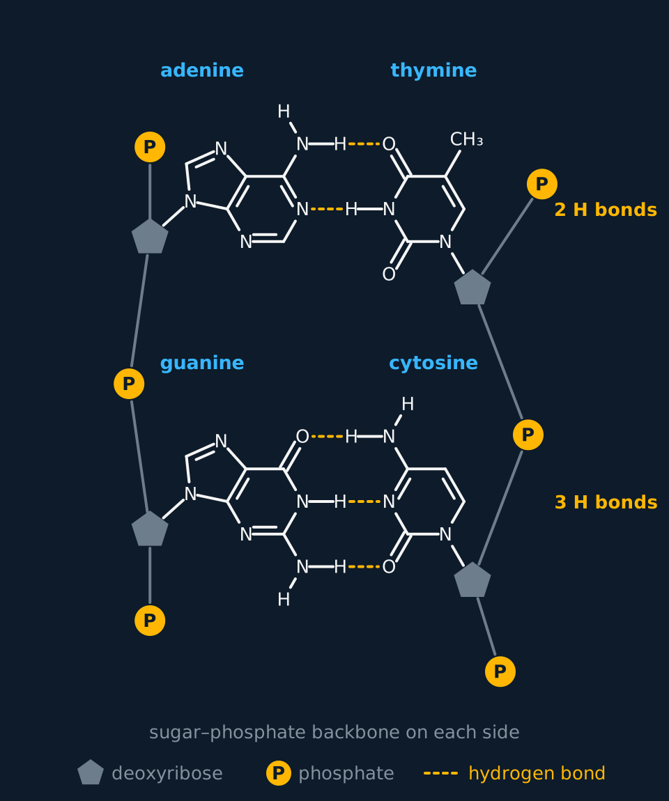
Describe the drawing
Two base pairs, one above the other, each held between a grey sugar–phosphate backbone on the left and another on the right, deoxyribose drawn as pentagons and phosphate as discs marked P. At the top adenine, a fused six- and five-membered ring, faces thymine, a six-membered ring, and two dashed hydrogen bonds join them: the N–H of adenine's NH₂ group to a C=O of thymine, and the N–H of thymine to a ring nitrogen of adenine. Below, guanine faces cytosine across three dashed hydrogen bonds: guanine's C=O to an N–H of cytosine's NH₂, guanine's ring N–H to a ring nitrogen of cytosine, and an N–H of guanine's NH₂ to cytosine's C=O. Each base is bonded through a ring nitrogen to its deoxyribose.
Cisplatin is the square planar complex cis-[Pt(NH₃)₂Cl₂]. Blood contains a high concentration of chloride ions, so the chloride ligands stay in place as the drug travels. Inside a cell the chloride concentration is far lower, and the two chloride ligands are replaced, first by water and then by nitrogen atoms on DNA. The platinum bonds to a nitrogen on each of two neighbouring guanine bases on the same strand, in a ligand substitution reaction.
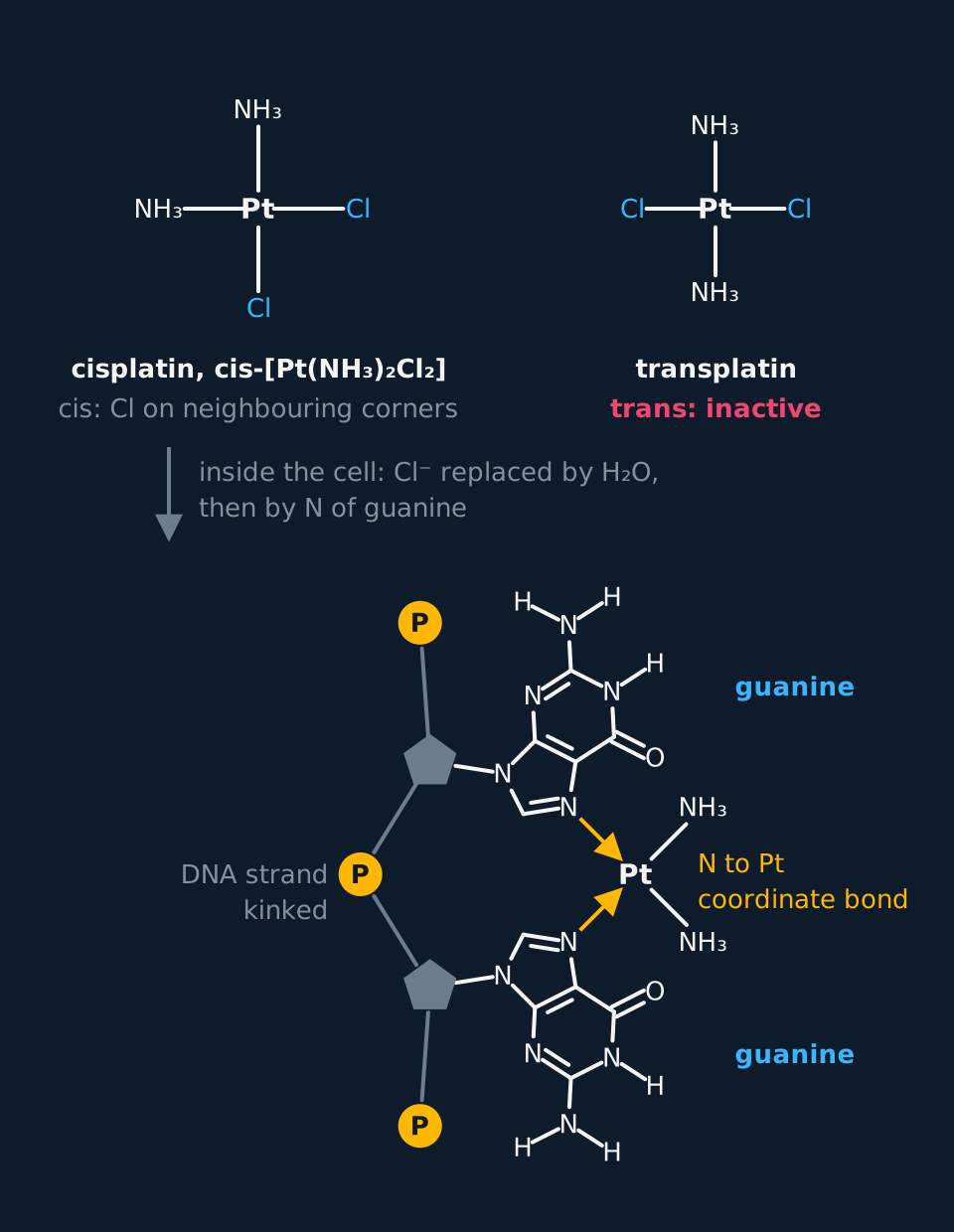
Describe the drawing
Two panels. Above, cisplatin drawn square planar with platinum at the centre, two ammonia ligands on neighbouring corners and two chloride ligands on the other two, and beside it transplatin with its chlorides on opposite corners, marked trans: inactive. An arrow notes that inside the cell the chlorides are replaced by water and then by a nitrogen of guanine. Below, a short length of one DNA strand with two neighbouring guanines, each joined to a deoxyribose on a sugar–phosphate backbone that bends sharply between them; the platinum, still carrying its two ammonia ligands, is bonded by amber coordinate bonds to nitrogen N7 of each guanine at neighbouring corners of its square.
The bound platinum kinks the DNA strand, so the strands cannot be separated and copied properly. The cell cannot replicate its DNA, cannot divide, and dies. Cancer cells divide rapidly, so they are affected most. The trans isomer has its two chlorides opposite each other and cannot reach two neighbouring guanines on one strand, so only the cis isomer is used as a drug.
Cisplatin also acts on healthy cells, worst on those that divide quickly, such as hair follicles and the lining of the gut, and it damages the kidneys. Patients lose hair and suffer sickness. The dose is kept as low as will treat the cancer, given in courses with breaks between them, and the risk of the side effects is weighed against the much greater risk of the cancer left untreated.
- Nucleotide
- The monomer of DNA, made of a phosphate group, the sugar 2-deoxyribose and one of four bases.
Other boards: electrophoresis, esters and the trans isomer
Cambridge examines a second separation of amino acids, which uses the charge on each one instead of how strongly it is held by a TLC plate. In electrophoresis a strip of paper or a slab of gel is soaked in a buffer solution of fixed pH, the mixture is spotted in the middle, and a potential difference is applied across it. An amino acid that is positive at that pH moves towards the negative electrode; one that is negative moves towards the positive electrode; one at its isoelectric point has no overall charge and stays where it was spotted.
How far each moves depends on its charge and its size. A larger charge moves faster, and a larger ion, such as a peptide, moves more slowly than a small amino acid with the same charge. Choosing the pH of the buffer sets the charges, so a pH between the isoelectric points of two amino acids sends them in opposite directions. The strip is dried and sprayed with ninhydrin, as a TLC plate is, and the same method separates the fragments of DNA in genetic fingerprinting.
| Thin-layer chromatography | Electrophoresis | |
|---|---|---|
| What separates them | how strongly each is held by the stationary phase against the solvent | overall charge at the buffer's pH, and size |
| What moves them | the solvent rising up the plate | a potential difference across the strip |
| Which way they move | all in one direction | positive ions to the negative electrode, negative ions to the positive, neutral ones not at all |
| How spots are shown | ninhydrin or ultraviolet light | ninhydrin |
TRY IT: Three amino acids at pH 6.0
Glycine (isoelectric point 6.0), aspartic acid (2.8) and lysine (9.7) are separated by electrophoresis in a buffer at pH 6.0. Say where each is found afterwards.
Check your answer
Glycine is at its isoelectric point, so it is a zwitterion with no overall charge and stays at the starting line.
The pH is above aspartic acid's isoelectric point, so its second COOH has also lost a proton; its charge is −1 and it moves towards the positive electrode.
The pH is below lysine's isoelectric point, so its second NH₂ is protonated; its charge is +1 and it moves towards the negative electrode.
- Electrophoresis
- The separation of charged species, such as amino acids in a buffer of fixed pH, by their movement towards the oppositely charged electrode when a potential difference is applied.
OCR also examines the ester of an amino acid. The carboxylic acid group keeps the reactions of its family, so glycine heated with ethanol and concentrated sulfuric acid gives ethyl aminoethanoate. In that acid mixture the amine group is protonated, so the ester forms as its salt, ⁺H₃NCH₂COOCH₂CH₃, and the free ester is released by adding alkali.
H₂NCH₂COOH(s) + CH₃CH₂OH(l) ⇌ H₂NCH₂COOCH₂CH₃(l) + H₂O(l)With concentrated sulfuric acid as the catalyst, written for the neutral molecules.
Edexcel asks why the drug is supplied as the cis isomer alone rather than as a mixture with the trans. Transplatin does not stop the cancer cells dividing, but it is still a platinum compound that cells take up and the kidneys must clear. In a mixture it would add to the side effects without adding to the treatment, and the dose of the active isomer would be uncertain. The synthesis is designed to give the cis isomer, and each batch is checked for the trans form.
In the exam
- For a species at a given pH, low pH means NH₃⁺ and COOH, high pH means NH₂ and COO⁻. The scheme does not accept a charge drawn on the wrong atom, such as a positive charge on a carbon or an H left on COO⁻.
- Each level of protein structure earns its mark with what holds it: hydrogen bonds between peptide links for secondary; named interactions between side chains, including the S–S bond, for tertiary. 'Bonds' unnamed is not credited.
- For cisplatin, the mark scheme credits the nitrogen on guanine as the binding site, the chloride ligands being replaced, and the result: DNA replication prevented, so the cell cannot divide.
- An evaluation of cisplatin needs both sides: the benefit of stopping cancer cells dividing weighed against the harm to healthy, rapidly dividing cells, and how the dose limits that harm.
- In drug design, the answer must say the drug binds the active site and blocks the substrate; 'it stops the enzyme working' restates the question and is not credited.
Check yourself
Glycine dissolves in water and decomposes above 230 °C without melting. Explain these properties, and give the formula of the species glycine forms in hydrochloric acid.
Answer
In the solid, each glycine molecule exists as a zwitterion, ⁺H₃NCH₂COO⁻, made when the COOH group donates a proton to the NH₂ group.
The zwitterions form an ionic lattice held by strong attractions between opposite charges, which need a lot of energy to overcome, and the charged groups are strongly attracted to polar water molecules.
In hydrochloric acid the COO⁻ group accepts a proton, giving ⁺H₃NCH₂COOH, a positive ion.
Questions
Question 16 marksAQA · OCR A
Evaluate the use of cisplatin, cis-[Pt(NH₃)₂Cl₂], as an anticancer drug, explaining how it stops cancer cells dividing.
Mark scheme
Indicative content
- inside the cell the chloride concentration is low, so the two chloride ligands are replaced, first by water and then by DNA
- the platinum forms coordinate bonds to a nitrogen atom on each of two neighbouring guanine bases on one strand of DNA
- this kinks the DNA so the strands cannot be separated and copied, replication is prevented and the cell cannot divide
- cancer cells divide rapidly, so they are affected most, which is the benefit that makes cisplatin an effective treatment
- it also acts on healthy cells that divide quickly, causing hair loss and sickness, and it damages the kidneys
- the harm is limited by the lowest effective dose given in courses with breaks, and is judged worth the risk against an untreated cancer
Levels
- Level 3 (5–6 marks) both the mechanism and the evaluation are complete: ligand replacement of chloride by water and then by DNA, coordinate bonds to the nitrogen atoms of two guanine bases, the prevention of replication and division, the benefit to rapidly dividing cancer cells, the harm to healthy dividing cells, and a judgement on balance
- Level 2 (3–4 marks) platinum binding to DNA bases and the halt to replication are described with a benefit and a side effect; the ligand substitution, water and then guanine replacing chloride, or the final judgement is not given
- Level 1 (1–2 marks) cisplatin is said to kill cancer cells, often by 'attacking' them or their membranes, with no binding to DNA and no harm to healthy cells weighed
Question 24 marks
Give the type of reaction and the other product when glycine, H₂NCH₂COOH, and serine, H₂NCH(CH₂OH)COOH, join to form a dipeptide, and the condensed formulae of the two dipeptides that contain one of each.
Mark scheme
- B1 a condensation reaction, forming a peptide link, –CONH–
- B1 the other product is water
- B1 H₂NCH₂CONHCH(CH₂OH)COOH, with glycine's carbonyl joined to serine's nitrogen
- B1 H₂NCH(CH₂OH)CONHCH₂COOH, with serine's carbonyl joined to glycine's nitrogen
Question 34 marksAQA
Describe the primary, secondary and tertiary structure of a protein, and the bonds that maintain the secondary and tertiary structure.
Mark scheme
- B1 the primary structure is the sequence of amino acids in the polypeptide chain
- B1 the secondary structure is an α-helix or a β-pleated sheet
- B1 the secondary structure is held by hydrogen bonds between the N–H of one peptide link and the C=O of another
- B1 the tertiary structure is the folding of the whole chain, held by hydrogen bonds, ionic bonds and disulfide bridges between side chains
All 20 questions with mark schemes, the other 17 set out in full on the question page.
Worth remembering
- Leave glycine out of any list of chiral amino acids: its central carbon carries two hydrogens.
- A zwitterion drawn in strong acid is wrong: at low pH both groups carry H, NH₃⁺ and COOH; in alkali neither does.
- Two different amino acids give two different dipeptides: write both, with the peptide link the right way round.
- Secondary structure is held by hydrogen bonds between peptide links, not between side chains.
- Cisplatin drawn bonded to a phosphate or a sugar is at the wrong site: it bonds to a nitrogen on guanine.
CHECK YOUR PROGRESS
Rate how confident you are with each objective for this lesson. Ratings are saved in this browser, on this device, unless you sign in.
- Describe the structure of an amino acid, identify its chiral centre, and draw the zwitterion and the ions it forms in acid and in alkali.
- Explain how a peptide link forms by condensation, and give the two dipeptides made from two different amino acids.
- Describe the primary, secondary and tertiary structure of a protein and the bonds that hold each in place.
- Describe how a protein is hydrolysed and its amino acids identified by thin-layer chromatography, calculate Rf values, and predict how amino acids move in electrophoresis.
- Explain how enzymes act as stereospecific catalysts and how a drug can act as an enzyme inhibitor.
- Describe the structure of DNA, and explain how cisplatin prevents DNA replication and why its use carries risks.
Open the full revision checklist to see every objective in the curriculum in one place.
Practise this lesson
WORKBOOK
The written questions from this topic, on paper with room to work, and a separate book of mark schemes. The question that is set on a diagram stays on this page, where the diagram can be drawn. Free to use; please do not redistribute or sell.