Physics › Medical physics › The ear and the transmission of sound
The ear and the transmission of sound
The ossicles act as a lever and the oval window has a fraction of the eardrum's area, so the middle ear raises pressure without adding energy. That step-up is what drives the vibration into the fluid of the cochlea. Along the basilar membrane each frequency disturbs its own region, so position codes pitch.
Choose your exam board to show the notes and questions relevant to your specification. You can change this setting at any time, and content for other boards remains available.
The physics of the ear, part 1 of 2. Part 2 is Intensity, loudness and the decibel scale.
Builds on Progressive waves and Longitudinal and transverse waves and Stationary waves in air columns.
IN THIS TOPIC
- Describe the transmission of sound through the outer, middle and inner ear.
- Explain why the middle ear raises the pressure before the sound reaches the cochlea.
COMMON MISCONCEPTION
The middle ear amplifies sound by adding energy to the wave.
The middle ear adds no energy: the ossicles are a passive lever, and the pressure rises because roughly the same force is delivered from the large eardrum to the much smaller oval window. Amplification of pressure, not of power.
Air, bone, fluid
Everything the brain will ever learn about a sound leaves the ear as nerve impulses from an organ full of fluid, and the sound itself arrives as a pressure variation in air. Getting from one to the other is a transmission problem, and the structure of the ear is the solution to it.
The ear is a chain of three sections, and each one hands the vibration to the next in a different medium. The outer ear is the visible pinna, which collects sound and funnels it into the ear canal. The canal is a tube closed at the far end by the eardrum, and like any closed pipe it resonates. Its first resonance falls near 3 kHz, which is one reason the ear is at its most sensitive there: a canal about 2.5 cm long fits a quarter of a wavelength between the open air and the drum, so it picks out λ = 4L ≈ 10 cm, and sound of that wavelength in air is a frequency of roughly 3 kHz.
At the end of the canal the eardrum vibrates with the arriving pressure variation. That is the boundary of the middle ear, an air-filled cavity spanned by three small linked bones, the ossicles, which carry the motion across to a much smaller membrane called the oval window.
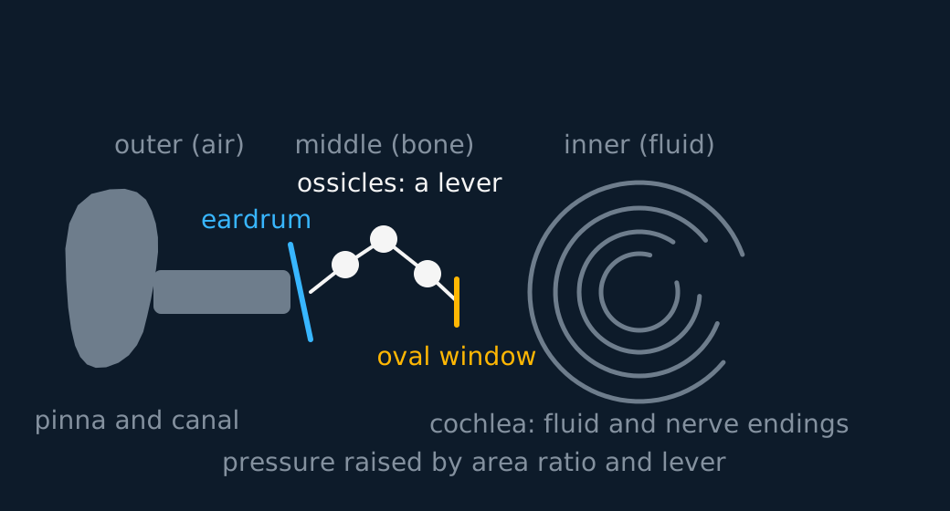
The pressure step-up in the middle ear
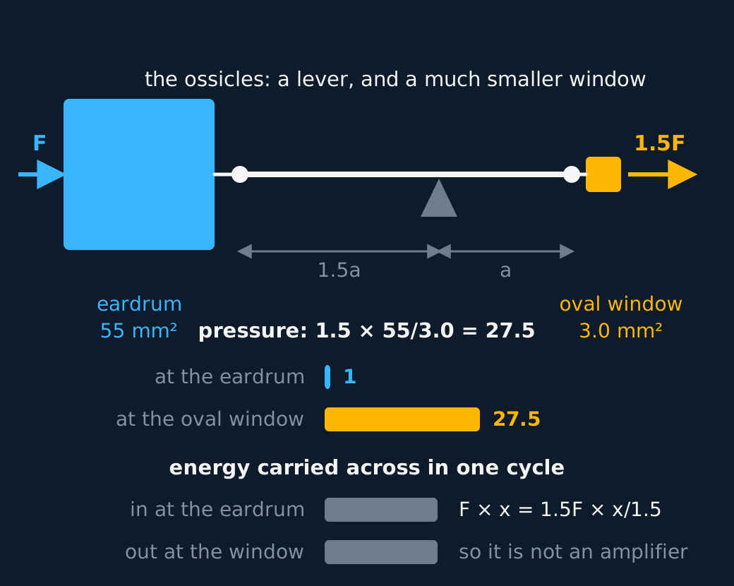
Two things happen on the way across, and both raise the pressure. The ossicles are linked as a lever, so the force at the far end is larger than the force at the near end. More importantly the eardrum has perhaps twenty times the area of the oval window, and the same force spread over a twentieth of the area is twenty times the pressure. Together they lift the pressure by a factor of the order of twenty or thirty.
That pressure increase is what the next stage needs. Beyond the oval window lies the inner ear, and the cochlea inside it is full of fluid. Sound passing straight from air to fluid would meet a huge mismatch and almost all of it would reflect, exactly as an ultrasound pulse reflects at an air gap. The middle ear is the matching stage that gets the energy across.
The mismatch has a name this option meets again. In ultrasound imaging the same problem is written as a difference in acoustic impedance, and the answer there, the coupling gel between probe and skin, does the same job the middle ear does here: it stops the wave meeting an air-to-something boundary that would send it straight back.
The middle ear does not supply energy: in the idealised account, it increases pressure at the expense of displacement amplitude and improves energy transfer from air to the fluid of the cochlea. Describe this as pressure transformation rather than as active power amplification.
Describe the drawing
The middle ear drawn from the lesson's own numbers. On the left the eardrum, on the right the oval window, the two squares drawn in the true ratio of their areas, 55 square millimetres against 3.0. Between them the ossicles are drawn as a lever whose arms are in the ratio 1.5 to 1, so the force arriving as F leaves as 1.5F, and the two arrows are drawn in that ratio too. Beneath, the pressure at each membrane as a bar: the second is 27.5 times the first, which is the lever's 1.5 multiplied by the area ratio of 55 to 3.0. Beneath that, the energy carried across in one cycle at each end, drawn as two bars of exactly the same length, because the larger force moves through a proportionately smaller distance. A large, easily moved membrane concentrating its force onto a small one raises the pressure and adds no energy at all, so the middle ear steps up pressure and is not an amplifier.
WORKED EXAMPLE
The pressure gain, from the geometry
An eardrum has an area of 55 mm2 and the oval window an area of 3.0 mm2. The lever action of the ossicles multiplies the force by 1.5. Estimate the factor by which the pressure of the vibration is raised across the middle ear.
The sound exerts a force F on the eardrum, and the ossicles deliver 1.5F to the oval window.
Pressure is force over area, so the gain is the force factor times the area ratio: 1.5 × 55/3.0 = 28.
The area ratio contributes most of the factor and the lever the rest. Both are geometrical: no energy is added, and the same calculation carried out on the energy gives no gain.
Inside the cochlea
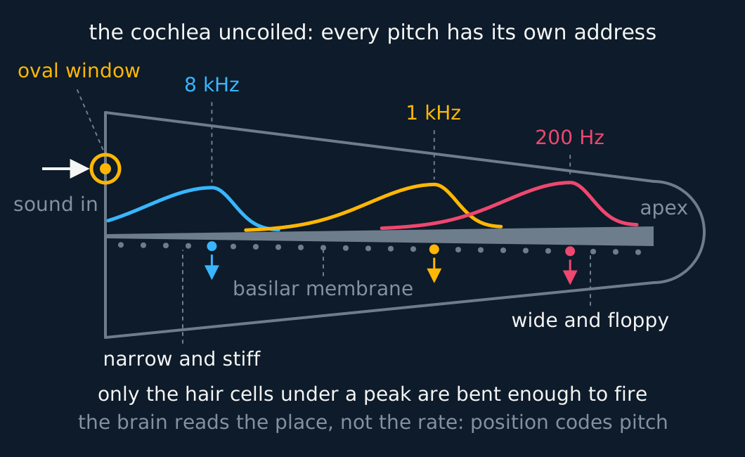
Once through the oval window, the vibration travels in the cochlea's fluid and along the basilar membrane that runs the length of its coil. The membrane is not uniform: it is narrow and stiff near the oval window and becomes wider and floppier towards the far end, so each frequency disturbs its own region of the membrane most, high frequencies peaking near the entrance and low frequencies far along.
Describe the drawing
The cochlea drawn uncoiled as a channel tapering from the oval window at the left to the apex at the right, with the basilar membrane running along it as a wedge, narrow at the window and wide at the apex. Three computed response envelopes sit on the membrane: an 8 kHz tone peaking close to the entrance, a 1 kHz tone near the middle and a 200 Hz tone far along by the apex. A row of hair cells lines the membrane, and at each peak the cell beneath is enlarged with an impulse arrow leaving it for the brain: pitch is read off from which place fired, not from any arithmetic on the wave.
Hair cells sit along the membrane, and the ones at the disturbed region are bent by the motion and fire nerve impulses to the brain. Position along the cochlea is therefore how the ear codes pitch: the brain does no frequency arithmetic; it reads an address. A pure tone excites one small stretch of membrane, a chord excites several at once, and the nerve reports which stretches were busy.
That completes the chain the specification asks for, air to bone to fluid to nerve: pinna, canal, eardrum, ossicles, oval window, cochlea, hair cells, brain.
Intensity, and why the scale is logarithmic has moved to the next part of this lesson.
Equal loudness curves has moved to the next part of this lesson.
What damage does to the curves has moved to the next part of this lesson.
ASSESSMENT FOCUS
- Give the transmission chain in order, with the medium at each stage. Pinna and canal in air, eardrum, three ossicles across the middle ear, oval window, fluid in the cochlea, hair cells, nerve impulses.
- Give both reasons for the pressure gain. The ossicles act as a lever, and the eardrum's area is far larger than the oval window's, so the same force acts over a much smaller area.
- Say what the pressure gain is for. The cochlea is fluid-filled, and without the middle ear almost all the sound would reflect at the air-to-fluid boundary instead of entering.
- The gain is a pressure gain, not an energy gain. Nothing in the middle ear adds energy to the wave, so describe it as pressure transformation rather than as active power amplification.
- Pitch answers turn on position. Each frequency disturbs its own region of the basilar membrane most, and the brain reads which hair cells fired, so position along the cochlea codes the pitch.
CHECK YOURSELF
The eardrum of a particular ear has an area of 60 mm2 and its oval window an area of 3.2 mm2, and the ossicles multiply the force by 1.3. Calculate the factor by which the pressure is raised across the middle ear, and explain why the increase is necessary.
Show a hint
Pressure is force per area: the lever multiplies the force, and the smaller window divides the area.
Show the answer
The area ratio alone raises the pressure by 60/3.2 = 19 times, because the ossicles deliver the eardrum's force to a much smaller area.
The lever multiplies the force by 1.3 on the way across, so the overall gain is 1.3 × 60/3.2 = 24.
The cochlea beyond the oval window is filled with fluid, and sound arriving from air at a fluid boundary would almost all reflect. The pressure step-up is what drives the vibration into the fluid, so without it almost none of the sound's energy would enter the inner ear.
Sound crosses the ear in order: pinna, canal, eardrum, ossicles, oval window, cochlea, hair cells, nerve, passing from air to bone to fluid.
The middle ear raises the pressure by a lever and an area ratio, so the sound can get into the fluid of the cochlea.
Position along the cochlea codes pitch: each frequency disturbs its own stretch of the basilar membrane most.
WORKBOOK
Printable practice for this topic: original exam-style questions with room to work, and a fully worked answer book. Free to use; please do not redistribute or sell.
Or read them with their mark schemes on the ear and the transmission of sound questions page.
CHECK YOUR PROGRESS
Rate how confident you feel with each objective for this lesson. Ratings are saved in this browser, on this device, unless you sign in.
- Describe the transmission of sound through the outer, middle and inner ear.
- Explain why the middle ear raises the pressure before the sound reaches the cochlea.
Open the full revision checklist to track your progress across the whole unit.